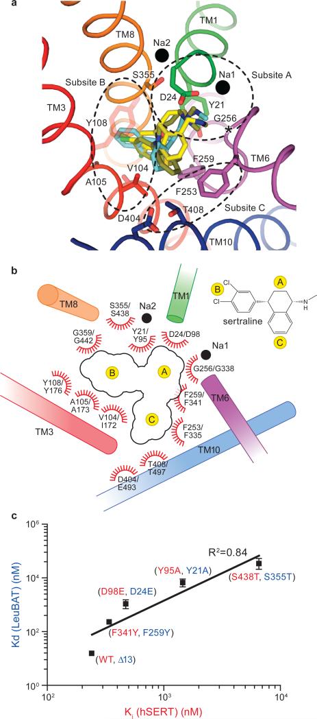
Fig 4. Implication for drug binding in hSERT and validation by mutational studies.
(a) Superposition of SSRI (sertraline, yellow), SNRI (duloxetine, cyan) and TCA (CMI, olive) in the primary binding pocket of LeuBAT, viewed from extracellular side. The key residues in the pockets and the two sodium ions are shown as sticks and black spheres, respectively. The regions enclosed by dashed lines define subsites A, B and C in the primary drug binding pocket. (b) Schematic representation of drug interactions in the primary binding pockets of LeuBAT/hSERT. The transmembrane helices are shown as cylinders. Residue numbering follows LeuBAT and hSERT, respectively. (c) Plot of sertraline binding constants for Y21A, D24E, F259Y and S355T mutants of the Δ13 LeuBAT against the inhibition constants for the corresponding mutants Y95A, D98E, F341Y and S438T in hSERT, respectively. Error bars, s.e.m, n = 3

