DNA strand breaks are a prerequisite for many cancer-associated genomic alterations, including amplifications, deletions, inversions, and structural rearrangements. High-throughput technologies that measure genome-wide DNA quantities, such as DNA sequencing and single nucleotide polymorphism or comparative genomic hybridization arrays, enable researchers to accurately detect such lesions.1
Recently we reported a computational analysis of intragenic DNA breakpoints in glioblastoma (GBM), the most common adult tumor in the central nervous system.2 Using a cohort of more than 500 samples, we identified an expected increase in DNA strand breaks near genes commonly targeted by copy number gain and losses in GBM, such as EGFR on 7p, CDKN2A/B on 9q, and PTEN on 10q. Less anticipated was the enrichment of DNA breakpoints on an 18 megabase region encompassing cytoband 12q14–15.
The 12q region, which we referred to as 12q14–15 breakpoint enriched region or BER, contains the well-known oncogenes murine double minute 2 (MDM2) and cyclin-dependent kinase 4 (CDK4), which are frequently amplified in GBM (7.6% and 14%, respectively).3 Detected in 24 of 493 (4.9%) GBMs, all BER samples showed a dense pattern of interspersed local copy number gains and losses. Among these small “copy number islands”, CDK4 and MDM2 were almost always amplified simultaneously (87.5% of BERs). Interestingly, each DNA gain appeared at similar dosage levels, suggesting that they were conjunctly amplified. Using matched RNA sequencing (RNAseq) data we observed direct gene fusions among the BER-related amplicons in 7 of 9 RNAseq available BER cases. This finding raises the question whether the frequent breaks in this relatively small region represent a mechanism aimed at simultaneous amplification of the CDK4 and MDM2 oncogenes. In our data set, the 21 of 33 (64%) of GBM with dual amplification of CDK4 and MDM2 also displayed a BER pattern, and this percentage may represent a lower boundary due to a stringent statistical threshold.
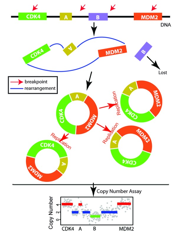
Based on the pattern of revolving copy number gains and corroborating transcript fusions, we propose a hypothetical model that involves double minutes (Fig. 1), an extrachromosomal circular structure formed by DNA end-to-end joining.4 Using whole-genome sequencing data, more than 20 12q14–15 segments in a BER were shown to be connected and suggestive of a double minute-like structure.3 Double minutes have been reported as a means to amplify the epithelial growth factor receptor gene (EGFR) in gliomas.5 We speculate that glioma cells can similarly leverage this structure to jointly amplify CDK4 and MDM2. An unresolved issue is the mechanism by which this short DNA region is fragmented prior to the segments being joined.

Figure 1. A double minute model to explain the 12q14–15 breakpoint enriched region (BER).
Our survey of intragenic breakpoints identified a second unanticipated outlier of breakpoint enrichment in chromosome 1p. In this instance, the increased breakage frequency was related to the gene FAS1-associated factor 1 (FAF1). Intragenic breakpoints in FAF1, which we found to lead to mRNA depletion, is an epiphenomenon of the focal deletion of the 5′ adjacent tumor suppressor gene CDKN2C. However, transfection experiments demonstrated a proapoptotic role for FAF1 in cell culture and tumor-sphere formation. Our results suggest that converging breakpoints in one gene may provide increased cellular fitness, and thereby lead to proliferative advantages.
In summary, our analysis revealed DNA breakpoints as a fingerprint of localized genome instability. In particular, we found a focal shattering pattern on 12q that was associated with adverse outcomes in a subset of glioblastoma patients.2 Related to the pattern, we reported orchestrated amplification of CDK4 and MDM2. Further studies are required to determine that the BER pattern relates to a synergistic interaction between the 2 oncogenes.
Zheng S, et al. Genes Dev. 2013;27:1462–72. doi: 10.1101/gad.213686.113.
Footnotes
Previously published online: www.landesbioscience.com/journals/cc/article/26874
References
- 1.Chin L, et al. Genes Dev. 2011;25:534–55. doi: 10.1101/gad.2017311. [DOI] [PMC free article] [PubMed] [Google Scholar]
- 2.Zheng S, et al. Genes Dev. 2013;27:1462–72. doi: 10.1101/gad.213686.113. [DOI] [PMC free article] [PubMed] [Google Scholar]
- 3.Brennan CW, et al. TCGA Research Network Cell. 2013;155:462–77. doi: 10.1016/j.cell.2013.09.034. [DOI] [PMC free article] [PubMed] [Google Scholar]
- 4.Storlazzi CT, et al. Genome Res. 2010;20:1198–206. doi: 10.1101/gr.106252.110. [DOI] [PMC free article] [PubMed] [Google Scholar]
- 5.Vogt N, et al. Proc Natl Acad Sci U S A. 2004;101:11368–73. doi: 10.1073/pnas.0402979101. [DOI] [PMC free article] [PubMed] [Google Scholar]


