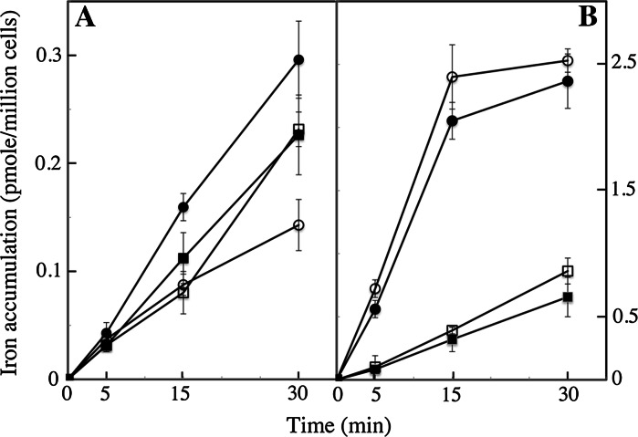
Fig. 5.
Copper-dependence of iron uptake. O. tauri (a) and E. huxleyi (b) cells were grown for 3 d under a 12:12 light–dark regime in Mf medium containing 0.1 μM ferric citrate and either 0.1 μM CuSO4 (closed symbols) or 0.1 mM of the copper-chelating agent, bathocuproin sulfonate (open symbols). Cells were harvested 2 h after dawn, washed once with iron-free and copper-free Mf medium, and tested for iron uptake from 1 μM ferrous ascorbate (circles) or 1 μM ferric citrate (squares) in microtiter plates (see Sect. 2). Mean±SE from three experiments

