Abstract
Two taxa, Hericium yumthangense (Russulales, Agaricomycotina) and Mycoleptodonoides sharmae (Polyporales, Agaricomycotina) are described as new to science from the Shingba Rhododendron sanctuary located in the northern district of Sikkim, India. Macro- and micromorphological characters are described and illustrated for both species, which are compared with allied taxa. ITS rDNA sequences supported H. yumthangense as a rather isolated species within Hericium, the species complexes of which were not resolved due to low interspecific sequence divergence. In the case of M. sharmae, 28S rDNA (D1/D2) data rendered this poorly known genus among well-known taxa of the core-polyporoid clade.
Keywords: Abies, Hericiaceae, Himalaya, Meruliaceae, Rhododendron, Sikkim, taxonomy
INTRODUCTION
Covering an area of 43 sq. km, the Shingba Rhododendron sanctuary is located in the Yumthang valley of the northern district of Sikkim, India, in eastern Himalaya, surrounded by China, Bhutan, and Nepal. It is characterized by temperate silver fir and Rhododendron forest and is a native habitat for Rhododendron niveum. Apart from the trees like Abies densa, Larix griffithiana, Picea spinulosa, Magnolia globosa, M. campbellii, Acer pectinatum, Betula utilis, and more than 40 species of Rhododendron, this natural area is also rich in rare herbaceous flowering plants from various genera, including Primula, Potentilla, Ranunculus, Euphorbia, Roscoea, Heracleum, Rubus, and Aconitum.
During a macrofungal survey of the Shingba Rhododendron sanctuary and adjacent areas by K.D. between August and September 2011, various saprotrophic wood-decaying and mycorrhizal macromycetes were collected. Macro- and micromorphological studies revealed some of them were new taxa including two hydnoid fungi. These are described here as Hericium yumthangense sp. nov. (Hericiaceae) and Mycoleptodonoides sharmae sp. nov. (Meruliaceae). Macro-, micro- and ultrastructural illustrations, together with detailed descriptions and standard reference rDNA (ITS, partial 28S), and mtSSU DNA barcodes are provided.
MATERIALS AND METHODS
Morphological studies
Macromorphological characters were observed and recorded from fresh basidiomata in the field. Colour codes and terms follow the chart in the Flora of British Fungi (Anon. 1969; “a” in the descriptions), except that those of spore prints follows Kränzlin (2005; “b” in the descriptions). Field photographs were taken with a Nikon D300s camera.
Micromorphological features were studied from dried samples mounted in a mixture of 5% KOH, 1% phloxin, Congo red and 30% glycerol, and Melzer’s reagent. Drawings were made with a drawing tube (attached to an Olympus CX41 microscope) at 1000×. Basidium length excludes sterigmata-length, and spore-dimensions exclude the dimension of the ornamentations (if any). Measurements are based on 20 examples. Basidiospores are measured in side view and given as KDa-KDc-KDb × KDx-KDz-KDy in which KDa is the minimum value for the length, KDb the maximum value for the length, KDc the mean value for the length, and KDx refers the minimum value for the width, KDy the maximum value for the width, and KDz the mean value for the width. The quotient of spore indicates the length-width ratio (Q = L/W) and is shown as Qa-Qc-Qb, where Qa refers the minimum quotient value, Qb the maximum quotient value amongst the measured collections, and Qc the mean quotient value.
Scanning electron micrographs of basidiospores were obtained from dry spore prints directly mounted on double-sided adhesive tape, pasted on a metallic specimen-stub, gold coated, and subsequently scanned in high vacuum mode at different magnifications to observe spore-ornamentations. The electron microscopy was carried out with a FEI’s Quanta 200 model scanning electron microscope (SEM) at the S.N. Bose National Centre for Basic Sciences, Kolkata (India).
DNA isolation, PCR, and sequencing
Total genomic DNA was extracted from herbarium specimens using the Jetquick general DNA clean up kit (Genomed), following the given protocols. The PCR for amplification of the ITS1-5.8S-ITS2 nuclear ribosomal DNA operon using ITS5/ITS1 and ITS4 was performed under standard conditions (White et al. 1990, Stielow et al. 2010). PCR conditions for amplifying the partial mtSSU and 28S rDNA using the standard primers MS1, MS2, LR0R and LR5 only differed in their annealing temperature (55 °C instead of 60 °C) and increased cycle extension time (90s/cycle). PCR products were directly purified using FastAP thermosensitive alkaline phosphatase and shrimp alkaline phosphatase (Thermo Scientific). The cycle-sequencing reaction was set up using ABI big dye terminator v. 3.1, using a quarter of the suggested volumes (manufacturer’s protocol), followed by bidirectional sequencing with a laboratory capillary electrophoresis system (Lifetechnologies, 3730XL DNA analyzer). Sequences were stored and manually corrected for sequencing artifacts and forward and reverse sequences assembled using the Biolomics database (www.bio-aware.com). Sequences were deposited at NCBI Genbank under the accession numbers provided in the section “Taxonomy”.
Phylogenetic inference
A search for respective Hericium ITS sequences yielded >100 sequences, which were downloaded from the international nucleotide sequence database collaboration (INSDC) database and examined regarding sufficient length, possible alignment ambiguities, and the resulting phylogenies. Since almost no sequence data is yet available for the genus Mycoleptodonoides (INSDC contained a single ITS sequence at the time of analysis), and its generic affiliation was unclear, partial 28S (D1/D2) sequences with high similarity to the query were retrieved via a BLAST search, manually filtered in respect to the quality criteria described above, and the dataset was further supplemented with sequences chosen according to Binder et al. (2005). Sequences for both datasets were subsequently aligned, using the EMBL-EBI MAFFT server (http://www.ebi.ac.uk/Tools/msa/mafft/) in standard, default setup. As previously described (Guevara-Guerrero et al. 2011), phylogenies inferred under the maximum-likelihood (ML) criterion (Felsenstein 1981) were conducted with RAxML v. 7.2.7, with search for the best tree under the GTR-MIX approach (Stamatakis et al. 2008). Maximum-likelihood trees for the Hericium dataset were rooted with Laxitextum bicolor, chosen as outgroup according to Larsson et al. (2004). The phlebioid outgroup (Phlebia spp.) for the Mycoleptodonoides dataset and clade names were depicted according to Binder et al. (2005). For depicting the trees, clades comprising at least twenty sequences were collapsed if they were either taxonomically homogeneous or contained only environmental samples. Sequence alignments are included in the online supplementary material.
RESULTS
Phylogenetic inference
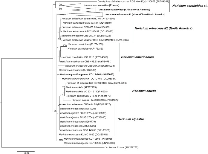
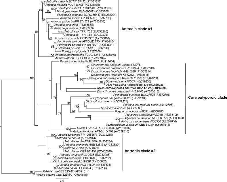
The final ITS rDNA alignment of the Hericium dataset contained 100 sequences and 633 positions, for which 276 were considered ambiguously aligned and therefore excluded from the primary alignment. The optimal final ML tree had a log-likelihood of −2687.19 and is shown in Fig. 1. The 28S (D1/D2) rDNA alignment of the Mycoleptodonoides dataset contained 53 sequences and had a total length of 971 positions. The final ML tree is given in Fig. 2; optimal log-likelihood was −4094.56; both alignments are available as supplementary material.
Fig. 1.
Phylogenetic tree inferred under the maximum-likelihood (ML) criterion from the ITS rDNA alignment of the Hericium dataset. Numbers on the branches represent support values from 1000 bootstrap replicates if at least 70%, with branches scaled in terms of the expected number of substitutions per sites. Leaf names are from their original GenBank annotations.
Fig. 2.
Phylogenetic tree inferred under the maximum-likelihood (ML) criterion from the LSU (28S)rDNA alignment of the Mycoleptodonoides dataset. Numbers on the branches represent support values from 1000 bootstrap replicates if at least 70%, with branches scaled in terms of the expected number of substitutions per sites. Terminal names are from their original GenBank annotations.
The monophyletic Hericium ingroup was separated into two subtrees. The basal branch comprising the core-subtree with H. erinaceus, H. americanum, H. alpestre, and H. abietis, received strong bootstrap branch support (BS) (86%), while the H. coralloides subtree was not supported by BS. Internal branches for the two “European” and “Asian/North American” H. coralloides clades were moderately (79%) to strongly supported (91%) respectively, however C. cirrhatus (EU78426) was resolved as a rather isolated taxon close to H. coralloides s.l. in our analysis. Several subclades (like e.g. H. alpestre, H. erinaceus, H. abietis) were not fully resolved and GenBank records inconsistently annotated. The specimen KD-11-146, representing the novel Hericium species, was neither close (with respect to low interspecific sequence variation) to the previously described Indian species (Das et al. 2011), H. bharengense, nor to one of the other subclades, and formed an isolated branch of its own.
For the Mycoleptodonoides dataset, the basal branch separating Antrodia clade 1 from Antrodia clade 2 and the core polyporoid clade received very strong support (100%). Besides the polyphyly of Antrodia among Fomitopsis species in clade 1 (82% BS), the branch inducing the split to clade 2 and the core polyporoid clade was not supported. Deeper internal branches within the three core clades received moderate to strong BS (70–100%). However, sequence annotations, therefore the original specimen identification, were moderately conflicting based on the publicly available 28S data. The hydnoid genus Mycoleptodonoides was nested, with respect to available sequences in the INSDC database, within the core polyporoid clade (Binder et al. 2005) close to the genera Diplomitoporus and Obba. The basal branch separating the subtrees, comprising Diplomitoporus, Obba, Gelatoporia and Mycoleptodonoides from several core polyporoid taxa (i.e. Polyporus, Ganoderma), received moderate support (78%), yet the isolation to Antrodia 2 was not resolved in our analysis (no BS).
Additional DNA barcodes, the ITS, partial LSU, and partial mtSSU sequences (if not used as described above) are deposited at the INSDC database and provide high quality sequence identifiers for the respective taxa. Sequences of the amplified loci were used in respect to availability of sufficient reference data only, i.e. the ITS for Hericium, and partial LSU data for positioning Mycoleptodonoides among the resupinate taxa, since its generic affiliation was unknown prior to sequencing. The insufficient quantity of mtSSU barcodes, currently available at the INSDC database, does not reach the same level of taxon coverage as when using partial D1/D2 alone. For the Mycoleptodonoides specimen it should be noted, that ITS sequence identity to Mycoleptodonoides aitchisonii HMJAU4527 (JF430078.1) was 98%, identity (588/602) bp, and gaps (0/602) bp.
TAXONOMY
Hericium yumthangense K. Das, Stalpers & Stielow sp. nov.
MycoBank MB800641
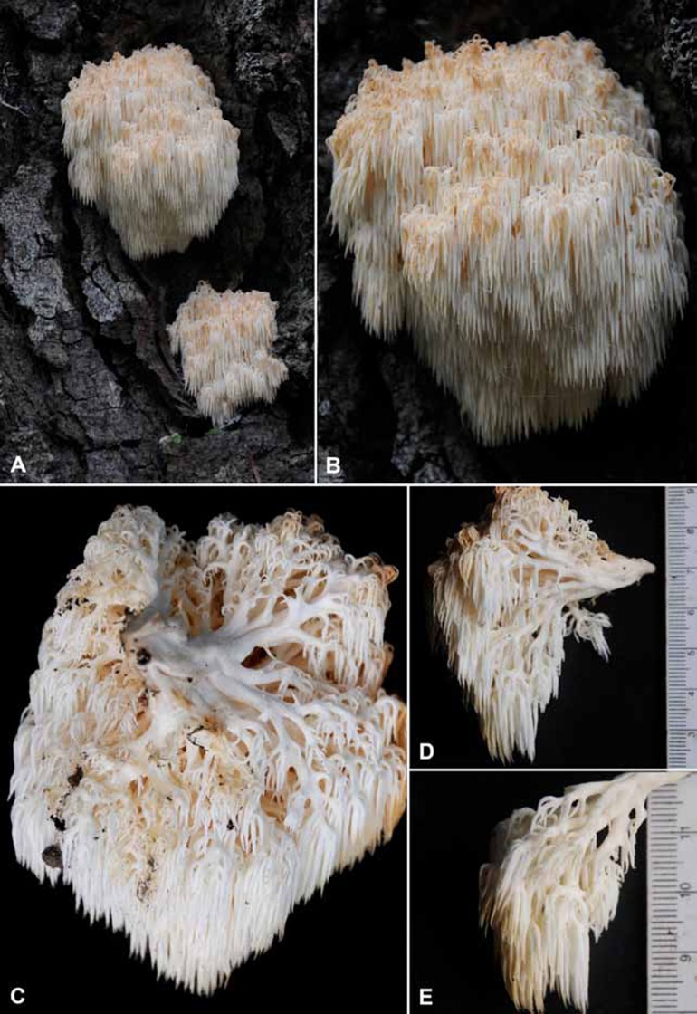
Fig. 3.
Hericium yumthangense (KD-11-146). A-B. Intricately branched basidiomata. C. Basidiomata showing the stipe-like base and branches. D. Primary branch bearing secondary, tertiary and quaternary branches and spines. E. Secondary branch bearing tertiary and quaternary branches and spines. Distances between two bars = 1 cm.
Fig. 5.
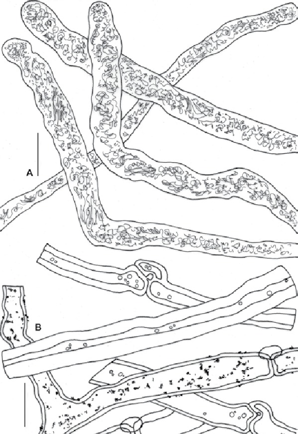
Hericium yumthangense (KD-11-146). A. Gloeoplerous hyphae. B. Contextual hyphae. Bars = 10 μm.
Fig. 9.
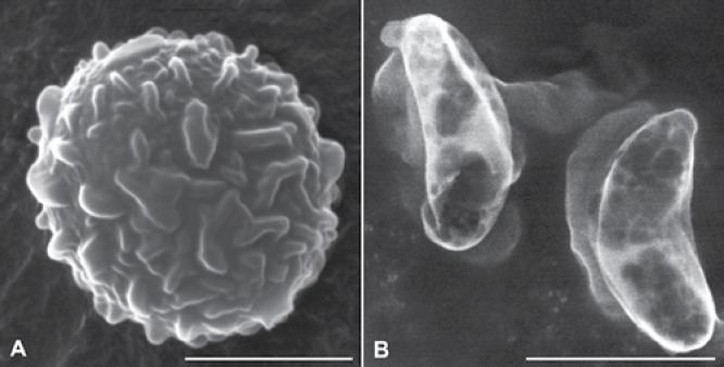
SEM micrographs of basidiospores. A. Hericium yumthangense B. Mycoleptodonoides sharmae. Bars = (A) 2 μm, and (B) 4 μm.
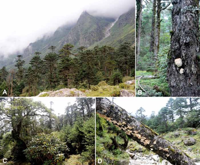
Fig. 10.
Habitat. A. Forested area dominated by Abies densa at Yumthang; collection site of Hericium yumthangense. B. Trunk of A. densa with basidiomata of H. yumthangense. C. Subalpine mixed broad-leaved and coniferous forest; collection site of Mycoleptodonoides sharmae. D. Decaying tree-trunk with basidiomata of M. sharmae.
Etymology: Named after Yumthang, the type locality.
Diagnosis: Basidiomata 70–100 × 50–80 mm, intricately branched with primary, secondary, tertiary and quaternary branches arising from a stipe-like rooting base. Spines 7–13 mm long, distributed irregularly throughout the branches. Spores 5.0–5.3–6.5 × 4.0–4.6–5.5 μm, subglobose to broadly ellipsoid, ornamentations are of broad ridges and larger isolated warts. Hyphal system monomitic, some contextual hyphae encrusted. Gloeocystidia to 8 μm wide, capitate to subcapitate or moniliform.
Type: India: Sikkim: Yumthang, alt. 3619 m, N 27°47′43.4″ E 88°42′20.3″, 30 Aug. 2011, K. Das KD-11-146 (BSHC – holotype; CBS herbarium – isotype); GenBank ITS (JX855033), LSU (JX855034), and mtSSU (JX855035).
Description: Basidiomata 70–100 × 50–80 mm, pendent, consisting mostly of five primary branches arising from a distinct rooting stipe-like base, being attached to the living host (Abies densa). Primary branches to 13 mm wide, ramified into progressively thinner (to 6 mm wide) fertile secondary branches that bear thinner fertile tertiary branches followed by thinnest fertile quaternary branches ending with clusters of 3–4 spines, white to pale yellow (a: 6F) or yellowish orange. Spines 7–13 mm long, moderately to densely distributed irregularly from all surface of secondary, tertiary and quaternary branches, pendent, concolorous with the branches when fresh, darker, yellow (a: 6F) to pale apricot to sienna (a: 11) or cinnamon (a: 10) when dry. Context yellowish white, unchanging when bruised or slowly becoming darker, but becoming straw (a: 50) with KOH, pale yellow (a: 4D–5E) with FeSO4. Odour pleasant. Taste mild or slightly bitter. Spore print white (b: 0 Y). Hyphal system monomitic; contextual hyphae generative, 4.5–11.5 μm wide, thick-walled (wall up to 3.5 μm thick), with frequent branching and conspicuous clamps of variable diam., some with incrustations; hymenial tramal hyphae 3–14 μm wide, colourless in KOH, comparatively thin-walled (wall to 2 μm thick), branched, with clamps of variable diam; gloeoplerous hyphae 4.5–8 μm wide, abundant with dense yellowish contents, apex slightly tapering or rounded sometimes moniliform, septa not found. Basidiospores 5.0–5.3–6.5 × 4.0–4.6–5.5 μm, subglobose to broadly ellipsoid (Q = 1.09–1.16–1.24), hyaline, amyloid, under light microscope almost smooth to slightly roughened with eccentric apiculus, composed of broad ridges and isolated warts (under SEM), ornamentations up to 0.4 μm high, blunt. Basidia 28–35 × 5–6 μm, clavate to subclavate, with basal clamps, 4-spored, sterigmata 4–5 × 1–1.5 μm. Gloeocystidia 5–8 μm wide, capitate to subcapitate or moniliform, emergent to 15 μm, content dense. Hymenium and subhymenium inamyloid, hyphae of the subhymenium thin-walled, to 5 μm wide.
Additional specimen examined: India: Sikkim-Yumthang, alt. 3644 m asl, N 27°47′54.8″ E 88°42′16.1″, on Abies densa in subalpine mixed forest, 30 Aug. 2011, K. Das KD-11-151 (BSHC).
Notes: Hericium yumthangense is characterized by the stipe-like small rooting base, intricate three tier branching system, 8–13 mm long spines, basidiospores with isolated warts and ridges, and the encrusted contextual hyphae. It can be distinguished from the other four known species of Hericium (H. bharengense, H. erinaceus, H. coralloides, and H. cirrhatum) reported from India, by the presence of encrusted hyphae in the context. Moreover, H. bharengense has a long distinctly narrow rooting base and spores with comparatively low ornamentation (0.2 μm high) giving a convoluted to brain-like appearance (Das et al. 2011). Hericium erinaceus is either unbranched, cushion-like or sparsely branched, bears long spines (to 5 cm long) and has larger basidiospores 4.0–7.0 × 4.0–5.5 μm (Thind & Khara 1975). Hericium coralloides has shorter spines (3–5 mm long; Thind & Khara 1975). While in H. cirrhatum, the basidioma consists of a regular cluster of pileoli united at the base and the spores are smooth (Das & Sharma 2010).
Hericium yumthangense appears somewhat close to a few other species, such as H. americanum and H. abietis. However, H. americanum bears spines only as terminal clusters of ultimate branches; the spines in H. yumthangense are distributed throughout the surfaces of secondary, tertiary, and quaternary branches. Furthermore, the spores are distinctly larger (5.5–7 μm diam) in H. americanum (Ginns 1984). The basidiomata of H. abietis are larger (750 × 250 mm), and without any stipe (represented by large solid tubercles) (Harrison 1973).
Mycoleptodonoides sharmae K. Das, Stalpers & Stielow sp. nov.
MycoBank MB800642
Fig. 6.
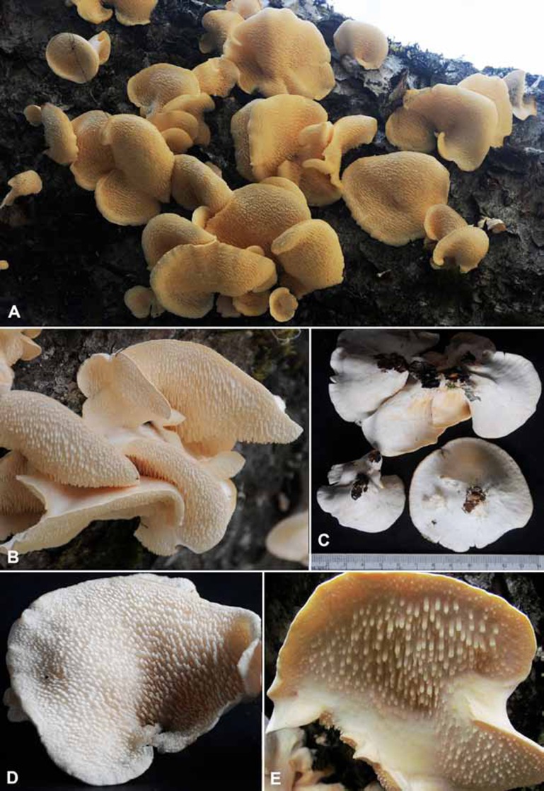
Mycoleptodonoides sharmae (KD-11-122). A. Single to concrescent basidiomata growing on decaying tree-trunk. B. Concrescent basidiomata. C. Dorsal view of single and concrescent basidiomata showing the rudimentary base, D–E. Ventral view of basidiomata showing spines.
Fig. 8.
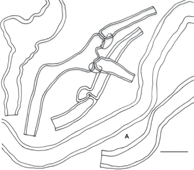
Mycoleptodonoides sharmae (KD-11-122), hyphae of pileus context. Bar = 10 μm.
Etymology: In recognition of Jai Ram Sharma for his contribution to the Indian mycobiota.
Diagnosis: Basidiomata 50–130 mm long, complex, pileate, pileus solitary to concrescent, single pileus 50–90 mm long (diam), fanshaped. Stipe absent or rudimentary. Spines to 6 mm long and 1.5 mm wide, present on the ventral surface of pileus, subulate to flattened. Spores 5.0–6.0–7.5 × 1.5–1.8–2.0 μm, cylindric to suballantoid or allantoid, smooth, inamyloid. Gloeocystidia-like hyphal elements to 5 μm wide, cylindrical, present at the tip of the spine.
Type: India: Sikkim: near Yumthang, 3522 m asl, N 27°46′42.4″ E 88°42′47.4″, on the decaying tree-trunk of a unidentified broad-leaf tree, subalpine mixed forest, 28 Aug. 2011, K. Das, KD-11-122 (BSHC – holotype; CBS herbarium – isotype); GenBank, ITS (JX855031), LSU (JX855030), and mtSSU (JX855032).
Description: Basidiomata to 130 mm long, complex, pileate, consisting of imbricate pilei arising from a rudimentary central base. Pileus single to concrescent, 50–90 mm diam, fan-shaped, convex, depressed towards the place of attachment, glabrous, fissured near the margin; margin regular to lobed, upturned with maturity, dorsal surface somewhat ribbed or veined but smooth towards margin, white when young and fresh, slowly yellowish with darker (a: 6F) margin; turning light yellow (a: 5E) with patches of yellow (a: 6F) and ochraceous (a: 9H) or saffron (a: 49) after drying, ventral surface spiny, light yellow (a: 5E) when young, becoming more distinctly yellow after drying with sienna (a: 11) at margin. Stipe absent or represented by the narrowest part of the pileus giving a rudimentary (never effused) base that attaches to the substrate; attachment never central. Spines to 6 mm long and 1.5 mm wide, decurrent, blunt, subulate to flattened, light yellow (a: 5E). Context fibrous to leathery, chalky white, becoming yellowish after drying, turning cream (a: 4D) with FeSO4 and KOH. Odour pleasant. Taste indistinctive. Spore print yellowish white (b: 2Y–5Y). Hyphal system monomitic; context of the pileus composed of generative hyphae; hyphae 3.5–15.5 μm wide, frequently branched, septate with clamps, mostly thick walled (to 2.5 μm thick), sometimes inflated, branches frequently form intricate knots. Context of spines composed of generative hyphae; hyphae to 5 μm wide, thin walled towards the tips of spine, slightly thick walled to solid towards the core. Basidia 18–50 × 3.5–6 μm, clavate, 4-spored, sterigmata to 5 μm long and 1.5 μm wide. Basidiospores 5.0–6.0–7.5 × 1.5–1.8–2.0 μm, cylindric to suballantoid or allantoid (Q = 2.77–3.24–3.84), smooth, inamyloid with oblique apiculus. Gloeocystidia-like hyphal elements up to 6 μm wide, cylindrical, dense, present at the tip of the spines.
Additional specimens examined: India: Sikkim: near Yumthang 3556 masl, N 27°46′42.4″ E 88°42′47.4″, on the decaying tree-trunk of an unidentified broad-leaf tree, subalpine mixed forest, 28 Aug. 2011, K. Das KD-11-130 (BSHC); ibid., 3556 m asl, N 27°46′46.2″ E 88°42′45.5″, 2 Sept. 2011, K. Das KD-11-187 (BSHC).
Notes: The solitary to concrescent fan-shaped pileus, blunt subulate to flattened spines, cylindrical to allantoid inamyloid spores (longer than 4 μm), monomitic hyphal system, thick-walled to solid generative hyphae in hymenial trama and absence of skeletal hyphae in spines, place the present taxon in the genus Mycoleptodonoides. Mycoleptodonoides sharmae can be distinguished by the larger spores. They are 5.5–6.5 × 2.0–2.5 μm in M. aitchisonii, 4–5 × 1.5–2 μm in M. vassiljevae, and 3.5–4.0 × 2.5–3 μm in M. tropicalis. In addition, the spores and rudimentary (neither effused nor stipitate) base and habitat, i.e. a decaying tree-trunk in subalpine forest, are diagnostic.
With large cylindrical to suballantoid spores and gloeocystidia-like hyphal elements at the spine tips, Mycoleptodonoides aitchisonii (also reported from India) appears morphologically close to M. sharmae. However, the presence of comparatively small spores (5.5–6.5 × 2.0–2.5 μm) and extensively effused base in M. aitchisonii (Maas Geesteranus 1961) separate it from the species presented here.
DISCUSSION
All taxa in Hericiaceae cause white rot of deciduous and coniferous trees, and the family currently comprises three genera; Hericium, Laxitextum, and Dentipelis. Hericium, has H. coralloides as the type species. The application of Hericium s.str. has been intensively debated due to the loss of the original specimens. Following several nomenclatural actions, including the neotypification of H. coralloides (Hallenberg 1983), the genus has been thoroughly investigated with respect to ecology, strain incompatibility, and very recently, species delimitation (Larsson & Larsson 2003, Hallenberg et al. 2012).
The authors of the latter study were investigating phylogenetic species boundaries in Hericium (Hallenberg et al. 2012), using the recently ratified universal fungal DNA barcode ITS (Schoch et al. 2012). Similar to our results, the obtained molecular clades were not fully resolved on the basis of ITS sequence data. The same observation was made by Das et al. (2011) when describing the new taxon H. bharengense from temperate mixed (broad-leaved and coniferous) Himalayan forests in the western district of Sikkim (India). Due to high interspecific ITS sequence divergence within Hericium, additional support from morphological and ecological characters is required to ensure a meaningful delimitation between known and new taxa in the genus. The overall poor phylogenetic resolution in our Hericium dataset, reflected by the inferred polytomies (Fig. 1), is in agreement with the analysis by Hallenberg et al. (2012). The core clade reflecting Hericium s.str., with H. americanum/H. erinaceus and H. alpestre/H. abietis was retrieved with poor statistical support (BS) as in the latter work, but with similar distances between species. However, given the very limited resolution of the ITS, protein coding genes, commonly used for the calibration of concatenated gene phylogenies in Agaricomycotina, could improve our limited understanding of speciation in Hericium. Beside the lack of phylogenetic resolution on the rDNA level, the new species H. yumthangense, can be delimitated from existing taxa occurring in India by morphological characters. In particular, the encrustated contextual hyphae, the rooting base and the comparably low spore-ornamentation, approximately 0.2 μm in height, characterize the species. ITS sequence data should not be regarded as absolutely decisive for interpreting this case, and as was the case in Hallenberg et al.’s (2012) study, our findings are well supported morphological characters. Since it was by beyond the scope of this study to revise Hericium s.str., other protein coding genes from additional specimens were not sequenced.
In contrast to the intensively studied genus Hericium, the number of taxonomic reports on Mycoleptodonoides is very limited. Since Nikolajeva (1952) described the type species M. vassiljevae from Ussuri, Russia, several recombinations from Mycoleptodonoides, such as M. adusta and M. pusilla into Mycorrhaphium (Maas Geesteranus 1962, Imazeki & Hongo 1989), have been made to clarify the generic delimitation. Mycoleptodonoides has a monomitic hyphal system, but thick-walled generative hyphae, and spines on the spores larger than 4 μm. In contrast, the morphologically closely related genus Mycorrhaphium is dimitic, with skeletal hyphae, and smaller spores. However, the nature of the skeletal hyphae resembles more closely the situation in Stereum than in Trametes, and molecular research is necessary to re-evaluate both the more exact taxonomic position and the value of mitism in the species of concern. While the type species, M. vassiljevae, is known only from the type locality and northern China, the second accepted species, M. aitchisonii, has a wider distribution from subtropical to boreal habitats. Yuan & Dai (2009) reported a third species from tropical southern China, M. tropicalis. The species of Mycoleptodonoides can be primarily differentiated by their basidiome morphology, which places those which are stipitate or sessile close together.
The relatively well resolved partial 28S phylogeny, but lack of additional sequence data (i.e. protein coding genes), does not allow any certain and well supported positioning of Mycoleptodonoides among the “core polyporoid” taxa. However, the high ITS sequence similarity to a Chinese specimen of M. aitchisonii (JF430078), verifies our additionally released DNA barcodes (ITS, mtSSU) for Mycoleptodonoides. M. sharmae can be delimitated from all known species by the basidiomata having a rudimentary base, a solitary to concrescent pileus, subulate to flattened spines, and large cylindrical to allantoid basidiospores (5–7.5 × 1.5–2 μm). Moreover, the exceptional subalpine habitat (collected between 3522–3556 m) in Eastern Himalaya, Sikkim-India, is also likely to be an ecological distinction and renders the new taxon unique within this poorly studied genus.
Revised key to Mycoleptodonoides species
-
1
Pileus stipitate to substipitate .................... 2
Pileus sessile or with a rudimentary base .................... 3
-
2(1)
Spores 4–5 × 1.5–2 μm, spines to 5 mm long, generative hyphae to 30 μm wide .................... M. vassiljevae
Spores 5.5–6.5 × 2.0–2.5 μm, spines to 7 mm long, generative hyphae to 15 μm wide .................... M. aitchisonii
-
3(1)
Spores cylindrical to suballantoid or allantoid (Q = 2.77-3.84), pileus solitary to concrescent; in subalpine forests .................... M. sharmae
Spores broadly ellipsoid to ellipsoid, never suballantoid to allantoid, (Q = 1.25-1.33), pileus never concrescent; in tropical forests .................... M. tropicalis
Fig. 4.
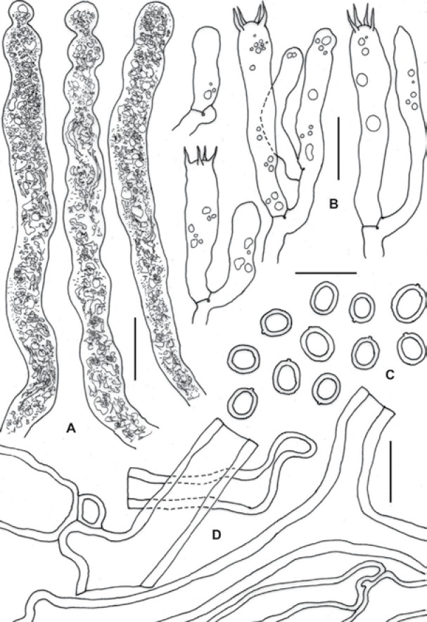
Hericium yumthangense (KD-11-146). A. Gloeocystidia. B. Basidia. C. Basidiospores. D. Hymenialtramal hyphae. Bars = 10 μm.
Fig. 7.
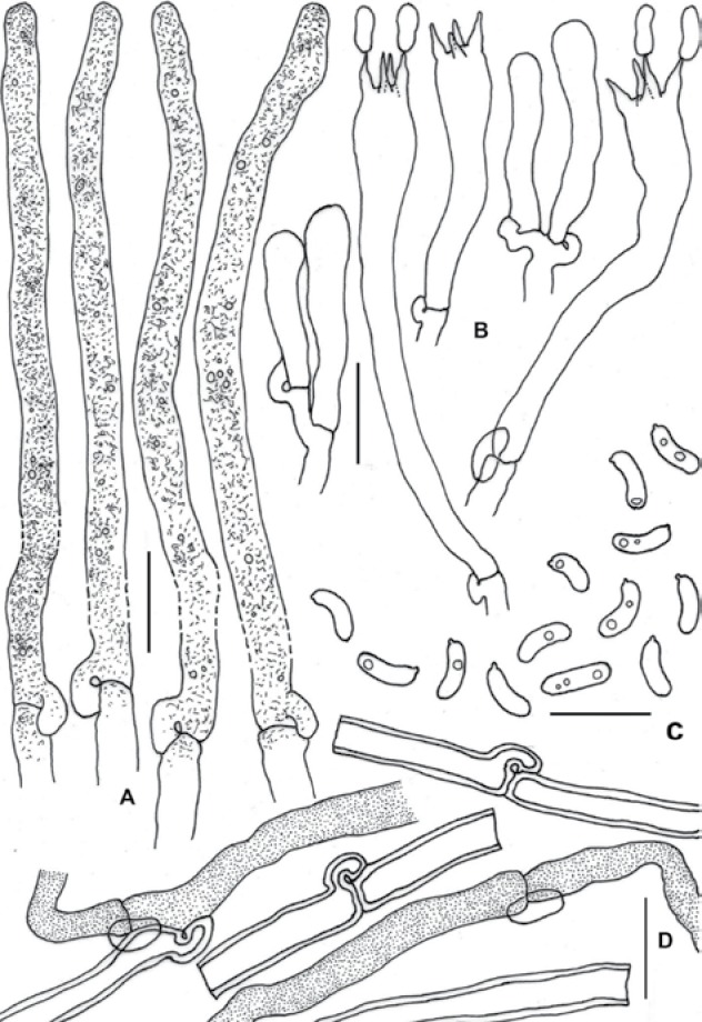
Mycoleptodonoides sharmae (KD-11-122). A. Gloeocystidia-like hyphal elements. B. Basidia. C. Basidiospores. D. Solid to slightly thick-walled hyphae of spine context. Bars = 10 μm.
Acknowledgments
We are grateful to the Director, Botanical Survey of India (Kolkata, India) for providing facilities during this study, and to the Department of Forest, Environment and Wildlife Management, Gangtok (India) for support. Sundar Kumar Rai (BSI, Gangtok) helped the first author in the field in many ways. The assistance rendered by Shakti Nath Das of the S.N. Bose National Centre for Basic Sciences, Kolkata, in supporting the SEM study is also duly acknowledged. Author J.B.S. would like to thank Michel de Vries for technical support with sequencing. Laboratory work at the CBS was financed by the Royal Dutch Academy of Arts and Science (Grant Stimuleringsfonds funded project “DNA-barcoding the CBS Collections”).
REFERENCES
- Anon. (1969) Flora of British Fungi: colour identification chart. Edinburgh: Her Majesty’s Stationery Office [Google Scholar]
- Binder M, Hibbett DS, Larsson KH, Larsson E, Langer E, Langer G. (2005) The phylogenetic distribution of resupinate forms across the major clades of mushroom-forming fungi (Homobasidiomycetes). Systematics and Biodiversity 3: 1–45 [Google Scholar]
- Das K, Sharma JR. (2010) Hericium cirrhatum (Pers.) Nikol., a new record to Indian mycoflora. Kavaka 37–38: 17–19 [Google Scholar]
- Das K, Stalpers J, Eberhardt U. (2011) A new species of Hericium from Sikkim, Himalaya (India). Cryptogamie, Mycologia 32: 285–293 [Google Scholar]
- Felsenstein J. (1981) Evolutionary trees from DNA sequences: a maximum likelihood approach. Journal of Molecular Evolution 17: 368–376 [DOI] [PubMed] [Google Scholar]
- Ginns J. (1984) Hericium coralloides N. Amer. auct. (= H. americanum sp. nov.) and the European H. alpestre and H. coralloides. Mycotaxon 20: 39–43 [Google Scholar]
- Guevara-Guerrero G, Stielow B, Tamm H, Cázares-Gonzales E, Göker M. (2011) Genea mexicana and Geopora tolucana, new sequestrate Pyronemataceae from Mexico, and the phylogeny of Geopora s. l. reevaluated. Mycological Progress 11: 711–724 [Google Scholar]
- Hallenberg N. (1983) Hericium coralloides and H. alpestre (basidiomycetes) in Europe. Mycotaxon 18: 181–189 [Google Scholar]
- Hallenberg N, Henrik Nilsson R, Robledo G. (2012) Species complexes in Hericium (Russulales, Agaricomycota) and a new species – Hericium rajchenbergii- from southern South America. Mycological Progress 12: 413–420 [Google Scholar]
- Harrison KA. (1973) The genus Hericium in North America. Michigan Botanist 12: 177–194 [Google Scholar]
- Imazeki R, Hongo T. (1989) Colored Illustrations of Mushrooms of Japan. Vol. 2 Osaka: Hoikusha [Google Scholar]
- Kraenzlin F. (2005) Die Pilze der Schweiz. Vol. 6. Russulaceae. Luzern: Verlag Mykologia [Google Scholar]
- Larsson E, Larsson KH. (2003) Phylogenetic relationships of russuloid basidiomycetes with emphasis on aphyllophoralean taxa. Mycologia 95: 1037–1065 [DOI] [PubMed] [Google Scholar]
- Larsson KH, Larsson E, Koljalg U. (2004) High phylogenetic diversity among corticioid homobasidiomycetes. Mycological Research 108: 983–1002 [DOI] [PubMed] [Google Scholar]
- Maas Geesteranus RA. (1961) A Hydnum from Kashmir. Persoonia 1: 409–413 [Google Scholar]
- Maas Geesteranus RA. (1962) Hyphal structures in hydnums. Persoonia 2: 77–405 [Google Scholar]
- Nikolajeva TL. (1952) Novyi rod ezhovikvykh (sem. Hydnaceae) gribov (A new genus of hydnaceous fungi). Botanicheskie Materialy Otdela Sporovykh Rastenii. Botanichescheskii Institut Akademia Nauk SSR 8: 117–121 [Google Scholar]
- Schoch C, Seifert KA, Huhndorf S, Robert V, Spouge JL, Levesque CA, Chen W, Fungal Barcoding Consortium (2012) Nuclear ribosomal internal transcribed spacer (ITS) region as a universal DNA barcode marker for Fungi. Proceedings of the National Academy of Sciences, USA 109: 6241–6246; rDOI:10.1073/pnas.1117018109. [DOI] [PMC free article] [PubMed] [Google Scholar]
- Stamatakis A, Hoover P, Rougemont J. (2008) A rapid bootstrap algorithm for the RAxML web servers. Systematic Biology 75: 758–771 [DOI] [PubMed] [Google Scholar]
- Stielow B, Bubner B, Hensel G, Münzenberger B, Hoffmann P, Klenk H-P, Göker M. (2010). The neglected hypogeous fungus Hydnotrya bailii Soehner (1959) is a widespread sister taxon of Hydnotrya tulasnei (Berk.) Berk. and Broome (1846). Mycological Progress 9: 195–203 [Google Scholar]
- Thind KS, Khara HS. (1975) The Hydnaceae of the North Western Himalayas–II. Indian Phytopathology 28: 57–65 [Google Scholar]
- White TJ, Bruns TD, Lee S, Taylor J. (1990) Amplification and direct sequencing of fungal ribosomal RNA genes for phylogenetics. In: PCR Protocols: a guide to methods and applications (Innis MA, Gelfand DH, Sninsky JJ, White TJ, eds): 315–322 San Diego: Academic Press [Google Scholar]
- Yuan HS, Dai YC. (2009) Hydnaceous fungi of China 4. Mycoleptodonoides tropicalis sp. nov., and a key to the species in China. Mycotaxon 110: 233–238 [Google Scholar]


