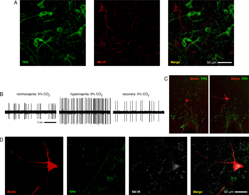
Figure 4. TPH and NK1R do not colocalize in the medullary raphé, and the medullary raphé contains CO2-stimulated non-5-HT cells that express NK1R.
(A) Shown are TPH (green) and NK1 receptor (red) immunostaining in the raphé magnus. Immunoreactivity of these two markers were never observed to colocalize in any of our tested sections. (B) Shown is a cell that increased firing frequency by 82% with hypercapnia. 10× views of two adjacent sections (C; ventral surface visible at bottom) show the juxtacellular fill and extensive dendritic processes of the recorded neuron (red) in raphé magnus and tissue staining for TPH-ir (green). 40× views of the filled cell (D) demonstrate that the recorded cell lacks TPH-ir seen in neighboring cells (green) but is NK1R-ir (white), indicating expression of NK1R by the recorded cell.

