
Fig. 2. Architecture and topology of MraY.
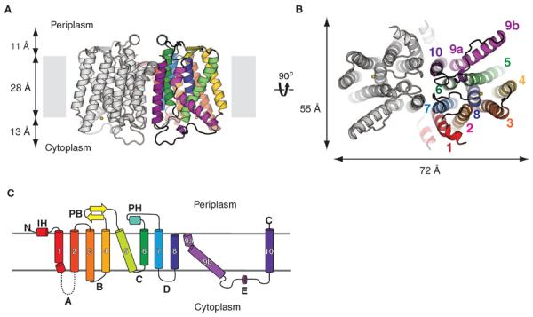
(A) View from within the membrane. Only transmembrane helices (TMs) from one protomer are colored. Loop E from only one protomer was present in the model, but loop E from both protomers are shown. The yellow sphere is Mg2+. (B) Cytoplasmic view. The yellow sphere is Mg2+. (C) Topology diagram of MraY protomer. Each TM is colored differently. TMs are given numbers, and cytoplasmic loops are given letters. Loop A is missing in the structure. The same colors are used for TMs in (A) through (C).