Abstract
Background
Neural habituation, the decrease in brain response to repeated stimulation, is a basic form of learning. There is strong evidence for behavioral and physiological habituation deficits in schizophrenia, and one previous study found reduced neural habituation within the hippocampus. However, it is unknown whether neural habituation deficits are specific to faces and limited to the hippocampus. Here we studied habituation of several brain regions in schizophrenia, using both face and object stimuli. Post-scan memory measures were administered to test for a link between hippocampal habituation and memory performance.
Methods
During an fMRI scan, 23 patients with schizophrenia and 21 control subjects viewed blocks of a repeated neutral face or neutral object, and blocks of different neutral faces and neutral objects. Habituation in the hippocampus, primary visual cortex and fusiform face area (FFA) was compared between groups. Memory for faces, words, and word pairs was assessed after the scan.
Results
Patients showed reduced habituation to faces in the hippocampus and primary visual cortex, but not the FFA. Healthy control subjects exhibited a pattern of hippocampal discrimination that distinguished between repeated and different images for both faces and objects, and schizophrenia patients did not. Hippocampal discrimination was positively correlated with memory for word pairs.
Conclusion
Patients with schizophrenia showed reduced habituation of the hippocampus and visual cortex, and a lack of neural discrimination between old and new images in the hippocampus. Hippocampal discrimination correlated with memory performance, suggesting reduced habituation may contribute to the memory deficits commonly observed in schizophrenia.
Keywords: schizophrenia, hippocampus, habituation, fMRI adaptation, repetition suppression, faces
1. Introduction
Habituation, the decrease in response to a stimulus following repeated exposure with no meaningful consequence, is a basic form of learning (Rankin et al., 2009). Habituation is adaptive because it allows for the allocation of limited cognitive resources towards novel events in the environment. Neural habituation can be measured with functional magnetic resonance imaging (fMRI) as a decrease in blood-oxygen level dependent (BOLD) response to repeated stimulation. Distributed brain areas exhibit habituation to visual cues, including visual processing areas (Summerfield et al., 2008; Weigelt et al., 2008), the prefrontal cortex (Wright et al., 2001; Yamaguchi et al., 2004), and the hippocampus and amygdala (Blackford et al., 2013; Blackford et al., 2010; Breiter et al., 1996; Fischer et al., 2003; Wright et al., 2001). In many previous studies, fMRI habituation (also called fMRI adaptation or repetition suppression) has been used to localize feature-specific processing in the brain (e.g. Grill-Spector et al., 2006). However, more recent investigations have linked habituation in the hippocampal formation with cognitive performance, finding that habituation in the parahippocampus is related to successful memory retrieval (Turk-Browne et al., 2006), and that patients with mild cognitive impairment, characterized by memory deficits, show reduced hippocampal habituation (Johnson et al., 2004).
Habituation deficits have been consistently observed in patients with schizophrenia, using both behavioral and electrophysiological measures. For example, patients with schizophrenia show reduced habituation of eye-blink startle to auditory or tactile cues (Bolino et al., 1994; Bolino et al., 1992; Braff et al., 1992b; Geyer and Braff, 1982), as well as reduced habituation of early evoked auditory responses (Olincy et al., 2010). Despite this wealth of evidence for disrupted habituation in schizophrenia, only one study has investigated fMRI habituation, finding reduced habituation in the right anterior hippocampus in response to repeated fearful faces (Holt et al., 2005). Many important questions remain regarding the integrity of fMRI habituation in schizophrenia, specifically whether habituation deficits are 1) present outside the hippocampus, 2) specific to social stimuli such as faces, or 3) related to memory impairments in schizophrenia. Considering the large body of previous research on sensory gating deficits (e.g. reduced pre-pulse inhibition and reduced mismatch negativity) (Braff et al., 1992a; Light and Braff, 2005), and specific disruptions in early visual processing in schizophrenia (Javitt, 2009), it is possible that habituation in sensory cortex is also reduced.
Here we extend studies of habituation in schizophrenia to test for reduced fMRI habituation in the hippocampus, early visual cortex (BA 17/18) and the fusiform face area, (FFA, Kanwisher et al., 1997), a major node in the face processing network found to be hypoactive in schizophrenia patients (Habel et al., 2010; Pinkham et al., 2008; Quintana et al., 2003; Seiferth et al., 2009). Hippocampal volumes were compared between groups to investigate whether reduced hippocampal habituation occurs in the context of hippocampal volume loss, a prominent finding in schizophrenia (Nelson et al., 1998; Velakoulis et al., 2006; Wright et al., 2000). We assessed habituation to both neutral faces and objects to probe whether reduced habituation is specific to social stimuli. Finally, post-scan memory measures were collected to test whether greater hippocampal habituation predicts better memory performance. We hypothesized that patients with schizophrenia would exhibit reduced habituation in the hippocampus and visual cortex for face and object stimuli, and that hippocampal habituation would be positively correlated with memory ability.
2. Methods and Materials
2.1 Subjects
Participants included 25 patients with schizophrenia (n=18) or schizoaffective disorder (n=7) and 23 healthy controls. Psychotic patients were recruited from the psychiatric inpatient unit and outpatient clinics of Vanderbilt University Medical Center. Healthy controls were recruited from the surrounding community via advertisements. The study protocol was approved by the Vanderbilt University Institutional Review Board, Nashville, TN, and written informed consent was obtained from all subjects.
All participants were administered the Structured Clinical Interview for DSM-IV (American Psychiatric, 2000) and the National Adult Reading Test as a measure of premorbid IQ (NART, Nelson, 1982). Psychotic patients were assessed with the Hamilton Depression Rating Scale (Hamilton, 1960), the Young Mania Rating Scale (Young et al., 1978), and the Positive and Negative Syndrome Scale (Kay et al., 1987). When available, research assessments were supplemented with clinical information from treating physicians. All patients with schizoaffective disorder met criteria A, B, and C for schizophrenia (i.e., symptoms lasted > 6 months and led to functional impairment) (American Psychiatric, 2000) and clinical variables did not differ between patients with schizophrenia and schizoaffective disorder. Participants were excluded for: significant medical or neurological illness, head injury, history of drug or alcohol dependence, substance abuse within the last 6 months, and/or uncorrected vision deficits. Prior to fMRI analysis 2 schizophrenia and 2 control participants were excluded for poor fMRI task performance (Supplementary Materials, section S1.2). The final sample included 23 schizophrenia patients and 21 controls who did not differ with respect to age, race, gender, premorbid IQ, or parental education, though control subjects had higher levels of education (Table 1). Chlorpromazine equivalent doses were calculated for patients taking antipsychotics (n=22) (Gardner et al., 2010) (Table 1).
Table 1.
Demographic and Clinical Characteristics of Participants
| Schizophrenia n = 23 |
Control n = 21 |
|
|---|---|---|
| Sex (male/female) | 12/11 | 11/10 |
| Race (black/white/other) | 10/13/0 | 7/13/1 |
| Age, Years | 43.35 (11.8) | 42.38 (10.0) |
| Participant’s Education, Years* | 13.70 (2.5) | 15.76 (2.3) |
| Parental Education, Years | 13.18 (2.7) | 13.40 (2.4) |
| IQ, NART | 106.17 (8.5) | 109.29 (8.3) |
| Ham-D | 3.44 (3.5) | - |
| YMRS | .97 (2.3) | - |
| PANSS- Total | 51.35 (13.0) | - |
| PANSS- Positive | 13.22 (5.6) | - |
| PANSS- Negative | 14.13 (6.7) | - |
| PANSS- General | 24.0 (5.5) | - |
| CPZ | 521.13 (261.8) | - |
| Duration of Illness, Years | 20.09 (10.8) | - |
| Number of Hospitalizations | 4.87 (4.3) | - |
CPZ, chlorpromazine equivalent doses; HAM-D, Hamilton Depression Rating Scale; NART, North American Adult Reading Test; PANSS, Positive and Negative Syndrome Scale; YMRS, Young Mania Rating Scale.
Control > Schizophrenia, p < .05
2.2 Experimental Paradigm
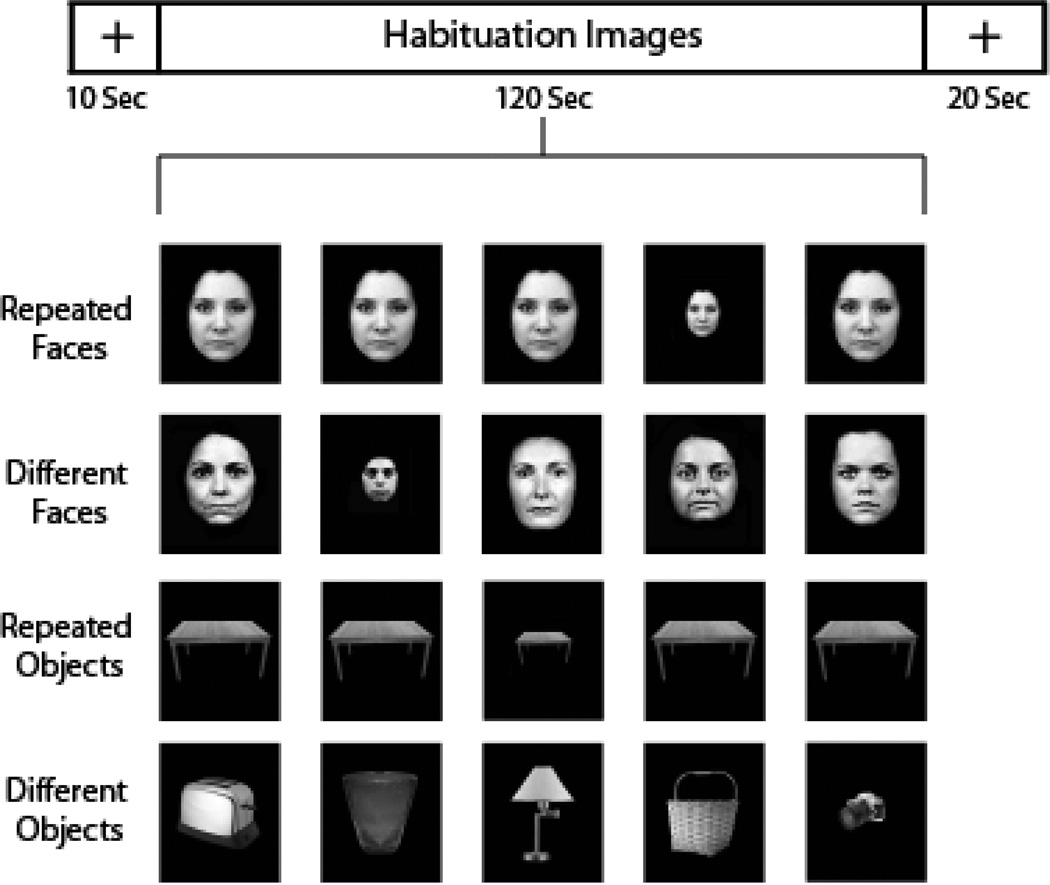
Participants completed an fMRI scan that assessed habituation to faces, habituation to objects, and a functional localizer for the FFA. Briefly, participants viewed eight 2-minute runs of images. Images were neutral faces (first 4 runs) or neutral objects (second 4 runs) and were presented for 500-ms followed by a 500-ms blank screen. On half the runs, the same image was presented (Repeated condition) and on half the runs a series of different images was presented (Different condition). To promote and quantify attention during the task, small versions of the face or object images (25% of original size, similar to Summerfield et al., 2008) were presented on 10% of trials, and detected by subjects with a button press (Figure 1). More details of the task, FFA localizer, and MRI parameters are in Supplementary Materials (sections S1, S2). Analyses were restricted to the first run of each condition because habituation was much reduced in the second run (Williams et al., In preparation).
Figure 1.
Task design – participants viewed alternating runs of Repeated and Novel images, 4 runs of face images followed by 4 object runs. During stimulus presentation participants pressed a button to detect rare (10% of trials) small images, which were excluded from habituation analyses.
2.3 Post-scan Memory Tests
After the scan participants completed three subscales of the Wechsler Memory Scale-III (Wechsler, 1997). They were tested on 1) memory for faces because the fMRI task used face images, 2) memory for word pairs (Verbal Paired Associates) because pair learning is impaired in schizophrenia (Achim and Lepage, 2003) and supported by the hippocampus, and 3) memory for single words (Word List) because verbal memory deficits are common in schizophrenia (Aleman et al., 1999; Fioravanti et al., 2005). One participant with schizophrenia did not complete memory tasks due to time constraints.
2.4 Data analysis
2.4.1 Target Detection and Memory Tests
Hit rate on the target detection task and all memory measures was compared between groups using 2-tailed, independent samples t-tests.
2.4.2 Structural Neuroimaging Data
Hippocampal segmentation used a previously established protocol (Pruessner et al., 2000) in the 3DSlicer program (version 3.4) (Pieper et al., 2004) by a single rater (AL) who was blind to diagnostic group. Reliability statistics were computed by segmenting 8 randomly selected subjects (4/group) at two different time points to determine Intraclass Correlation Coefficients for right (ICC=0.93) and left (ICC=0.93) hippocampi. Volumes were compared with a 2-tailed independent samples t-test.
2.4.3 Functional Neuroimaging Data
Analyses were performed in SPM8 (Wellcome Department of Cognitive Neurology, London, UK). Functional data were corrected for head motion, normalized to Montreal Neurological Institute template space (EPI template in SPM) and smoothed (5-mm full-width/half maximum kernel). Runs with >3mm of motion in any direction were excluded from analysis (one run excluded:1 control, 2 schizophrenia participants; two runs excluded: 1 control, 2 schizophrenia participants). First-level analyses modeled regressors for response to habituation images and small image targets. Habituation was modeled with a symmetrical linear regressor with a slope of −1 to capture voxels with gradual signal decrease across the run. Small images were modeled as separate events of 0 duration and included as regressors of no interest. The inverse of the habituation regressor (slope of 1) was modeled to isolate regions that show enhancement of BOLD response throughout the run. Fixation periods were implicitly modeled as the baseline. No temporal filter was applied because it is confounded with the linear decrease in BOLD that represents habituation. For the second-level analyses, 2 sample t-tests tested for between-group differences in habituation by comparing the beta weights associated with the habituation regressor within regions of interest (ROIs) selected a priori based on previous studies and hypotheses: the hippocampus, BA17/18 (primary visual cortex), and the FFA.
Habituation was assessed relative to the fixation baseline, consistent with previous studies (Fischer et al., 2003; Holt et al., 2005; Wright et al., 2001), and considered to be present in an ROI if a significant cluster was found at p < .05 corrected. A cluster-based threshold adjustment method based on Monte-Carlo simulations (AlphaSim, http://afni.nimh.gov/pub/dist/doc/manual/AlphaSim.pdf) was used to protect against Type I errors within ROIs. Percent signal change was extracted from significant clusters using Marsbar (Brett et al., 2002). To test the relation between hippocampal signal and memory performance we performed regression analyses using memory test score as a predictor, covarying for diagnosis.
2.4.4 Regions of Interest (ROIs)
Because hippocampal volume is often reduced in schizophrenia patients, we constructed a study-specific hippocampal ROI using hippocampal tracings (average of all subjects, 50% overlap across sample). Reductions in visual cortex volume in schizophrenia is not a consistent finding (though see (Donohoe et al., 2010); and visual cortex (BA17/18) ROIs were taken from a standard atlas (Maldjian et al., 2003). A functional localizer defined the FFA, which cannot be reliably isolated using purely anatomical landmarks. More ROI details are in Supplementary Materials (section S4).
3. Results
3.1 Target Detection
Performance on the target detection task was high (> 85% correct for all conditions) and did not differ between groups, indicating both groups were monitoring the stimuli (mean percent correct ± SD for Repeated faces, Different faces, Repeated objects, and Different objects in controls: 98±4, 96±4, 97±4, 87±10 and schizophrenia patients: 96±6, 92±9, 96±7, 86±10; all p>.17).
3.2 Habituation to Repeated Images
3.2.1 Hippocampus
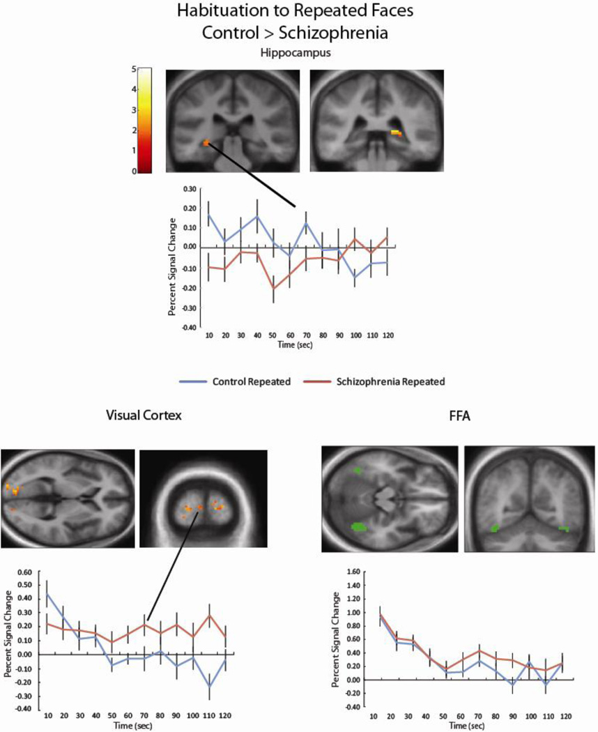
In both groups the hippocampus was activated in response to the initial presentation of Repeated faces (Supplementary Figure S1). Consistent with habituation, hippocampal response declined over time in the healthy control group. In contrast, patients with schizophrenia failed to show habituation (Figure 2). There were no differences between groups in response to Repeated objects, within the context of overall weaker habituation for objects relative to faces (Supplementary Figure S2).
Figure 2.
Patients with schizophrenia showed reduced habituation to Repeated faces in the left (k = 34; − 21, −22, −23) and right (k = 13; 18, −37, 4) hippocampus (cluster corrected p-value < .05). Extracting the percent signal change from these clusters shows that while control participants show decreasing hippocampal activity across the run, activity is more sustained for schizophrenia patients. A similar pattern was found in three clusters in the bilateral visual cortex (k = 111, −18, −79, 10; k = 22, 12, −76, 22; k = 19, 24, −97, 7, cluster-corrected p < .05). Percent signal change showed for a representative cluster. In contrast, both controls and schizophrenia patients show robust habituation to Repeated faces within the FFA (a representative single-subject FFA region of interest shown in green).
3.2.2 Visual Cortex
Primary visual cortex responded to the initial presentation of faces in both groups (Supplementary Figure S1). Similar to the hippocampus, controls showed habituation while schizophrenia patients did not (Figure 2). In contrast, within the FFA both schizophrenia patients and controls exhibited robust habituation to faces (non-significant group × time interaction, Right FFA F =.57, p=.53; Left FFA F=.70 p=.73) (Figure 2).
3.3 Response to Different Images
3.3.1 Hippocampus
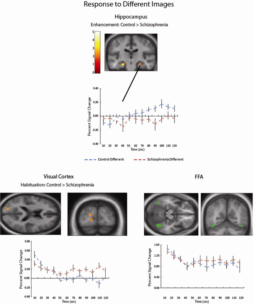
The inclusion of Different images allowed us to quantify responses to novelty, and to control for non-specific factors such as visual processing or neural fatigue. Within the hippocampus there was little habituation to Different faces for either group. Rather, hippocampal response to Different images was enhanced (e.g. increased) throughout the run for controls (Figure 3). Patients with schizophrenia did not show either pattern in response to Different images.
Figure 3.
In response to Different Images, control subjects showed increasing signal in the bilateral hippocampus (left k = 56, −27, −40, 1, k = 14, −24, −19, −26; right k = 27, 33, −37, −8, k = 17, 27, −22. −26 cluster-corrected p < .05); schizophrenia patients did not show significant hippocampal changes throughout the run. In the visual cortex, patients with schizophrenia showed reduced habituation to Different faces, relative to control participants (k = 53, −21, −88, 16 cluster-corrected p < .05). FFA habituation to Different faces was present for both groups.
3.3.2 Visual Cortex
In contrast, in the primary visual cortex habituation was observed to Different faces, consistent with habituation to faces as a category. Similar to the Repeated condition, schizophrenia patients showed reduced habituation to Different faces in the visual cortex, but groups did not differ in the FFA (Right FFA F=.81, p=.56; Left FFA F=1.1 p=.38) (Figure 3).
3.4 Hippocampal Discrimination
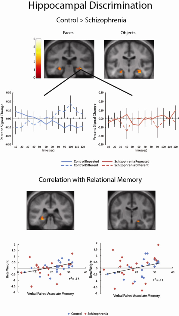
Given the somewhat unexpected pattern of enhancement, rather than habituation, of neural signal in the hippocampus in response to Different faces, we tested the interaction between Repeated and Different faces within the hippocampus. Healthy subjects showed hippocampal habituation to Repeated images and enhancement to Different images, consistent with comparator models of hippocampal function (see Discussion). This pattern of hippocampal discrimination, which appears to distinguish between Repeated and Different images, was not observed in schizophrenia patients (Figure 4), who showed relatively stable responses to both face types.
Figure 4.
In response to both Face and Object images, significant clusters in the bilateral hippocampus of healthy control subjects showed a pattern of hippocampal discrimination, exhibiting habituation in response to Repeated images and enhancement in response to Different images (Faces: left k = 20, −30, −28, −17; k = 18, −24, −40, −2 right k = 53; 21, −37, 4. Objects: left k = 33, −27, −25, −17; right k = 85, 33, −16, −32). The pattern was absent schizophrenia patients, who had similar responses to both stimulus types in these hippocampal regions. There was a positive correlation between hippocampal discrimination and verbal paired associate memory (total recall score) in the left (k = 24; −27, −34, −14) and right (k = 10; 21, 19, −20) hippocampus, such that greater hippocampal habituation was correlated with paired associate memory.
3.5 Memory Correlations
Controls performed better than schizophrenia patients on all memory measures (Table 2). To test the relationship between hippocampal habituation and memory performance, memory scores were used as a predictor variable within the hippocampal ROI, covarying for group. We found a positive correlation between hippocampal discrimination of face stimuli and memory for verbal paired associates (total recall) (Figure 4).
Table 2.
Performance on Post-Scan Memory Wechsler Memory Scales
| Schizophrenia n = 22 |
Normal Control n = 21 |
t and p values | ||
|---|---|---|---|---|
| Verbal Paired Associates | ||||
| Recall Total (sum 4 blocks) | 18.41 (8.47) | 22.86 (7.7) | t = 1.80, p = .08 | |
| Recall Delayed (single block) | 6.0 (2.0) | 7.24 (1.6) | t = 2.18, p = .035 | |
| Word List | ||||
| Recall Total (sum 4 blocks) | 28.64 (8.8) | 36.95 (6.3) | t = 3.54, p < .001 | |
| Short Delay Recall (single block) | 5.82 (3.8) | 8.67 (2.4) | t = 2.92, p = .006 | |
| Long Delay Recall (single block) | 5.45 (3.9) | 8.19 (2.5) | t = 2.70, p = .01 | |
| Faces | ||||
| Immediate Recognition | 36.50 (4.1) | 39.14 (4.1) | t = 2.10, p = .042 | |
| Delayed Recognition | 36.68 (3.4) | 40.10 (3.1) | t = 3.44, p < .001 |
3.6 Confounding variables
Hippocampal volume did not differ between groups, indicating that reduced hippocampal habituation in schizophrenia patients is not explained by smaller hippocampal volume (mean volume ± SD in mm3 for the left and right hippocampus in control subjects: 3235±436, 3337±474 and schizophrenia patients: 3336±406, 3551±435; all p > .12). Antipsychotic dose did not correlate with any habituation measure (all p > .23). Finally, reduced habituation in the visual cortex did not account for abnormalities in the hippocampus, as the strength of habituation was not correlated across ROIs.
4. Discussion
Here we used fMRI habituation to evaluate neural responses to repeated visual images in patients with schizophrenia, finding reduced habituation to repeated faces in the hippocampus and primary visual cortex. Including Different images allowed us to identify an additional deficit in schizophrenia. Healthy controls showed a pattern of hippocampal discrimination - habituation to Repeated images and enhancement to Different images - that was absent in schizophrenia patients. Hippocampal discrimination was correlated with relational memory, suggesting that disrupted hippocampal response to old and new images may relate to the well-documented relational memory deficits in schizophrenia (Armstrong et al., 2012a; Armstrong et al., 2012b; Coleman et al., 2010; Ragland et al., 2012; Titone et al., 2004; Williams et al., 2010). Crucially, schizophrenia patients showed neither habituation to Repeated faces nor enhancement to Different faces (Figure 4), and exhibited significantly reduced habituation relative to controls (Figure 2), indicating that detection of both item familiarity and item novelty are impaired in schizophrenia. It should be noted that initial response to faces in the hippocampus is reduced in schizophrenia patients (Figure 2). However, because this difference does not represent a floor effect, it does not preclude the interpretation of habituation differences between groups. Habituation to Repeated and Different faces was also reduced in the primary visual cortex of schizophrenia patients. However, both patients and controls showed robust habituation in the FFA, showing that not all visual areas are affected. The magnitude of habituation was not correlated across ROIs for individual subjects, which may suggest that separate habituation processes occurred within different brain regions, and that schizophrenia patients have habituation deficits in some (hippocampus, primary visual) but not other (FFA) systems.
Our findings for hippocampal response to Different images were unexpected, as we anticipated there would be habituation to faces as a category (Gauthier et al., 2000) or to novelty itself (Murty et al., 2013). However, the presence of hippocampal discrimination is consistent with the comparator model of hippocampal function (Hasselmo and Wyble, 1997; Lisman and Grace, 2005). According to this model, the CA1 region compares predictions based on previous experience, generated by dentate gyrus /CA3, with incoming sensory information from cortex. When predictions and sensory information are the same, a match signal is generated and stimuli are recognized as familiar. When sensory information does not match the prediction, a mismatch signal is generated and a stimulus is coded as novel. According to this model, in our control participants match signals in the Repeated blocks lead to decreasing hippocampal activation with repeated stimulation, whereas mismatch signals in the Different condition drove signal increase. The presence of distinct signals coding for different types of images (Repeated, Different) within the hippocampus likely contributes to its role in memory function, which requires the discrimination of familiar (e.g. remembered) and novel information. The absence of this mechanism in schizophrenia patients may contribute to memory deficits (Lisman et al., 2008), which is supported the observed correlation between hippocampal discrimination and relational memory performance (Figure 4). Habituation deficits across multiple brain regions are consistent with widespread neurotransmitter dysfunction in schizophrenia. Possible mechanisms include NMDA-receptor hypofunction (Javitt, 2009; Lisman et al., 2008; Tamminga et al., 2012; Tamminga et al., 2010) and reduced function of inhibitory GABAergic neurons (Heckers and Konradi, 2010) (Stephenson et al., 2003; Thiel et al., 2001).
Limitations include the study of a group of chronic, medicated schizophrenia patients. Future studies with patients in an early stage of their illness would help to elucidate the impact of antipsychotic treatment on habituation; however, habituation deficits were not global (normal habituation in FFA) and antipsychotic dose was not correlated with any habituation measure. Our study design did not counterbalance the order of face and object stimuli, nor the order of Repeated and Different images. Faces were always presented first to equate our design with previous studies using only face images, which may have contributed to overall weaker habituation to objects. Similarly, the first run always presented Repeated images, followed by Different images, leaving open the possibility that increased attention to Different images contributed to the hippocampal “enhancement” pattern observed in controls. However, similar habituation responses to Repeated and Different images in visual cortex and the FFA, which are subject to the same possible variations in attention, does not support the notion that attentional changes drove fundamentally different brain responses across all ROIs. Additional studies should counterbalance to address these limitations for interpretation.
fMRI habituation is a simple paradigm with low cognitive demands that elicits deficits in schizophrenia patients, some of which relate to memory performance. Future studies may test for links between habituation to faces and social/emotional deficits in schizophrenia, as inaccurate coding of face familiarity and novelty could impair emotion processing, disrupt social cognition, and contribute to social anxiety. In addition, previous work has shown that neural habituation is affected by serotonergic drugs (Stephenson et al., 2003; Thiel et al., 2001), indicating that in the future habituation may serve as a novel target for therapeutics.
Supplementary Material
Acknowledgements
The authors would like to thank Kristan Armstrong, Julia Sheffield, Dr. Baxter Rogers, and Dr. Neil Woodward for their assistance and feedback.
Role of the funding source
This work was supported by the National Institute of Mental Health (grant R01 MH070560 to SH; grant K01 MH083052 to JUB), and the National Center for Research Resources (grant UL1 RR024975-01, now at the National Center for Advancing Translational Sciences, Grant 2 UL1 TR000445-06, to LEW). The NIMH had no further role in study design; in the collection, analysis and interpretation of data; in the writing of the report; or in the decision to submit the paper for publication. The content is solely the responsibility of the authors and does not necessarily represent the official views of the NIH.
Footnotes
Publisher's Disclaimer: This is a PDF file of an unedited manuscript that has been accepted for publication. As a service to our customers we are providing this early version of the manuscript. The manuscript will undergo copyediting, typesetting, and review of the resulting proof before it is published in its final citable form. Please note that during the production process errors may be discovered which could affect the content, and all legal disclaimers that apply to the journal pertain.
Conflicts of interest
Dr. Williams has received funding from the National Center for Research Resources. Dr. Blackford has received funding from the National Institute of Mental Health. Andrew Luksik reports no actual potential conflicts of interest. Dr. Gauthier has received funding from the National Institute of Health, the National Science Foundation and the James S. McDonnell Foundation. Dr. Heckers has received funding from the National Institute of Mental Health and served as an unpaid consultant to DWM-5.
Contributors
LEW, JUB, IG, and SH designed the study. LEW assembled the stimuli, programmed the experiment, and collected the data. LEW and JUB analyzed the data. LEW, JUB, IG, and SH interpreted the data. AL did the manual hippocampal segmentation. LEW wrote the first draft of the manuscript. All authors contributed to and have approved the final manuscript.
Contributor Information
Jennifer Urbano Blackford, Email: jenni.blackford@vanderbilt.edu.
Andrew Luksik, Email: Andrew.s.luksik@vanderbilt.edu.
Isabel Gauthier, Email: i.gauthier@vanderbilt.edu.
Stephan Heckers, Email: Stephan.heckers@vanderbilt.edu.
References
- Achim AM, Lepage M. Is associative recognition more impaired than item recognition memory in Schizophrenia? A meta-analysis. Brain and cognition. 2003;53(2):121–124. doi: 10.1016/s0278-2626(03)00092-7. [DOI] [PubMed] [Google Scholar]
- Aleman A, Hijman R, de Haan EH, Kahn RS. Memory impairment in schizophrenia: a meta-analysis. The American Journal of Psychiatry. 1999;156(9):1358–1366. doi: 10.1176/ajp.156.9.1358. [DOI] [PubMed] [Google Scholar]
- American Psychiatric A. Diagnostic and Statistical Manual of Mental Disorders. Washington DC: American Psychiatric Association; 2000. [Google Scholar]
- Armstrong K, Kose S, Williams L, Woolard A, Heckers S. Impaired associative inference in patients with schizophrenia. Schizophrenia bulletin. 2012a;38(3):622–629. doi: 10.1093/schbul/sbq145. [DOI] [PMC free article] [PubMed] [Google Scholar]
- Armstrong K, Williams LE, Heckers S. Revised associative inference paradigm confirms relational memory impairment in schizophrenia. Neuropsychology. 2012b;26(4):451–458. doi: 10.1037/a0028667. [DOI] [PMC free article] [PubMed] [Google Scholar]
- Blackford JU, Allen AH, Cowan RL, Avery SN. Amygdala and hippocampus fail to habituate to faces in individuals with an inhibited temperament. Soc Cogn Affect Neurosci. 2013;8(2):143–150. doi: 10.1093/scan/nsr078. [DOI] [PMC free article] [PubMed] [Google Scholar]
- Blackford JU, Buckholtz JW, Avery SN, Zald DH. A unique role for the human amygdala in novelty detection. NeuroImage. 2010;50(3):1188–1193. doi: 10.1016/j.neuroimage.2009.12.083. [DOI] [PMC free article] [PubMed] [Google Scholar]
- Bolino F, Di Michele V, Di Cicco L, Manna V, Daneluzzo E, Casacchia M. Sensorimotor gating and habituation evoked by electro-cutaneous stimulation in schizophrenia. Biological Psychiatry. 1994;36(10):670–679. doi: 10.1016/0006-3223(94)91176-2. [DOI] [PubMed] [Google Scholar]
- Bolino F, Manna V, Di Cicco L, Di Michele V, Daneluzzo E, Rossi A, Casacchia M. Startle reflex habituation in functional psychoses: a controlled study. Neuroscience letters. 1992;145(2):126–128. doi: 10.1016/0304-3940(92)90002-o. [DOI] [PubMed] [Google Scholar]
- Braff DL, Grillon C, Geyer MA. Gating and habituation of the startle reflex in schizophrenic patients. Archives of General Psychiatry. 1992a;49(3):206–215. doi: 10.1001/archpsyc.1992.01820030038005. [DOI] [PubMed] [Google Scholar]
- Braff DL, Grillon C, Geyer MA. Gating and habituation of the startle reflex in schizophrenic patients. Archives of General Psychiatry. 1992b;49(3):206–215. doi: 10.1001/archpsyc.1992.01820030038005. [DOI] [PubMed] [Google Scholar]
- Breiter HC, Etcoff NL, Whalen PJ, Kennedy WA, Rauch SL, Buckner RL, Strauss MM, Hyman SE, Rosen BR. Response and habituation of the human amygdala during visual processing of facial expression. Neuron. 1996;17(5):875–887. doi: 10.1016/s0896-6273(00)80219-6. [DOI] [PubMed] [Google Scholar]
- Brett M, Anton JL, Valabregue R, Poline JB. Region of interest analysis using an SPM toolbox [abstract] NeuroImage. 2002;16(s497) [Google Scholar]
- Coleman MJ, Titone D, Krastoshevsky O, Krause V, Huang Z, Mendell NR, Eichenbaum H, Levy DL. Reinforcement ambiguity and novelty do not account for transitive inference deficits in schizophrenia. Schizophrenia bulletin. 2010;36(6):1187–1200. doi: 10.1093/schbul/sbp039. [DOI] [PMC free article] [PubMed] [Google Scholar]
- Donohoe G, Frodl T, Morris D, Spoletini I, Cannon DM, Cherubini A, Caltagirone C, Bossu P, McDonald C, Gill M, Corvin AP, Spalletta G. Reduced occipital and prefrontal brain volumes in dysbindin-associated schizophrenia. Neuropsychopharmacology : official publication of the American College of Neuropsychopharmacology. 2010;35(2):368–373. doi: 10.1038/npp.2009.140. [DOI] [PMC free article] [PubMed] [Google Scholar]
- Fioravanti M, Carlone O, Vitale B, Cinti ME, Clare L. A meta-analysis of cognitive deficits in adults with a diagnosis of schizophrenia. Neuropsychology review. 2005;15(2):73–95. doi: 10.1007/s11065-005-6254-9. [DOI] [PubMed] [Google Scholar]
- Fischer H, Wright CI, Whalen PJ, McInerney SC, Shin LM, Rauch SL. Brain habituation during repeated exposure to fearful and neutral faces: a functional MRI study. Brain Res Bull. 2003;59(5):387–392. doi: 10.1016/s0361-9230(02)00940-1. [DOI] [PubMed] [Google Scholar]
- Gardner DM, Murphy AL, O'Donnell H, Centorrino F, Baldessarini RJ. International consensus study of antipsychotic dosing. Am J Psychiatry. 2010;167(6):686–693. doi: 10.1176/appi.ajp.2009.09060802. [DOI] [PubMed] [Google Scholar]
- Gauthier I, Tarr MJ, Moylan J, Skudlarski P, Gore JC, Anderson AW. The fusiform "face area" is part of a network that processes faces at the individual level. Journal of cognitive neuroscience. 2000;12(3):495–504. doi: 10.1162/089892900562165. [DOI] [PubMed] [Google Scholar]
- Geyer MA, Braff DL. Habituation of the Blink reflex in normals and schizophrenic patients. Psychophysiology. 1982;19(1):1–6. doi: 10.1111/j.1469-8986.1982.tb02589.x. [DOI] [PubMed] [Google Scholar]
- Grill-Spector K, Henson R, Martin A. Repetition and the brain: neural models of stimulus-specific effects. Trends in cognitive sciences. 2006;10(1):14–23. doi: 10.1016/j.tics.2005.11.006. [DOI] [PubMed] [Google Scholar]
- Habel U, Chechko N, Pauly K, Koch K, Backes V, Seiferth N, Shah NJ, Stocker T, Schneider F, Kellermann T. Neural correlates of emotion recognition in schizophrenia. Schizophrenia Research. 2010;122(1–3):113–123. doi: 10.1016/j.schres.2010.06.009. [DOI] [PubMed] [Google Scholar]
- Hamilton M. A rating scale for depression. Journal of neurology, neurosurgery, and psychiatry. 1960;23:56–62. doi: 10.1136/jnnp.23.1.56. (Journal Article) [DOI] [PMC free article] [PubMed] [Google Scholar]
- Hasselmo ME, Wyble BP. Free recall and recognition in a network model of the hippocampus: simulating effects of scopolamine on human memory function. Behav Brain Res. 1997;89(1–2):1–34. doi: 10.1016/s0166-4328(97)00048-x. [DOI] [PubMed] [Google Scholar]
- Heckers S, Konradi C. Hippocampal pathology in schizophrenia. Curr Top Behav Neurosci. 2010;4:529–553. doi: 10.1007/7854_2010_43. [DOI] [PubMed] [Google Scholar]
- Holt DJ, Weiss AP, Rauch SL, Wright CI, Zalesak M, Goff DC, Ditman T, Welsh RC, Heckers S. Sustained activation of the hippocampus in response to fearful faces in schizophrenia. Biological psychiatry. 2005;57(9):1011–1019. doi: 10.1016/j.biopsych.2005.01.033. [DOI] [PubMed] [Google Scholar]
- Javitt DC. When doors of perception close: bottom-up models of disrupted cognition in schizophrenia. Annu Rev Clin Psychol. 2009;5:249–275. doi: 10.1146/annurev.clinpsy.032408.153502. [DOI] [PMC free article] [PubMed] [Google Scholar]
- Johnson SC, Baxter LC, Susskind-Wilder L, Connor DJ, Sabbagh MN, Caselli RJ. Hippocampal adaptation to face repetition in healthy elderly and mild cognitive impairment. Neuropsychologia. 2004;42(7):980–989. doi: 10.1016/j.neuropsychologia.2003.11.015. [DOI] [PubMed] [Google Scholar]
- Kanwisher N, McDermott J, Chun MM. The fusiform face area: a module in human extrastriate cortex specialized for face perception. The Journal of neuroscience : the official journal of the Society for Neuroscience. 1997;17(11):4302–4311. doi: 10.1523/JNEUROSCI.17-11-04302.1997. [DOI] [PMC free article] [PubMed] [Google Scholar]
- Kay SR, Fiszbein A, Opler LA. The positive and negative syndrome scale (PANSS) for schizophrenia. Schizophrenia bulletin. 1987;13(2):261–276. doi: 10.1093/schbul/13.2.261. [DOI] [PubMed] [Google Scholar]
- Light GA, Braff DL. Mismatch negativity deficits are associated with poor functioning in schizophrenia patients. Archives of General Psychiatry. 2005;62(2):127–136. doi: 10.1001/archpsyc.62.2.127. [DOI] [PubMed] [Google Scholar]
- Lisman JE, Coyle JT, Green RW, Javitt DC, Benes FM, Heckers S, Grace AA. Circuit-based framework for understanding neurotransmitter and risk gene interactions in schizophrenia. Trends Neurosci. 2008;31(5):234–242. doi: 10.1016/j.tins.2008.02.005. [DOI] [PMC free article] [PubMed] [Google Scholar]
- Lisman JE, Grace AA. The hippocampal-VTA loop: controlling the entry of information into long-term memory. Neuron. 2005;46(5):703–713. doi: 10.1016/j.neuron.2005.05.002. [DOI] [PubMed] [Google Scholar]
- Maldjian JA, Laurienti PJ, Kraft RA, Burdette JH. An automated method for neuroanatomic and cytoarchitectonic atlas-based interrogation of fMRI data sets. NeuroImage. 2003;19(3):1233–1239. doi: 10.1016/s1053-8119(03)00169-1. [DOI] [PubMed] [Google Scholar]
- Murty VP, Ballard IC, Macduffie KE, Krebs RM, Adcock RA. Hippocampal networks habituate as novelty accumulates. Learn Mem. 2013;20(4):229–235. doi: 10.1101/lm.029728.112. [DOI] [PMC free article] [PubMed] [Google Scholar]
- Nelson HE. National Adult Reading Test (NART): Test Manual. Windsor: NFER-Nelson; 1982. [Google Scholar]
- Nelson MD, Saykin AJ, Flashman LA, Riordan HJ. Hippocampal volume reduction in schizophrenia as assessed by magnetic resonance imaging: a meta-analytic study. Archives of General Psychiatry. 1998;55(5):433–440. doi: 10.1001/archpsyc.55.5.433. [DOI] [PubMed] [Google Scholar]
- Olincy A, Braff DL, Adler LE, Cadenhead KS, Calkins ME, Dobie DJ, Green MF, Greenwood TA, Gur RE, Gur RC, Light GA, Mintz J, Nuechterlein KH, Radant AD, Schork NJ, Seidman LJ, Siever LJ, Silverman JM, Stone WS, Swerdlow NR, Tsuang DW, Tsuang MT, Turetsky BI, Wagner BD, Freedman R. Inhibition of the P50 cerebral evoked response to repeated auditory stimuli: results from the Consortium on Genetics of Schizophrenia. Schizophrenia Research. 2010;119(1–3):175–182. doi: 10.1016/j.schres.2010.03.004. [DOI] [PMC free article] [PubMed] [Google Scholar]
- Pieper A, Halle M, Kikinis R. 3D slicer. Proceeding of IEEE international symposium on biomedical imaging: From nano to macro SLP technical report #. 2004;448:632–635. [Google Scholar]
- Pinkham AE, Hopfinger JB, Pelphrey KA, Piven J, Penn DL. Neural bases for impaired social cognition in schizophrenia and autism spectrum disorders. Schizophrenia Research. 2008;99(1–3):164–175. doi: 10.1016/j.schres.2007.10.024. [DOI] [PMC free article] [PubMed] [Google Scholar]
- Pruessner JC, Li LM, Serles W, Pruessner M, Collins DL, Kabani N, Lupien S, Evans AC. Volumetry of hippocampus and amygdala with high-resolution MRI and three-dimensional analysis software: minimizing the discrepancies between laboratories. Cereb Cortex. 2000;10(4):433–442. doi: 10.1093/cercor/10.4.433. [DOI] [PubMed] [Google Scholar]
- Quintana J, Wong T, Ortiz-Portillo E, Marder SR, Mazziotta JC. Right lateral fusiform gyrus dysfunction during facial information processing in schizophrenia. Biological Psychiatry. 2003;53(12):1099–1112. doi: 10.1016/s0006-3223(02)01784-5. [DOI] [PubMed] [Google Scholar]
- Ragland JD, Ranganath C, Barch DM, Gold JM, Haley B, MacDonald AW, 3rd, Silverstein SM, Strauss ME, Yonelinas AP, Carter CS. Relational and Item-Specific Encoding (RISE): task development and psychometric characteristics. Schizophrenia bulletin. 2012;38(1):114–124. doi: 10.1093/schbul/sbr146. [DOI] [PMC free article] [PubMed] [Google Scholar]
- Rankin CH, Abrams T, Barry RJ, Bhatnagar S, Clayton DF, Colombo J, Coppola G, Geyer MA, Glanzman DL, Marsland S, McSweeney FK, Wilson DA, Wu CF, Thompson RF. Habituation revisited: an updated and revised description of the behavioral characteristics of habituation. Neurobiol Learn Mem. 2009;92(2):135–138. doi: 10.1016/j.nlm.2008.09.012. [DOI] [PMC free article] [PubMed] [Google Scholar]
- Seiferth NY, Pauly K, Kellermann T, Shah NJ, Ott G, Herpertz-Dahlmann B, Kircher T, Schneider F, Habel U. Neuronal correlates of facial emotion discrimination in early onset schizophrenia. Neuropsychopharmacology : official publication of the American College of Neuropsychopharmacology. 2009;34(2):477–487. doi: 10.1038/npp.2008.93. [DOI] [PubMed] [Google Scholar]
- Stephenson CM, Suckling J, Dirckx SG, Ooi C, McKenna PJ, Bisbrown-Chippendale R, Kerwin RW, Pickard JD, Bullmore ET. GABAergic inhibitory mechanisms for repetition-adaptivity in large-scale brain systems. NeuroImage. 2003;19(4):1578–1588. doi: 10.1016/s1053-8119(03)00257-x. [DOI] [PubMed] [Google Scholar]
- Summerfield C, Trittschuh EH, Monti JM, Mesulam MM, Egner T. Neural repetition suppression reflects fulfilled perceptual expectations. Nature neuroscience. 2008;11(9):1004–1006. doi: 10.1038/nn.2163. [DOI] [PMC free article] [PubMed] [Google Scholar]
- Tamminga CA, Southcott S, Sacco C, Wagner AD, Ghose S. Glutamate Dysfunction in Hippocampus: Relevance of Dentate Gyrus and CA3 Signaling. Schizophrenia bulletin. 2012 doi: 10.1093/schbul/sbs062. [DOI] [PMC free article] [PubMed] [Google Scholar]
- Tamminga CA, Stan AD, Wagner AD. The hippocampal formation in schizophrenia. Am J Psychiatry. 2010;167(10):1178–1193. doi: 10.1176/appi.ajp.2010.09081187. [DOI] [PubMed] [Google Scholar]
- Thiel CM, Henson RN, Morris JS, Friston KJ, Dolan RJ. Pharmacological modulation of behavioral and neuronal correlates of repetition priming. The Journal of neuroscience : the official journal of the Society for Neuroscience. 2001;21(17):6846–6852. doi: 10.1523/JNEUROSCI.21-17-06846.2001. [DOI] [PMC free article] [PubMed] [Google Scholar]
- Titone D, Ditman T, Holzman PS, Eichenbaum H, Levy DL. Transitive inference in schizophrenia: impairments in relational memory organization. Schizophrenia research. 2004;68(2–3):235–247. doi: 10.1016/S0920-9964(03)00152-X. [DOI] [PubMed] [Google Scholar]
- Turk-Browne NB, Yi DJ, Chun MM. Linking implicit and explicit memory: common encoding factors and shared representations. Neuron. 2006;49(6):917–927. doi: 10.1016/j.neuron.2006.01.030. [DOI] [PubMed] [Google Scholar]
- Velakoulis D, Wood SJ, Wong MT, McGorry PD, Yung A, Phillips L, Smith D, Brewer W, Proffitt T, Desmond P, Pantelis C. Hippocampal and amygdala volumes according to psychosis stage and diagnosis: a magnetic resonance imaging study of chronic schizophrenia, first-episode psychosis, and ultra-high-risk individuals. Archives of General Psychiatry. 2006;63(2):139–149. doi: 10.1001/archpsyc.63.2.139. [DOI] [PubMed] [Google Scholar]
- Wechsler D. Wechsler Memory Scale-III. San Antonio: The Psychological Corporation; 1997. [Google Scholar]
- Weigelt S, Muckli L, Kohler A. Functional magnetic resonance adaptation in visual neuroscience. Rev Neurosci. 2008;19(4–5):363–380. doi: 10.1515/revneuro.2008.19.4-5.363. [DOI] [PubMed] [Google Scholar]
- Williams LE, Blackford JU, Heckers S. In preparation. Habituation to repeated visual image: A comparison of different models [Google Scholar]
- Williams LE, Must A, Avery S, Woolard A, Woodward ND, Cohen NJ, Heckers S. Eye Movement Behavior Reveals Relational Memory Impairment in Schizophrenia. Biological Psychiatry. 2010;68:617–624. doi: 10.1016/j.biopsych.2010.05.035. (Journal Article) [DOI] [PMC free article] [PubMed] [Google Scholar]
- Wright CI, Fischer H, Whalen PJ, McInerney SC, Shin LM, Rauch SL. Differential prefrontal cortex and amygdala habituation to repeatedly presented emotional stimuli. Neuroreport. 2001;12(2):379–383. doi: 10.1097/00001756-200102120-00039. [DOI] [PubMed] [Google Scholar]
- Wright IC, Rabe-Hesketh S, Woodruff PW, David AS, Murray RM, Bullmore ET. Meta-analysis of regional brain volumes in schizophrenia. The American Journal of Psychiatry. 2000;157(1):16–25. doi: 10.1176/ajp.157.1.16. [DOI] [PubMed] [Google Scholar]
- Yamaguchi S, Hale LA, D'Esposito M, Knight RT. Rapid prefrontal-hippocampal habituation to novel events. The Journal of neuroscience : the official journal of the Society for Neuroscience. 2004;24(23):5356–5363. doi: 10.1523/JNEUROSCI.4587-03.2004. [DOI] [PMC free article] [PubMed] [Google Scholar]
- Young RC, Biggs JT, Ziegler VE, Meyer DA. A rating scale for mania: reliability, validity and sensitivity. The British journal of psychiatry : the journal of mental science. 1978;133:429–435. doi: 10.1192/bjp.133.5.429. (Journal Article) [DOI] [PubMed] [Google Scholar]
Associated Data
This section collects any data citations, data availability statements, or supplementary materials included in this article.


