Summary
We define a novel Bcl-x isoform, Bcl-xγ, that is generated by alternative splicing and characterized by a unique 47 amino acid C-terminus. Bcl-xγ is expressed primarily in thymocytes, where it may depend on an interaction between the TCR and host MHC products, and in mature T cells, where its expression is associated with ligation of the T cell receptor. Overexpression of Bcl-xγ in T cells inhibits activation-induced apoptosis; inhibition of Bcl-xγ, after stable expression of Bcl-xγ antisense cDNA, enhances activation-induced apoptosis. In contrast to other Bcl-x isoforms, cells that fail to express Bcl-xγ after CD3 ligation undergo programmed cell death, while activated T cells that express Bcl-xγ are spared. Identification of Bcl-xγ helps provide amolecular explanation of T cell activation and death after antigen engagement.
Introduction
Ligation of the T cell receptor (TCR) on thymocytes and mature T cells leads to either successful cellular activation and proliferation or to apoptosis. Until recently, apoptosis following cellular activation was thought to be limited to thymocytes, where it represents the major mechanism of negative selection in developing T cell clones. More recently, it became clear that the response of peripheral T cells is also regulated by activation-induced cell death (Weber et al., 1995).
Immunization with high doses of peptide (Pircher et al., 1989; Rocha et al., 1991; Zhang et al.,1994) or retroviral superantigens (Webb et al., 1990; Ignatowicz et al., 1992) leads to transient clonal activation followed by widespread T cell elimination. Infection with high doses of lymphocytic choriomeningitis virus also results in rapid T cell activation followed by massive clonal deletion within 1–2 weeks (Zinkernagel et al., 1993). These observations led to deliberate induction of apoptosis of autoreactive T cells by in vivo administration of high doses of autoantigen (Critchfield et al., 1994). The implication of these and other studies is that T cell activation followed by apoptosis is a common response phenotype that can regulate both primary T cell responses and the development of immunological memory. However, we do not understand the TCR-linked signaling events responsible for T cell activation on the one hand, and for apoptosis on the other.
An important advance in our understanding of genetic regulation of T cell apoptosis has come from the description of the Bcl gene family (reviewed by Akbar et al., 1993; Nunez et al., 1994; Reed, 1994; Cory, 1995; Hockenberry, 1995). Within this group, the Bcl-x gene encodes several alternatively spliced protein isoforms that can enhance or diminish T cell apoptosis after transfection (Boise et al., 1993; Hockenberry et al., 1993; Nunez et al., 1994; Ma et al., 1995), and studies of mice deficient in Bcl-x have suggested that at least some members of this family can prolong the lifespan of double-positive (DP) thymocytes by inhibition of apoptosis, although the precise contribution of these isoforms to this process has not been established (Ma et al., 1995; Motoyama et al., 1995).
The Bcl-xL isoform can inhibit cell death upon growth factor withdrawal (Boise et al., 1993), and overexpression leads to increased numbers of mature thymocytes (Chao et al., 1995; Grillot et al., 1995), although apparently it does not affect negative selection (Grillot et al., 1995; Takahashi et al., 1997). A second Bcl-x isoform, Bcl-xS, encodes a smaller protein of 170 amino acids (aa) that may enhance apoptosis in non-T cells under certain conditions (Clarke et al., 1995; Minn et al., 1996). Recently, the murine Bcl-x gene family has been expanded to include two additional isoforms, Bcl-xΔTM and Bcl-xβ, which may inhibit apoptosis in B cells (Fang et al., 1994) and neurons (Gonzalez-Garcia et al., 1995), respectively. Although these and other isoforms of Bcl-x may also regulate T cell growth and development, the full complement of Bcl-x isoforms that participate in this process is not yet clear.
All of the murine Bcl-x isoforms described so far are expressed in virtually all tissues, and there has been no evidence for a T cell–specific isoform whose expression is tightly connected to TCR ligation. Here we show that alternative splicing of Bcl-x generates a novel Bcl-x isoform, termed Bcl-xγ, which carries a unique C-terminus and is expressed primarily in T cells. Unlike previously described isoforms, Bcl-xγ is specifically connected to TCR ligation and is essential for resistance to TCR-dependent apoptosis.
Results
Structural Characteristics of Bcl-xγ
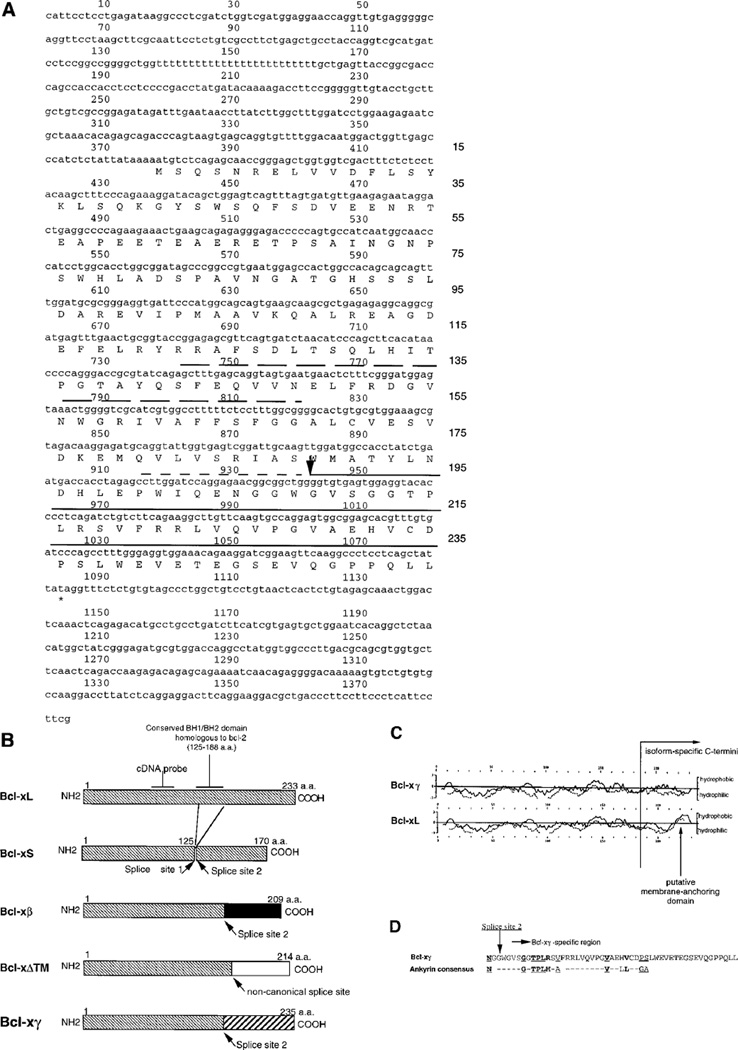
Since previously isolated Bcl-x isoforms that display ubiquitous expression were derived from non-T cell cDNA libraries, we screened a mouse thymus λ Zap II cDNA library using a 60-mer synthetic oligonucleotide probe specific for a conserved sequence of human Bcl-x (423–483 base pairs [bp]) (Boise et al., 1993).We isolated and purified six clones (Bcl-x5, Bcl-x6, Bcl-x7, Bcl-x8, Bcl-x10, and Bcl-x11); four contained inserts of the same length and sequence and corresponded to Bcl-xL cDNA and a fifth to Bcl-xβ (GenBank accession numbers U51279 and U51278) (Fang et al., 1994; Gonzalez-Garcia et al., 1994). One clone, termed Bcl-x7 (GenBank accession number U51277), contained a 1384 bp insert composed of a 5′ noncoding region of 377 nucleotides, an open reading frame (ORF) of 708 nucleotides, and a 3′ noncoding region of 299 nucleotides (Figure 1A). This represented a novel isoform of the Bcl-x gene in which the 3′ coding region of Bcl-xL was replaced by a 144 bp sequence that predicts a unique C-terminus of 47 aa (Figure 1A). This insert did not represent a cloning artifact because the novel 144 bp subsequence begins precisely at a conserved donor/acceptor splice site used by murine and human Bcl-x isoforms (Boise et al., 1993; Fang et al., 1994; Gonzalez-Garcia et al., 1994) (Figure 1B), and the sequence was independently cloned from thymocyte RNA (using a primer specific for a conserved region of murine Bcl-x and a Bcl-x7–specific primer). The recovered thymic sequence was identical to the cDNA insert isolated from the λ Zap II cDNA library, and the new isoform was designated Bcl-xγ.
Figure 1. Molecular Features of Bcl-xγ.
(A) cDNA sequence and predicted protein sequence of Bcl-xγ (GenBank accession number U51277). The position of the ORF encoding Bcl-xγ protein is indicated, and the predicted amino acid sequence (numbered to the right of each sequence) is given below the nucleotide sequence. Arrow, the splice site at which the unique Bcl-xγ C-terminus (continuously underlined) begins; long-dashed underline and a short-dashed underline, the conserved BH1 and BH2 domains, respectively.
(B) Schematic comparison of murine Bcl-x isoforms: Bcl-xL, Bcl-xS, Bcl-xβ, Bcl-xΔTM, and Bcl-xγ. Murine Bcl-x isoforms share a long N-terminal region (hatched). The BH1/BH2 region between splice sites 1 and 2 of Bcl-xS is shown. Splice site 2, used to generate Bcl-xβ and Bcl-xγ, is compared to the noncanonical splice site used to generate Bcl-xΔTM (located five residues downstream from splice site 2). The distinct C-termini of Bcl-xβ, Bcl-xΔTM, and Bcl-xγ, which show no homology with Bcl-xL and Bcl-xS C-terminal regions, also are shown, along with the positions of conserved BH1/BH2 domains. The position of the oligonucleotide probe used here to screen a mouse thymus cDNA library is indicated.
(C) Hydrophobicity plot of Bcl-xL and Bcl-xγ. The hydrophobicity of Bcl-xL and Bcl-xγ was calculated using the GCG program based on Goldman’s (solid line) or Kyte-Doolittle’s (dashed line) algorithm. The amino acid sequence of Bcl-xL and Bcl-xγ is numbered above and below the hydrophobicity plots. Vertical line, the beginning of the isoform-specific C-terminal sequences of Bcl-xγ and Bcl-xL; vertical arrow, the hydrophobic segment representing a putative membrane anchoring domain of Bcl-xL.
(D) Alignment of the specific region of mouse Bcl-xγ with the consensus sequence of the ankyrin-like domain sequence. The consensus sequence of ankyrin-like domain is shown. Amino acid symbols: bold and underlined, the six residues in Bcl-xγ that are identical to ankyrin consensus residues; bold, the two highly conserved residues in Bcl-xγ underlined, three residues in Bcl-xγ with homology to the ankyrin consensus residues. Dashed lines, the correct spacing of the ankyrin-like motif.
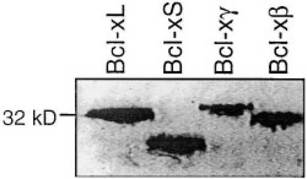
In vitro transcription and translation assays using linearized recombinant Bcl-x plasmids were performed to confirm the length of the ORF deduced from the cDNA sequence of Bcl-xγ (Figure 2). As expected from the predicted Bcl-xγ ORF of 708 nucleotides/235 aa, the apparent size of the translated Bcl-xγ protein was slightly larger than the (233 aa) Bcl-xL protein product and considerably larger than both the (170 aa) Bcl-xS (Fang et al., 1994) and the (209 aa) Bcl-xβ proteins (Gonzalez-Garcia et al., 1994). Analysis of the hydrophobicity of the unique C-terminus of the Bcl-xγ protein indicated that Bcl-xγ lacks an obvious hydrophobic domain flanked by charged residues (Figure 1C),which are present in human and murine Bcl-xL and Bcl-xS (Hockenberry et al., 1990; Boise et al., 1993; Nguyen et al., 1993). Differential centrifugation of Bcl-xγ–transfected cellular extracts (Yang et al., 1995) has indicated that Bcl-xγ protein is present mainly in the cytosol rather than membrane subcellular fraction (manuscript in preparation), similar to Bcl-xΔTM (Fang et al., 1994). A 33 aa region within the C-terminal domain of Bcl-xγ shows strong homology with the consensus sequence of the ankyrin-like domains (Figure 1D) that are embedded in a number of intracellular proteins, including Bcl-3, which uses this subsequence to bind to NF-κBp50 (Hatada et al., 1992); the p53 binding protein 53BP2, which uses the ankyrin-like domains to bind to L2 loop of p53 (Gorina and Pavletich, 1996); and bHLH (basic–helix-loop-helix), in which insertion of an ankyrin-like domain affects dimerization and DNA binding (Klein et al., 1993). In the first two cases, four to seven ankyrin repeats are present and may be required for physiological function, while in the latter case, tissue-specific RNA splicing of a single ankyrin-like domain regulates basic–helix-loop-helix dimerization and DNA binding (Klein et al., 1993).
Figure 2. Analysis of Protein Products of Bcl-xL, Bcl-xS, Bcl-xβ, and Bcl-xγ on 12%SDS-PAGE after In Vitro Transcription and Translation.
The apparent size of Bcl-xγ protein after in vitro transcription/translation is consistent with the size predicted from its ORF, since the Bcl-xγ protein product migrates at a position similar to Bcl-xL (233 aa residues) (see Figure 1B) and more slowly than the Bcl-xβ protein (209 aa) and Bcl-xS (170 aa). Molecular weight standards (kilodaltons) are indicated at the left margin.
Expression of Bcl-xγ
To ensure accurate and semiquantitative measurement, by reverse transcriptase polymerase chain reaction (RT-PCR), expression of Bcl-xγ and other Bcl-x isoforms, equal amounts of total RNA from each cellular sample was reverse transcribed, and amplification curves for each primer pair (for each Bcl-x isoform, interferon-γ [IFNγ], interleukin-2 [IL-2], and β-actin) were plotted against increasing cycles of PCR. This analysis showed that the slopes of these curves were similar (data not shown), suggesting that a potential bias reflecting differing amplification efficiencies of different primer pairs was minimal.
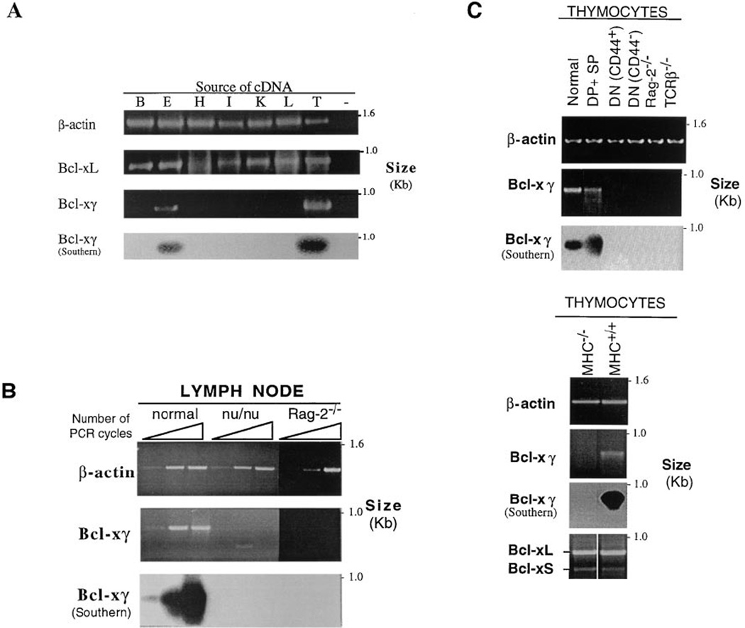
Samples of the reaction mix were taken from cycles in the exponential portion of the PCR reaction for measurement and comparison of gene expression, as previously described (Moore et al, 1994; Zheng and Flavell, 1997). According to RT-PCR, Bcl-xL (as well as Bcl-xβ and Bcl-xΔTM [data not shown]) was expressed in all tissues tested, including brain, eyes, heart, intestine, kidney, liver, and thymus (Figure 3A), consistent with previous reports (Boise et al., 1993; Gonzalez-Garcia et al., 1994). In contrast, Bcl-xγ expression was detected in thymus, lymph node, and eye, but not brain, heart, intestine, kidney, or liver (Figure 3A). The specificity of the amplified Bcl-xγ fragments in these tissues was confirmed by Southern blotting with a Bcl-xγ–specific probe that did not overlap with the sequences of the primers (Figure 3A). Failure to detect Bcl-xγ in tissues such as brain, heart, intestine, kidney, and liver by RT-PCR did not result from degradation of preparations of RNA from these tissues, since the ratio of ethidium bromide–stained 28S to 18S rRNA bands in agarose gels was the same for all tissues (data not shown), and since Bcl-xL, the β-actin gene, and other genes were successfully amplified by RT-PCR from the same RNA samples that were negative for Bcl-xγ. These results indicate that expression of the Bcl-xγ isoform in murine tissues is more restricted than is expression of other members of the Bcl-x family (Fang et al., 1994; Gonzalez-Garcia et al., 1994).
Figure 3. Tissue Expression of Bcl-xγ.
(A) Expression of Bcl-xγ in different murine tissues. RT-PCR products were analyzed on 1% agarose gels stained with ethidium bromide. Molecular weight markers are indicated at the right margin of the gels. The amount of amplified Bcl-xγ fragment and amplified β-actin fragment both increased in proportion to the number of thermal cycles, and the PCR amplification efficiency of Bcl-xγ primer pair was similar to that of the β-actin primer pair used as an internal control for RT-PCR for each tissue cDNA. Levels of β-actin from these tissues in the linear phase of PCR before saturation of amplification indicated that the actual amount of cDNA from each tissue used in this analysis was similar. A Bcl-xL fragment was also amplified to determine potential differences in tissue expression of Bcl-x isoforms. As reported previously, Bcl-xL was detected in every tissue examined. In contrast, the Bcl-xγ fragment was detected in thymus (T) and eye (E) but not in brain (B), heart (H), intestine (I), kidney (K), or liver (L). The specificity of the Bcl-xγ fragments from the tissues scoring as positive was confirmed by a Southern blot hybridization using a [32P]dCTP-labeled Bcl-xγ–specific probe that did not overlap with either primer of Bcl-xγ used in the RT-PCR.
(B) Expression of Bcl-xγ in lymph nodes of normal, nu/nu, and Rag2−/− mice. Analysis of cDNA indicated that Bcl-xγ is amplified in lymph nodes of normal but not nu/nu donors (PCR amplification of β-actin fragment served as control). To measure the linear phase of PCR amplification, aliquots of reaction mix were tested at 27, 31, and 35 cycles for β-actin and at 37, 41, and 45 cycles for Bcl-xγ and were visualized by agarose gel electrophoresis and ethidium bromide staining. The specificity of Bcl-xγ fragments amplified was confirmed by a Southern blot hybridization as described in (A); the blot was overexposed to detect any weak hybridization.
(C) Expression of Bcl-xγ in thymocyte subpopulations. Expression of Bcl-xγ was not detected in DN (CD4−CD8−) thymocytes (either CD44+ or CD44−) from normal, Rag2−/−, or TCRβ−/− donors. Bcl-xγ was detected in normal thymocytes, and DP (90%) and single-positive thymocytes (after depletion of DN cells) according to RT-PCR. PCR analysis of thymocytes from MHC−/− mice also showed that expression of MHC products are required for expression of Bcl-xγ but not Bcl-xL and Bcl-xS. The specificity of Bcl-xγ fragments amplified was confirmed by Southern blot hybridization, as described (A); the blot was overexposed to detect weak hybridization in PCR lanes that did not show apparent hybridization (negative groups).
We asked whether expression of Bcl-xγ in lymphoid tissues was restricted to T cells, B cells, or monocytes. Bcl-xγ was not expressed in peripheral lymphoid tissues of mice deficient in recombinase-activating gene-2 (Rag2), consistent with its selective expression in lymphocytes. Furthermore, Bcl-xγ was expressed in lymph nodes of BALB/c but not BALB/c nu/nu mice, suggesting that its expression is confined to T lymphocytes (Figure 3B).
Additional analysis of Bcl-xγ expression in the thymus indicated that it is not detectable in double-negative cells from normal or Rag2−/− donors, nor in thymocytes from mice that are deficient in the TCR β chain and fail to undergo TCR-dependent maturation into DP thymocytes. Bcl-xγ is expressed by DP thymocytes, since preparations that contained approximately 90% DP cells expressed Bcl-xγ (Figure 3C), while purified single-positive thymocytes did not express detectable Bcl-xγ (data not shown). These results suggest that Bcl-xγ expression in thymocytes is similar to that of Bcl-xL, as reported by others using different approaches (Boise et al., 1993; Ma et al., 1995; data not shown).
Bcl-xγ expression in DP thymocytes may depend on engagement of the TCR by major histocompatibility complex (MHC)/peptide ligands in the thymus, since Bcl-xγ was not detectable in thymocytes from mutant mice deficient in MHC class I/II (MHC double-deficient mice), which we have shown consist of approximately 90% DP cells (Crump et al., 1993) (Figure 3C). By contrast, expression of Bcl-xL and Bcl-xS (and Bcl-xβ and Bcl-ΔTM; data not shown) was unchanged in thymocytes from MHC double-deficient mice, indicating that, unlike Bcl-xγ, expression of Bcl-xL and Bcl-S isoforms in the thymus is not obviously dependent on MHC products.
Association with the TCR
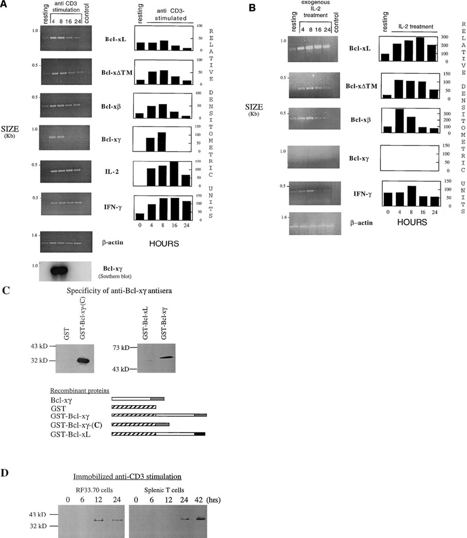
Bcl-xγ expression was not detectable in the resting murine T helper 1 (Th1) clone O3 but increased substantially by 4 hr after CD3 ligation. In contrast, Bcl-xγ was not expressed after IL-2 activation of these cells and remained undetectable even after the number of PCR cycles was increased to the maximal number (50 cycles) before polymerase activity becomes limiting (Coen, 1994), although [3H] TdR incorporation (data not shown) was similar in both stimulation protocols (Figures 4A and 4B). By comparison, all other murine Bcl-x isoforms (Bcl-xL, Bcl-xβ, Bcl-xΔTM, and Bcl-xS) were expressed in resting O3 cells and displayed similar increments 8 hr after either IL-2R or CD3 ligation (Figures 4A and 4B), similar to the reported increases in these isoforms using different techniques (Fang et al., 1994). TCR-dependent expression of Bcl-xγ was not limited to nontransformed primary T cell clones: neither of the T cell hybridomas AF3. G7 or RF33.70, nor the lymphoma cell line EL4, expressed detectable Bcl-xγ mRNA unless CD3 was ligated (data not shown).
Figure 4. Expression of Bcl-x Isoforms in Activated T Cells.
(A) Expression of Bcl-x isoforms in activated T cells after CD3 ligation. (Left) After exposure of O3 T cells to plate-bound anti-CD3 antibody for the indicated intervals, total RNA was extracted, and RT-PCR amplification by an IL-2 and IFNγ fragment indicated activation as early as 4 hr. Expression of all of Bcl-x isoforms was up-regulated between 4 and 8 hr after stimulation, but the increase in Bcl-xγ expression was the most dramatic and returned to normalized levels more quickly. Because of variations in the levels of expression among the different Bcl-x isoforms, the number of PCR cycles used to measure the linear phase of PCR amplification varied among the isoforms but was constant at all time points for each given isoform. The specificity of Bcl-xγ fragments amplified was confirmed by a Southern blot hybridization as described in Figure 3A; the blot was overexposed to detect weak hybridization in negative PCRlanes (negative groups). (Right) Agarose gels were scanned and quantitated using an IS-1000 digital imaging system, adjusting for exposure times so that the intensity of DNA fragment signals corresponded to the linear range of densitometric detection. To ensure that comparisons of cDNA levels in different samples were based upon the same amount of cDNA in each sample, the area under the densitometric peak of each sample was divided by the area under the β-actin densitometric peak for the corresponding sample. The ratios of Bcl-x isoform and control (IL-2, IFNγ) cDNA to β-actin cDNA for each sample are shown as relative densitometric units.
(B) Expression of Bcl-x isoforms in activated T cells after IL-2 stimulation. (Left) After incubation of O3 T cells with 25 U/ml IL-2 resulting in levels of [3H]TdR incorporation that were similar to that obtained after CD3 ligation, total RNA was extracted. Bcl-xL, Bcl-xβ, and Bcl-xΔTM were up-regulated 4–24 hr after IL-2 treatment, but Bcl-xγ expression was not detectable. (Right) The PCR-amplified fragments on agarose gels were scanned, quantitated using the IS-1000 digital imaging system, and normalized as described above. The results indicate that signaling through IL-2 receptor does not up-regulate the expression of Bcl-xγ.
(C) Specificity of a rabbit anti-Bcl-xγ antisera. (Top) Western blot analysis was performed to define the specificity of an antiserum raised in rabbits immunized with a GST-fusion protein containing the Bcl-xγ–specific C-terminus (outlined in bottom panel) (GST-Bcl-xγ-C). After absorption of antisera with a GST column, the IgG fraction reacts with GST-Bcl-xγ-C but not GST (left) and with GST-Bcl-xγ but not GST-Bcl-xL (right) at a 1:2500 final dilution. (Bottom) Schematic representation of three GST fusion proteins used in these studies (see Experimental Procedures and [B] for details).
(D) Expression of Bcl-xγ protein in activated T cells after CD3 ligation detected by Western blotting Western blots were performed using the anti-Bcl-xγ IgG characterized in (C) at a final dilution of 1:2500 after 12% SDS-PAGE analysis of 10 mg/each of cell lysates prepared at the indicated times after exposure of RF33.70 cells (left) or splenic T cells (right). A protein that migrated at approximately 34 kDa (consistent with the apparent size of Bcl-xγ in Figure 5A) was detected within 12 hr of activation of a T cell hybridoma RF33.70 and within 24 hr of activation of purified splenic T cells.
We also documented that up-regulation of Bcl-xγ at the RNA level after T cell activation was accompanied by enhanced expression at the protein level. Since Bcl-xγ is similar in size to the more abundantly expressed Bcl-xL, it is difficult to detect Bcl-xγ in unmanipulated T cells using antibodies to the common region of Bcl-x. We therefore used a glutathione S-transferase (GST) fusion protein containing the Bcl-xγ–specific C-terminus to immunizerabbits. The resulting antisera, after absorption on GST columns, specifically reacted with Bcl-xγ and not with Bcl-xL or GST by Western blot analysis (Figure 4C). This antibody detected Bcl-xγ as a single species that comigrated with in vitro–translated Bcl-xγ (34 kDa) within 12 hr after anti-CD3 stimulation of a T cell hybridoma and within 24 hr after stimulation of purified splenic T cells (Figure 4D).
Effect on T Cell Apoptosis
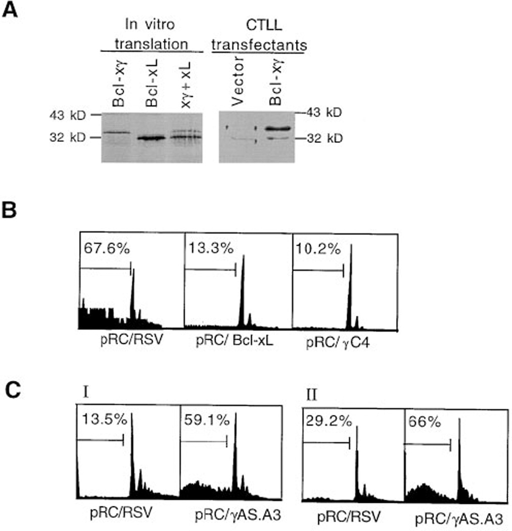
Since studies of previously described Bcl-x isoforms have indicated that they either enhance (Boise et al., 1993; Clarke et al., 1995; Minn et al., 1996) or inhibit (Boise et al., 1993; Fang et al., 1994; Gonzalez-Garcia et al., 1994, 1995) apoptosis, we asked whether stable Bcl-xγ expression after transfection into a T cell line might regulate programmed cell death following TCR ligation. CTLL-2 transfectants that stably overexpressed Bcl-xγ (pRC/γC4) expressed increased constitutive levels of Bcl-xγ protein (Figure 5A) and, after CD3 ligation, displayed almost complete inhibition of apoptosis compared to vector control transfectants (pRC/RSV). For example, CD3 ligation leading to approximately 70% apoptosis of pRC/RSV transfectants led to about 10% apoptosis of pRC/γC4 transfectants (Figure 5B).
Figure 5. The Effect of Overexpression of Bcl-xγ Sense or Antisense cDNA on T Cell Apoptosis.
(A) Overexpression of Bcl-xγ protein in stable Bcl-xγ transfectants. Western blot analysis (right) using a rabbit anti-Bcl-xcommon region (1:1000 dilution) on blots of cell lysate (10 µg) from pRC/RSV (vector control) or pRC/Bcl-xγ CTLL-2 transfectants after 12% SDS-PAGE. Autoradiographed (left) in vitro–translated Bcl-xL and Bcl-xγ alone or together on the same 12% SDS-PAGE were used as migration markers for cellular Bcl-xL and Bcl-xγ. pRC/Bcl-xγ transfectants but not vector control transfectants display a band that reacts with anti-Bcl-x and comigrates with translated Bcl-xγ at approximately 34 kDa.
(B) Influence of Bcl-xγ overexpression on T cell apoptosis. The cell cycle profile represents CTLL-2 cells that stably express the indicated constructs pRC/RSV (vector control), pRC/Bcl-xL, and antipRC/γC4 (Bcl-xγ) after activation by plate-bound anti-CD3. The distribution of cells between the G1, S, and G2/M phases of the cell cycle are shown; the abscissa indicates the relative cell number and the ordinate indicates DNA content based on PI staining. The numbers at top left represent the percentage of cells that display apparent DNA contents of less than diploid (subdiploid), corresponding to the subpopulation of apoptotic cells. These stable transfectants expressed similar levels of CD3 according to immunofluoresence, and all had similar baseline levels of apoptosis (4%– 10%; data not shown). Each panel represents one of triplicate wells; results are representative of three experiments.
(C) Comparison of apoptosis of Bcl-xγ antisense (pRC/ γAS.A3) transfectants with vector control (pRC/RSV) transfectants. In experiments in which relatively low levels of apoptosis were obtained after incubation of transfectants on anti-CD3 coated plates, the degree of apoptosis was substantially elevated in activated pRCγAS.A3 antisense transfectants, compared to vector-transfected controls (pRC/RSV). Each panel represents one of triplicate wells; results are representative of three experiments.
We examined the effects of deliberate inhibition of Bcl-xγ on activated-induced apoptosis. CTLL-2 cells were stably transfected with a Bcl-xγ antisense vector pRC/γAS.A3 containing a 0.4 kb Bcl-xγ fragment in reverse orientation, so that the complementary (antisense) strand of Bcl-xγ would be expressed. Overexpression of Bcl-xγ–specific antisense transcripts in pRCγAS.A3 transfectants had no detectable nonspecific toxic effects, as judged by 3H-labeled thymidine incorporation or thesurvival rate of nonactivated cells according to propidium iodide (PI) staining (data not shown). However, expression of pRCγAS.A3 markedly enhanced receptor-induced apoptosis (Figure 5C), presumably reflecting specific inhibition of efficient translation of Bcl-xγ mRNA transcripts in activated T cells (Robinson-Benion and Holt, 1995; Fakhrai et al., 1996).
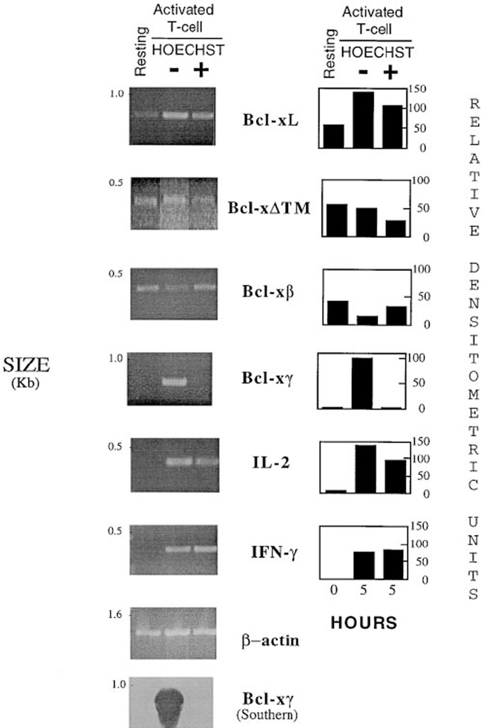
These experiments indicate that T cells that do not express Bcl-xγ after activation might undergo apoptosis. To address this question using nontransformed T cells, we exploited the finding that activated T cells (including Th1 clone O3) undergoing apoptosis after CD3 ligation stain intensely with Hoechst 33342 dye within 4–8 hr after activation, while the Hoechst-negative subpopulation of activated T cells goes on to divide and produce cytokines (Weber et al., 1995). Activated O3 cells were analyzed for Bcl-x isoform expression after Hoechst-based sorting of blast cells 5 hr after CD3 ligation. Bcl-xγ was strongly expressed in the successfully activated fraction but was not detectable in the Hoechst-positive fraction destined to undergo apoptosis (Figure 4C), even after maximum runs of 50 PCR cycles. In contrast, the Bcl-xL, Bcl-xβ and Bcl-ΔTM isoforms were equally expressed in both the viable and apoptotic fractions of activated T cells (Figure 6).
Figure 6. Expression of Bcl-x Isoformsin O3 T Cell Clone Stimulated by Plate-Bound Anti-CD3 Antibody for 5 hr and Sorted by Flow Cytometry.
(Left) After stimulation of the O3 T cell clone by plate-bound anti-CD3 antibody for 5 hr, cells were subjected to staining with Hoechst 33342 dye and PI. After dead cells were gated out, activated cells were sorted into two subpopulations by flow cytometry, Hoechst-high (apoptotic) and Hoechst-low. Total RNA from both Hoechst-positive and Hoechst-negative subpopulations was extracted as described in Experimental Procedures. RT-PCR amplification of IFNγ fragment and IL-2 fragment after stimulation by anti-CD3 in both Hoechst-high and Hoechst-low cells confirmed activation of O3. Bcl-xγ was selectively expressed in Hoechst-low cells but not in Hoechst-high (apoptotic) cells, while all other Bcl-x isoforms are expressed in both forms. Failure to detect Bcl-xγ in Hoechst-high cells did not result from degraded preparations of total RNA or cDNA, since β-actin and other Bcl-x isoforms were detected in these samples.
(Right) The PCR-amplified fragments analyzed on agarose gel were scanned and quantitated using an IS-1000 digital imaging system, followed by normalization, as described above.
Discussion
Differential splicing of individual genes can be an important mechanism for generating lineage-specific proteins that carry out distinct but related biological functions, as exemplified by the expression of CD44 and CD45 isoforms by different cell types (Kincade et al., 1993; Novak et al., 1994). However, despite the potential importance of differentially spliced Bcl-x gene products to thymocyte development, none of the isoforms discovered to date is selectively expressed in thymocytes and T cells. Instead, the known Bcl-x products are expressed in virtually all tissues, can be induced by a variety of stimuli (Fang et al., 1994; Gonzalez-Garcia et al., 1994), and may not regulate TCR-dependent selection in the thymus (Grillot et al., 1995). Here we show that murine Bcl-x generates a 235 aa protein isoform, marked by a unique C-terminus, that is expressed primarily in activated T cells.
Bcl-xγ, cloned and sequenced independently from two different cDNA libraries, contains a functional ORF as judged by sequence analysis, in vitro transcription and translation, and overexpression as a GST fusion protein in Escherichia coli. Transcription of Bcl-xγ is detected by RT-PCR followed by Southern blotting in immature DP thymocytes and may require MHC products for its intrathymic expression. It is expressed at both the mRNA and the protein levels shortly after CD3-mediated activation of T cells, and, unlike other Bcl-x isoforms, expression of Bcl-xγ in peripheral T cells depends on signaling initiated by the TCR but not the IL-2 cytokine receptor. Overexpression of this Bcl-x isoform in a T cell line inhibits anti-CD3–induced apoptosis, while overexpression of a specific antisense transcript of Bcl-xγ enhances apoptosis after TCR ligation. These results strongly argue that expression of Bcl-xγ plays a critical role in T cell survival after TCR ligation.
Although other Bcl-x isoforms can be up-regulated in T cells after TCR and IL-2R ligation, and overexpression of the Bcl-xL isoform can enhance T cell survival (Fang et al., 1994; Boise et al., 1995; Mueller et al., 1996), physiologic expression of these Bcl-x isoforms is not sufficient to confer resistance to apoptosis following TCR ligation, since they are expressed equally well in apoptotic and nonapoptotic T cell blasts. In contrast, failure of Bcl-xγ expression after CD3 ligation represents a genetic marker of programmed cell death, while activated T cells that express Bcl-xγ are spared. The tight coupling of Bcl-xγ expression to the TCR may ensure that survival of activated T cells is governed by the nature of TCR engagement rather than by nonspecific cytokine stimuli. The observation that Bcl-xγ, but not Bcl-xL, expression by immature (DP) thymocytes requires host MHC products suggests that TCR ligation is also necessary for Bcl-xγ expression in this tissue. It is possible that expression of other isoforms such as Bcl-xL may be important to guarantee survival of immature DP thymocytes long enough to provide a cellular substrate for positive and negative selection. Expression of Bcl-xγ after TCR engagement but not other stimuli may represent a genetic mechanism that converts the interaction between peptide ligand and the TCR into a signal leading to successful activation and positive selection. According to this view, failure to induce Bcl-xγ would lead to apoptosis and deletion.
The factors that induce Bcl-xγ expression following TCR ligation are not addressed here. We have reported earlier that successful activation or apoptosis is determined by the relative levels of two distinct signaling pathways connected to the TCR (Weber and Cantor, 1994; Weber et al., 1996). One pathway, marked by an increase in intracellular reactive oxygen intermediates, leads to apoptosis. A second signaling pathway, associated with phosphatidylinositol turnover, leads to successful activation and, according to the studies shown here, may selectively induce Bcl-xγ expression. Although additional experiments are required to test this hypothesis directly, the definition of a tissue-restricted Bcl-x isoform and its role in T cell apoptosis after TCR ligation represents an important step toward a molecular explanation of T cell activation and death.
Experimental Procedures
Mice
C57BL/6J, BALB/c, TCRβ knockout, and BALB/c nu/nu mice were purchased from the Jackson Laboratory (Bar Harbor, ME). MHC class I/II–deficient mice were kindly provided by L. H. Glimcher (Harvard School of Public Health, Boston, MA). Rag2-deficient mice were kindly provided by F.W. Alt (Children’s Hospital, Harvard Medical School, Boston, MA).
T Cells
Splenic T cells were isolated from BALB/c mice using rat anti-mouse B220, Ly-6G, CD11b (Pharmingen, San Diego, CA), along with Dynabeads M450 sheep anti-rat IgG (Dynal), according to a modification of the manufacturer’s protocol. In brief, T cells were negatively isolated from spleens of several mice and suspended in DMEM-2 (Dulbecco’s modified Eagle’s medium supplemented with 2%fetal calf serum) at 1 × 10⁷ cells/ml. The lymphocytes were mixed with a pool of specific antibodies including anti-mouse B220, anti-mouse Ly-6G, and anti-mouse CD11b at 4°C for 40 min. Lymphocytes were rinsed twice with DMEM-2 followed by incubation with Dynabeads M450 sheep anti-rat IgG at 4°C for 30min. After magnetic separation for 2 min, splenic T cells remaining in the supernatants were negatively selected.
CTLL-2 is a mouse T cell line derived from C57BL/6 (Broome et al, 1995). O3 is a murine CD4+ Th1 clone derived from BALB/c mice after in vitro selection for proliferation to OVA in association with antigen-presenting cells of BALB/c (Friedman et al., 1987). The AF3.G7 hybridoma, generated by fusing cow insulin–immune C57BL/6 lymph node cells with the BW5147 thymoma line, expresses a Vβ6+/Vα3.2+ TCR and responds to both cow insulin peptide and to MTV-7, according to IL-2 production (Spinella et al., 1987). EL4 is a mouse lymphoma cell line established in C57BL/6N mice that produces high titers of murine IL-2 (Wein and Roberts, 1965). The RF33.70 CD8 T cell hybridoma was kindly provided by K. Rock (University of Massaschusetts Medical Center, Worcester, MA).
T Cell Activation after CD3 Ligation
Plates precoated with anti-CD3-ε (145.2C11, Pharmingen) by overnight incubation (5 µg/ml in phosphate-buffered saline [PBS] [pH 8.5], 37°C) were given O3 T cell clone (1 × 10⁶/ml) and incubated (37°C) in DMEM plus 5% fetal bovine serum for the indicated time frames before staining with Hoechst 33342 dye and PI and analysis by flow cytometry, as described (Ormerod et al., 1993;Weber et al., 1997). After dead cells had been gated out, activated T cell blasts were sorted into Hoechst-negative (nonapoptotic) and Hoechst-positive (apoptotic) subpopulations on a Becton Dickinson flow cytometer. In experiments with CTLL-2 transfectants, two sets of conditions were used: one to achieve high levels and one to achieve low levels of of CD3-dependent activation and apoptosis. In the first case, 5–10 µg/ml anti-CD3 was used to coat the plates, and for low-level stimulation and apoptosis, reduced concentrations of anti-CD3 were used and cells were grown in IL-2 until just prior to the assay.
Overexpression of Bcl-xγ Sense and Antisense cDNAs in CTLL-2 Cells
The plasmid pRC/RSV containing enhancer–promoter sequences from the Rous sarcoma virus (RSV) long terminal repeat (Invitrogen, San Diego, CA) was used to construct pRC/RSV-Bcl-xL by inserting 0.75 kb fragment that contained a full-length ORF of Bcl-xL. The pRC/RSV-Bcl-xγ vector was constructed by inserting a 1.0 kb fragment containing the full-length ORF of Bcl-xγ. The pRC/RSV-Bcl-xγ antisense vector was constructed by cloning a 0.4 kb XbaI–HindIII Bcl-xγ–specific fragment in reverse orientation in a HindIII–XbaI linearized pRC/RSV vector. This 0.4 kb Bcl-xγ– specific fragment was produced by PCR amplification with an XbaI- containing primer specific for Bcl-xγ coding region 5′-GGCCTCTAGAGGTGTGAGTGGAGGTACACCC-3′ and a HindIII- containing primer specific for Bcl-xγ noncoding region 5′-GGCCAAGCTTCGTCCTTCCTGAAGTCCTCCT-3′. Correct orientation of Bcl-xL, Bcl-xγ, and Bcl-xγ antisense inserts in the recombinant vector was confirmed by restriction enzyme digestion and DNA sequencing. Stable expression of the CTLL-2 T cell line was achieved after transfection with 10 µg of XbaI-linearized vector by electroporation in a Gene-Pulser II (Bio-Rad, Hercules, CA) at 270 V and 950 µF for 20 ms.
Two days after transfection, T cells were diluted into 96-well plates at 5 × 10³ cells/0.1 ml or 1 × 10⁴ cells/0.1 ml/well in media containing 750 µg/ml G418. After 2 weeks, individual clones resistant to G418 were selected, expanded, and maintained in medium containing 250 mg/ml G418. In addition, the empty vector pRC/RSV was used to simultaneously transfect and expand CTLL-2 cells according to the same protocol. RT-PCR was performed to confirm efficient expression of Bcl-x genes in the transfected clones using RNA after digestion with RNase-free DNase before RT to avoid contamination of RNA preparations. Total RNA from Bcl-xL and Bcl-xγ transfectants and cells transfected with the pRC/RSV control vector was amplified by RT-PCR, separated on agarose gels, and confirmed by Southern blot hybridization with a 32P-labeled DNA probe prepared from the cDNA coding for the Bcl-x common region. Bcl-xγ antisense transfectants were verified by RT-PCR amplification of total RNA with an upstream primer specific for vector sequence between promoter and insert and a downstream Bcl-xγ antisense–specific primer.
Isolation of cDNA Clones and DNA Sequencing
A mouse thymus λ ZapII cDNA library derived from BALB/cJ mice (Stratagene, La Jolla, CA) was screened with a 32P-labeled 60-meroligonucleotide derived from a human Bcl-x cDNA sequence that is homologous to a conserved region of chicken Bcl-x (bp 423–483) (Boise et al. 1993) and hybridized to approximately 106 phages blotted on 20 filters according to Wood et al. (1985) and Jacobs et al. (1988). Six positive clones were identified and subcloned into Bluescript plasmids, and positive DNA inserts were sequenced (twice each) by a double-stranded DNA dideoxy chain termination method using T7 DNA polymerase (US Biochem).
In Vitro Transcription and Translation
Recombinant Bluescript plasmids containing cDNA from Bcl-xL, Bcl-xS, Bcl-xβ, and Bcl-xγ (1 µg/each) were linearized with a unique PstI restriction enzyme at polycloning sites. In vitro transcription and translation were then performed at 30°C for 90 min using a TNT T7/T3-coupled reticulocyte lysate system for a total reaction volume of 50 µl each, according to the manufacturer’s protocol (Promega). Newly synthesized and [35S]methionine- labeled proteins (5 µl/each) were then analyzed by a 12% sodium dodecyl sulfate polyacrylamide gel electrophoresis (SDS-PAGE) and subjected to autoradiography.
PCR Cloning
Total RNA from murine thymus (BALB/cJ) was reverse transcribed on a GeneAmp PCR 9600 (Perkin-Elmer) using a GeneAmp RNA-PCR kit (Perkin-Elmer Cetus) at 42°C for 30 min, 99°C for 5 min, and 4°C for 5 min, according to the manufacturer’s protocols. We used a primer specific for the 5′ upstream Bcl-x common region (5′-TCGCTCGCCCACATCCCAGCTTCACATAACCCC-3′) and a second primer specific for the 3′ downstream Bcl-xγ region (5′-CTGGTTCGGCCCACGTCCTTCCTGAAGT-CCTCC-3′) for PCR cloning. Amplification products were separated by agarose gel, purified with Geneclean II kit (Bio101), subcloned into the PCR-Direct vector, and sequenced by dideoxy chain termination.
Gene Expression Measured by RT-PCR
Total RNA extracted from murine tissues was homogenized using RNAzol B (Tel-Test) before RT-PCR was performed. In addition to the primers used, a primer containing a 3′ region unique to Bcl-xL and Bcl-xS (5′-CCACCAACAAGACAGGCT-3′) was used to pair with the 5′ primer from the Bcl-x common region for amplification of the Bcl-xL fragment. Similarly, a primer complementary to the 3′ specific region of Bcl-xΔTM (5′-CTCTCCTCCCTCA-CACACCCCTCTC-3′) and primer 3ep (5′-AAGATACAGGTCCCTTAAA-3′) complementary to the 3′ specific region of Bcl-xβ were used to pair with a 5′ primer for the Bcl-x common region for amplification of Bcl-xΔTM and Bcl-xβ fragments, respectively. A pair of primers of mouse β-actin was used as an internal control for RT-PCR to ensure the use of similar amounts of total RNA and cDNA in every experiment. Oligonucleotides used as primers for PCR amplification of the mouse β-actin fragment were 5′-ATGGATGACGATATCGCTGC-3′ and 5′-CTAGAAGCACTTGCGGTGCAC-3′. Primers specific for IL-2, IL-3, and IFNγ were used as positive controls for T cell activation in RT-PCR. Oligonucleotides used as primers for PCR amplification of the mouse IL-2 fragment were 5′-TTCAAGCTCCACTTCAAGCTC-3′ and 5′-GACAGAAGGCTATCCATCTCC-3′. Primer sequences for PCR amplification of the mouse IFNγ fragment were 5′-TGCATCTTGGCTTTGCAGCTCTTCCTCATG-3′ and 5′-GGACCTGTGGGT-TGTTGACCTCAAACTTG-3′. PCR-amplified fragments were analyzed on agarose gels followed by scanning and quantitation using an IS-1000 digital imaging system (Alpha Innotech), with exposure times adjusted so that the intensity of DNA fragment signals corresponded to the linear range of densitometric detection. To ensure that comparisons of cDNA levels in different samples were based on the same amount of cDNA in each sample, the area under the densitometric peak of each sample was divided by the area under the β-actin densitometric peak for the corresponding sample. The ratios of Bcl-x isoform and control (IL-2 or IFN γ) cDNA to β-actin cDNA for each sample were expressed as relative densitometric units. PCR reactions were typically performed through 35–45 cycles using Taq DNA polymerase (Perkin-Elmer) supplemented with TaqStart antibody to maintain the specificity of amplified fragments (Clon-tech). Each three-step thermal cycle consisted of 30 s at 94°C, 30 s at 60°C, and 30 s at 72°C. To encompass the exponential phase of the amplification, 25 µl of the reaction mix was removed at regular intervals during PCR, as previously described (Moore et al., 1994). PCR products in 10 µl for each sample visualized by agarose gel electrophoresis and ethidium bromide staining were positively identified by size, and Bcl-xγ fragments were further confirmed by Southern blot hybridization. A negative control containing all reagents except cDNA was included in each PCR analysis.
Southern Blot Hybridization
The RT-PCR products amplified with Bcl-xγ–specific primers were separated on agarose gels, blotted onto a nylon filters (Micron Separations), and subjected to ultraviolet-crosslinking. A 360 bp Bcl-xγ–specific probe prepared by PCR amplification from the recombinant plasmid Bcl-x7 encoding Bcl-xγ using primers that do not overlap with the primers was used to detect gene expression, followed by labeling with [α-32P]dCTP (3000 Ci/mmol, New England Nuclear) by random oligomer priming (Oligolabeling kit, Pharmacia). Radiolabeled probes were purified using Nick-spin columns (Pharmacia) to remove excess unincorporated radioactive nucleotides before hybridization overnight at 42°C in 6× sodium chloride/sodium citrate (SSC) buffer containing 50% formamide. The filters were washed in 2× SSC containing 0.1% SDS at 42°C for 30 min and in 0.2× SSC containing 0.1% SDS at 65°C for 30 min, followed by autoradiography.
Apoptosis Assays
Plates precoated with anti-mouse CD3 antibody (1–10 µg/ml, 145.2C11 Pharmingen) were washed three times before addition of CTLL-2 clones that had been washed three times with IL-2–free RPMI 1640 medium supplemented with 5% FCS and incubated at 2 ml/well at a concentration of 1.25 × 10⁵ /ml at 37°C for 24 hr. In some experiments intended to maximize CD3-induced apoptosis, incubation medium was replaced, at 6 and 12 hr after cells were plated, with fresh RPMI 1640 medium supplemented with 5% FCS to reduce secondary responses to potential growth factors secreted by cells activated after TCR ligation. The percentage of cells undergoing apoptosis for each transfected clone was analyzed by PI staining (Broome et al., 1995; McGahon et al., 1995). In brief, 18–24 hr after activation by plate-bound anti-CD3, cells were harvested, rinsed twice with cold PBS containing 5 mM EDTA, fixed with 50% ethanol in PBS containing 5 mM EDTA for 30 min at room temperature, treated with 40 µg/ml of DNase-free RNase A in PBS for 30 minutes, and stained with 50 µg/ml of PI in PBS before analysis in an Epics XL flow cytometry system using a standard setting in semilogarithmic mode (Coulter). Since partial loss of DNA from apoptotic cells due to activation of endogenous endonuclease(s) and/or marked condensation of the chromatin accompanies apoptosis and renders these areas of DNA inaccessible to PI staining, subdiploid cells with fluorescence lower than that of G0/G1 cells were considered to be apoptotic (McGahon et al., 1995), while cells in G0/G1, S, and G2/M phases were scored as viable.
Generation of GST Fusion Proteins
A cDNA fragment encoding full-length Bcl-xγ with EcoRI on both ends was generated by PCR using a primer (Fu-5) specific for the N-terminus of the Bcl-x common region (5′-CCGGGAATTCATCTCAGAGCAACCGGGAGCTGGTG-3′) and a primer (Fu-3γ) specific for the Bcl-xγ–specific 3′ noncoding region (5′-CCGGGAATTCGGATCCCGTCCTTCCTGAAGTCCTCCT-3′). A cDNA fragment encoding full-length Bcl-xL was produced by PCR using the common region primer Fu-5 and the primer Fu-3 (5′-CCGGGAATTCTCAGTTGAAGCCCTCCTTGCCTTTCCG-3′) for the Bcl-xL–specific 3′ region except the hydrophobic transmembrane domain on the C-terminus, to avoid the potential problem of inclusion bodies during overexpression in bacteria. Similarly, a cDNA fragment encoding the 47 aa residue of the Bcl-xγ specific C-terminus was generated by PCR with a primer specific for the N-terminus ofBcl-xγ–specific region 5′-CCGGGAATTCAGGTGTGAGTGGAGGTACACCC-3′ and primer Fu-3γ. The underlined sequences in these primers were designed for subcloning. All of these Bcl-x fragments were fused in frame to the C-terminus of GST protein after cloning into the EcoRI site of the GST expression vector pGEX-3X and were further examined by restriction digestion and DNA sequencing before transformation into the BL-21 strain of E. coli.
The GST and the fusion proteins GST-Bcl-xγ, GST-Bcl-xL, and GST-Bcl-xγ-C containing the unique C-terminus of Bcl-xγ were purified according to the manufacturer’s protocol (Pharmacia). In brief, 2 ml aliquots of bacterial cultures freshly grown overnight and containing plasmids pGEX-3X, pGEX-3X-Bcl-xγ, pGEX-3X-Bcl-xL, or pGEX-3X-Bcl-xγ-C were respectively inoculated into 200 ml of 2YT medium with 50 µg/ml of ampicillin. Expression of the fusion proteins was induced by adding isopropylthiogalactoside (final concentration, 0.1mM). Samples were then centrifuged and ultrasonified (Sonifier 450, Branson), digested with Triton X-100, and centrifuged (10,000 rpm for 10 min). Supernatants were incubated with 2 ml of glutathione-agarose beads (Sigma) and eluted with 4 ml of 10 mM reduced glutathione before analysis on 12% SDS-PAGE and determination of protein concentration by Bradford’s reagent (Bio-Rad).
Antibody Preparation
Since several isoforms of Bcl-x, such as Bcl-xL, Bcl-xβ, Bcl-xΔTM, and Bcl-xγ share the N-terminal common region of Bcl-x, fusion protein GST-Bcl-xγ cannot be used as an immunogen to raise antibody specific to Bcl-xγ. To raise Bcl-xγ–specific antibody, the 47 aa protein of the Bcl-xγ–specific C-terminal region was fused in frame to the C-terminus of GST, resulting in production of the fusion protein GST-Bcl-xγ-C. A polyclonal antiserum to GST-Bcl-xγ-C was raised according to standard procedures (Bailey, 1994). Polyclonal antibody was purified on immobilized GST columns (Pierce) to remove the antibody to GST, and the immunoglobulin fraction in the antiserum was purified using E-Z-SEP (Pharmacia). The specificity and titer of the antibody were analyzed using serial dilution of antibody to stain Western blots with an equal amount of GST, GST-Bcl-xγ-C, or GST-Bcl-xL and GST-Bcl-xγ.
Western Blotting
Proteins overexpressed in transformed E. coli or in stably transfected clones or prepared from T cells were subjected to 12%SDS-PAGE with Tris-glycine buffer and transferred onto nitrocellulose filters in 20% methanol in Tris-glycine buffer, using an enhanced chemiluminescence kit (Amersham). In brief, protein blots were incubated in 5%bovine serum albumin, 0.2% Tween 20 in Tris-buffered saline (TBST) for 1 hr at room temperature, incubated with the first antibody in TBST at room temperature for 1 hr, rinsed, and incubated with an anti-rabbit immunoglobulin peroxidase conjugate (Boehringer Mannheim). After washing, blots were subjected to fluorography.
Acknowledgments
We are grateful to Linda K. Clayton and Antonio J. da Silva for mouse thymus cDNA; to YaLing Zhou for computer analysis; and to XiaoYan Ke, Alistair Noble, Gary Pestano, and the members of our laboratory for helpful discussions and assistance. We thank Alison Angel for assistance in the preparation of this manuscript. This work was supported in part by National Institutes of Health research grants AI37833, AI12184, and AI13600 to H. C.
References
- Akbar AN, Salmon M, Savil J, Janossy G. A possible role for bcl-2 in regulating T cell memory: a ‘balancing act’ between cell death and survival. Immunol. Today. 1993;14:526–532. doi: 10.1016/0167-5699(93)90181-J. [DOI] [PubMed] [Google Scholar]
- Bailey GS. The raising of a polyclonal antiserum to a protein. In: Walker JM, editor. Methods in Molecular Biology. vol. 32. Totowa, NJ: Humana Press; 1994. pp. 381–388. [DOI] [PubMed] [Google Scholar]
- Boise LH, Gonzalez-Garcia M, Postema CE, Ding LY, Lindsten T, Turka LA, Mao XH, Nunez G, Thompson CB. Bcl-x, a Bcl-2-related gene that functions as a dominant regulator of apoptotic cell death. Cell. 1993;74:597–608. doi: 10.1016/0092-8674(93)90508-n. [DOI] [PubMed] [Google Scholar]
- Boise LH, Minn AJ, Noel PJ, June CH, Accavitti MA, Lindsten T, Thompson CB. CD28 costimulation can promote T cell survival by enhancing the expression of Bcl-xL. Immunity. 1995;3:87–98. doi: 10.1016/1074-7613(95)90161-2. [DOI] [PubMed] [Google Scholar]
- Broome HE, Dargan CM, Krajewski S, Reed JC. Expression of Bcl-2, Bcl-x and Bax after T cell activation and IL-2 withdrawal. J. Immunol. 1995;155:2311–2317. [PubMed] [Google Scholar]
- Chao DT, Linette GP, Boise LH, White LS, Thompson CB, Korsmeyer SJ. Bcl-xL and Bcl-2 repress a common pathway of cell death. J. Exp. Med. 1995;182:821–828. doi: 10.1084/jem.182.3.821. [DOI] [PMC free article] [PubMed] [Google Scholar]
- Clarke MF, Apel IJ, Benedict MA, Eipers PG, Sumantran V, Gonzalez-Garcia M, Doedens M, Fukunaga N, Davidson B, Dick JE, et al. A recombinant bcl-xs adenovirus selectively induces apoptosis in cancer cells but not in normal bone marrow cells. Proc. Natl. Acad. Sci. USA. 1995;92:11024–11028. doi: 10.1073/pnas.92.24.11024. [DOI] [PMC free article] [PubMed] [Google Scholar]
- Coen DM. The polymerase chain reaction. In: Ausubel FM, Brent R, Kingston RE, Moore DD, Seidman JG, Smith JA, Struhl K, Albright LM, Coen DM, Varki A, editors. Current Protocols in Molecular Biology. vol. 2. New York: Green Publishing and John Wiley & Sons; 1994. p. 15.01. [Google Scholar]
- Cory S. Regulation of lymphocyte survival by the Bcl-2 gene family. Annu. Rev. Immunol. 1995;13:513–543. doi: 10.1146/annurev.iy.13.040195.002501. [DOI] [PubMed] [Google Scholar]
- Critchfield JM, Racke MK, Zuniga-Pflucker JC, Cannella B, Raine CS, Goverman J, Lenardo MJ. T cell deletion in high antigen dose therapy of autoimmune encephalomyelitis. Science. 1994;263:1139–1143. doi: 10.1126/science.7509084. [DOI] [PubMed] [Google Scholar]
- Crump AL, Markowitz JS, Grusby MJ, Glimcher LH, Canmovetor H. Thymocyte development in MHC-deficient mice: Evidence for stochastic commitment to the CD4 and CD8 lineages. Proc. Natl. Acad. Sci. USA. 1993;90:10739. doi: 10.1073/pnas.90.22.10739. [DOI] [PMC free article] [PubMed] [Google Scholar]
- Fakhrai H, Dorigo O, Shawler DL, Lin H, Mercola D, Black KL, Royston I, Sobol RE. Eradication of established intracranial rat gliomas by transforming growth factor b antisense gene therapy. Proc. Natl. Acad. Sci. USA. 1996;93:2909–2914. doi: 10.1073/pnas.93.7.2909. [DOI] [PMC free article] [PubMed] [Google Scholar]
- Fang W, Rivard JJ, Mueller DL, Behrens TW. Bcl-xL rescues WEHI 231 B-lymphocytes from oxidant-mediated death following diverse apoptotic stimuli. J. Immunol. 1994;153:4388–4398. [PubMed] [Google Scholar]
- Friedman S, Sillcocks D, Cantor H. Alloreactivity of an OVA-specific T cell clone. I. Stimulation by class II MHC and novel non-MHC B-cell determinants. Immuogenetics. 1987;26:193–203. doi: 10.1007/BF00346512. [DOI] [PubMed] [Google Scholar]
- Gonzalez-Garcia M, Perez-Ballestero R, Ding LY, Duan L, Boise LH, Thompson CB, Nunez G. Bcl-xL is the major mRNA form expressed during murine development and its product localizes to mitochondria. Development. 1994;120:3033–3042. doi: 10.1242/dev.120.10.3033. [DOI] [PubMed] [Google Scholar]
- Gonzalez-Garcia M, Garcia I, Ding L, O’Shea S, Boise LH, Thompson CB, Nunez G. Bcl-x is expressed in embryonic and postnatal neural tissues and functions to prevent neurional cell death. Proc. Natl. Acad. Sci. USA. 1995;92:4304–4308. doi: 10.1073/pnas.92.10.4304. [DOI] [PMC free article] [PubMed] [Google Scholar]
- Gorina S, Pavletich NP. Structure of the p53 tumor suppressor bound to the ankyrin and SH3 domains of 53BP2. Science. 1996;274:1001–1005. doi: 10.1126/science.274.5289.1001. [DOI] [PubMed] [Google Scholar]
- Grillot DAM, Merino R, Nunez G. Bcl-xL displays restricted distribution during T cell development and inhibits multiple forms of apoptosis but not clonal deletion in transgenic mice. J. Exp. Med. 1995;182:1973–1983. doi: 10.1084/jem.182.6.1973. [DOI] [PMC free article] [PubMed] [Google Scholar]
- Hatada EN, Niters A, Wulezyu FG, Naumann M, Meyer R, Nucifora G, McKeithan TW, Scheidereit C. The ankyrin repeat domains of the NF-κB precursor p105 and the protooncogene cl-3 act as specific inhibitors of NF-κB DNA binding. Proc. Natl. Acad. Sci. USA. 1992;89:2489–2493. doi: 10.1073/pnas.89.6.2489. [DOI] [PMC free article] [PubMed] [Google Scholar]
- Hockenberry DM. bcl-2, a novel regulator of cell death. Bioessays. 1995;17:631–638. doi: 10.1002/bies.950170709. [DOI] [PubMed] [Google Scholar]
- Hockenberry D, Nunez G, Milliman C, Schreiber RD, Korsmeyer SJ. Bcl-2 is an inner mitochondrial membrane protein that blocks programmed cell death. Nature. 1990;348:334–336. doi: 10.1038/348334a0. [DOI] [PubMed] [Google Scholar]
- Hockenberry DM, Oltvai ZN, Yiu XM, Milliman CL, Korsmeyer SJ. Bcl-2 functions in an antioxidant pathway to prevent apoptosis. Cell. 1993;75:241–251. doi: 10.1016/0092-8674(93)80066-n. [DOI] [PubMed] [Google Scholar]
- Ignatowicz L, Kappler JW, Marrack P. The effects of chronic infection with a superantigen producing virus. J. Exp. Med. 1992;175:917–923. doi: 10.1084/jem.175.4.917. [DOI] [PMC free article] [PubMed] [Google Scholar]
- Jacobs KA, Rudersdorf R, Neill SD, Dougherty JP, Brown EL, Fritsch EF. The thermal stability of oligonucleotide duplexes is sequence-independent in tetraalkylammonium salt solutions: application to identifying recombinant DNA clones. Nucleic Acids Res. 1988;16:4637–4650. doi: 10.1093/nar/16.10.4637. [DOI] [PMC free article] [PubMed] [Google Scholar]
- Kincade PW, He Q, Ishihara K, Miyake K, Lesley J, Hyman R. CD44 and other cell interaction molecules contributing to B lymphopoiesis. Curr. Topics Microbiol. Immunol. 1993;184:215–222. doi: 10.1007/978-3-642-78253-4_17. [DOI] [PubMed] [Google Scholar]
- Klein ES, Simmons DM, Swanson LW, Rosenfeld MG. Tissue-specific RNA splicing generates an ankyrin-like domain that affects the dimerization and DNA-binding properties of a bHLH protein. Genes Dev. 1993;7:55–71. doi: 10.1101/gad.7.1.55. [DOI] [PubMed] [Google Scholar]
- Ma A, Pena J, Chang B, Margosian E, Davidson L, Alt FW, Thompson CB. Bcl-x regulates the survival of double-positive thymocytes. Proc. Natl. Acad. Sci. USA. 1995;92:4763–4767. doi: 10.1073/pnas.92.11.4763. [DOI] [PMC free article] [PubMed] [Google Scholar]
- McGahon AJ, Martin SJ, Bissonnette RP, Mahboubi A, Shi Y, Mogil RJ, Nishioka WK, Green DR. The end of the (cell) line: methods for the study of apoptosis in vitro. Meth. Cell Biol. 1995;46:153–185. doi: 10.1016/s0091-679x(08)61929-9. [DOI] [PubMed] [Google Scholar]
- Minn AJ, Boise LH, Thomspon CB. Bcl-xS antagonizes the protective effects of Bcl-xL. J. Biol. Chem. 1996;271:6306–6312. doi: 10.1074/jbc.271.11.6306. [DOI] [PubMed] [Google Scholar]
- Moore NC, Anderson G, Williams GT, Owen JJT, Jenkinson EJ. Developmental regulation of bcl-2 expression in the thymus. Immunology. 1994;81:115–119. [PMC free article] [PubMed] [Google Scholar]
- Motoyama N, Wang F, Roth KA, Sawa H, Nakayama K-I, Nakayama K, Negishi I, Senju S, Zhang Q, Fujii S, et al. Massive cell death of immature hematopoietic cells and neurons in Bcl-x deficient mice. Science. 1995;267:1506–1510. doi: 10.1126/science.7878471. [DOI] [PubMed] [Google Scholar]
- Mueller DL, Seiffert S, Fang W, Behrens TW. Differential regulation of Bcl-2 and Bcl-x by CD3, CD28, and the IL-2 receptor in cloned CD41helper T cells. J. Immunol. 1996;156:1764–1771. [PubMed] [Google Scholar]
- Nguyen M, Millar DG, Yong VW, Korsmeyer SJ, Shore GC. Targeting of Bcl-2 to the mitochondrial outer membrane by a COOH-terminal signal anchor sequence. J. Biol. Chem. 1993;268:25265–25268. [PubMed] [Google Scholar]
- Novak TJ, Ferber D, Leitenberg D, Hong S-C, Johnson P, Bottomly K. Isoforms of the transmembrane tyrosine phosphatase CD45 differentially affect T cell recognition. Immunity. 1994;1:109–119. doi: 10.1016/1074-7613(94)90104-x. [DOI] [PubMed] [Google Scholar]
- Nunez G, Merino R, Grillot D, Gonzalez-Garcia M. Bcl-2 and Bcl-x: regulatory switches for lymphoid death and survival. Immunol. Today. 1994;15:582–588. doi: 10.1016/0167-5699(94)90221-6. [DOI] [PubMed] [Google Scholar]
- Ormerod MG, Sun XM, Snowden RT, Davies R, Fearnhead H, Cohen GM. Increased membrane permeability of apoptotic thymocytes: a flow cytometry study. Cytometry. 1993;14:595–602. doi: 10.1002/cyto.990140603. [DOI] [PubMed] [Google Scholar]
- Pircher HP, Burki K, Lang R, Hengartner H, Zinkernagel RM. Tolerance induction in double specific T cell receptor transgenic mice varies with antigen. Nature. 1989;342:559–561. doi: 10.1038/342559a0. [DOI] [PubMed] [Google Scholar]
- Reed JC. Bcl-2 and the regulation of programmed cell death. J. cell Biol. 1994;124:1–6. doi: 10.1083/jcb.124.1.1. [DOI] [PMC free article] [PubMed] [Google Scholar]
- Robinson-Benion C, Holt JT. Antisense techniques. Meth. Enzymol. 1995;254:3–23. doi: 10.1016/0076-6879(95)54025-3. [DOI] [PubMed] [Google Scholar]
- Rocha B, von Boehmer H. Peripheral selection of the T cell repertoire. Science. 1991;251:1225–1228. doi: 10.1126/science.1900951. [DOI] [PubMed] [Google Scholar]
- Spinella DG, Hansen TH, Walsh WD, Behlke MA, Tillinghast JP, Chou HS, Whiteley PJ, Kapp JA, Pierce CW, Shevach EM, et al. Receptor diversity of insulin-specific T cell lines from C57BL (H-2b) mice. J. Immunol. 1987;138:3991–3995. [PubMed] [Google Scholar]
- Takahashi T, Honda H, Hirai H, Tsujimoto Y. Overexpressed Bcl-xL prevents bacterial superantigen-induced apoptosis of thymocytes in vitro. Cell Death Differentiation. 1997;4:159–165. doi: 10.1038/sj.cdd.4400214. [DOI] [PubMed] [Google Scholar]
- Webb S, Morris C, Sprent J. Extrathymic tolerance of mature T cells: clonal elimination as a consequence of immunity. Cell. 1990;63:1249–1256. doi: 10.1016/0092-8674(90)90420-j. [DOI] [PubMed] [Google Scholar]
- Weber GF, Cantor H. Phosphatidylinositol synthesis is a proximal event in intracellular signalling coupled to TCR ligation: differential induction by conventional antigen and retroviral superantigen. J. Immunol. 1994;152:4433–4443. [PubMed] [Google Scholar]
- Weber GF, Abromson-Leeman S, Cantor H. A signalling pathway coupled to T cell receptor ligation by superantigens which leads to programmed cell death. Immunity. 1995;2:363–372. doi: 10.1016/1074-7613(95)90144-2. [DOI] [PubMed] [Google Scholar]
- Weber GF, Ashkar S, Glimcher MJ, Cantor H. Receptor-ligand interaction between CD44 and osteopontin (Eta-1) Science. 1996;271:509–511. doi: 10.1126/science.271.5248.509. [DOI] [PubMed] [Google Scholar]
- Weber GF, Daley J, Kraeft S-K, Chen LB, Cantor H. Measurement of apoptosis in heterogenous cell populations. Cytometry. 1997;27:136–144. doi: 10.1002/(sici)1097-0320(19970201)27:2<136::aid-cyto5>3.0.co;2-e. [DOI] [PubMed] [Google Scholar]
- Wein J, Roberts E. Influence of H-2 isoantibody on electrophoretic behavior of EL4 lymphoma cells. Cancer Res. 1965;25:1753–1758. [PubMed] [Google Scholar]
- Wood WI, Gitschier J, Lasky LA, Lawn RM. Base composition-independent hybridization in tetramethylammonium chloride: a method for oligonecleotide screening of highly complex gene libraries. Proc. Natl. Acad. Sci. USA. 1985;82:1585–1588. doi: 10.1073/pnas.82.6.1585. [DOI] [PMC free article] [PubMed] [Google Scholar]
- Yang X-F, Crine P, Boileau G. The nature of topogenic sequences determines the transport competence of topological mutants of neutral endopeptidase-24.11. Biochemical J. 1995;312:99–105. doi: 10.1042/bj3120099. [DOI] [PMC free article] [PubMed] [Google Scholar]
- Zhang L, Shannon J, Sheldon J, Teh H-S, Mak TW, Miller RG. Role of infused CD8+ cells in the induction of peripheral tolerance. J. Immunol. 1994;152:2222–2228. [PubMed] [Google Scholar]
- Zheng W-P, Flavell RA. The transcription factor GATA-3 is necessary and sufficient for Th2 cytokine gene expression in CD4 T cells. Cell. 1997;89:587–596. doi: 10.1016/s0092-8674(00)80240-8. [DOI] [PubMed] [Google Scholar]
- Zinkernagel RM, Moskophidis D, Kundig T, Oehen S, Pircher H, Hengartner H. Effector T cell induction and T cell memory versus peripheral deletion of T cells. Immunol. Rev. 1993;131:199–223. doi: 10.1111/j.1600-065x.1993.tb01517.x. [DOI] [PubMed] [Google Scholar]


