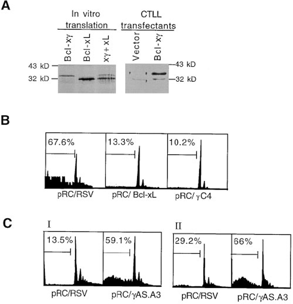
Figure 5. The Effect of Overexpression of Bcl-xγ Sense or Antisense cDNA on T Cell Apoptosis.
(A) Overexpression of Bcl-xγ protein in stable Bcl-xγ transfectants. Western blot analysis (right) using a rabbit anti-Bcl-xcommon region (1:1000 dilution) on blots of cell lysate (10 µg) from pRC/RSV (vector control) or pRC/Bcl-xγ CTLL-2 transfectants after 12% SDS-PAGE. Autoradiographed (left) in vitro–translated Bcl-xL and Bcl-xγ alone or together on the same 12% SDS-PAGE were used as migration markers for cellular Bcl-xL and Bcl-xγ. pRC/Bcl-xγ transfectants but not vector control transfectants display a band that reacts with anti-Bcl-x and comigrates with translated Bcl-xγ at approximately 34 kDa.
(B) Influence of Bcl-xγ overexpression on T cell apoptosis. The cell cycle profile represents CTLL-2 cells that stably express the indicated constructs pRC/RSV (vector control), pRC/Bcl-xL, and antipRC/γC4 (Bcl-xγ) after activation by plate-bound anti-CD3. The distribution of cells between the G1, S, and G2/M phases of the cell cycle are shown; the abscissa indicates the relative cell number and the ordinate indicates DNA content based on PI staining. The numbers at top left represent the percentage of cells that display apparent DNA contents of less than diploid (subdiploid), corresponding to the subpopulation of apoptotic cells. These stable transfectants expressed similar levels of CD3 according to immunofluoresence, and all had similar baseline levels of apoptosis (4%– 10%; data not shown). Each panel represents one of triplicate wells; results are representative of three experiments.
(C) Comparison of apoptosis of Bcl-xγ antisense (pRC/ γAS.A3) transfectants with vector control (pRC/RSV) transfectants. In experiments in which relatively low levels of apoptosis were obtained after incubation of transfectants on anti-CD3 coated plates, the degree of apoptosis was substantially elevated in activated pRCγAS.A3 antisense transfectants, compared to vector-transfected controls (pRC/RSV). Each panel represents one of triplicate wells; results are representative of three experiments.

