Abstract
Introduction
Operating on an enlarged spleen via the laparoscopic approach presents several challenges. A homemade bag may facilitate retrieval of the enlarged spleen assisted by a laparoscope and save medical expense.
Aim
To assess the feasibility and safety of laparoscopic splenectomy for moderate or massive splenomegaly using our technique and a homemade retrieval bag.
Material and methods
Fifty patients underwent laparoscopic splenectomy for moderate or massive splenomegaly which was defined as the major axis exceeding 17 cm by abdominal computed tomography. A homemade retrieval bag made from a commercial sterile infusion container which costs about US$ 1–2 per piece was used for spleen retrieval. Two transabdominal sutures for suspension of the retrieval bag were made to aid specimen removal in this technique.
Results
There were 31 males and 19 females with mean age of 56 ±11 years. Laparoscopic splenectomy was successfully completed in 49 of these 50 patients. Overall, mean operative time was 149 ±31 min (range: 100–252 min). Median estimated blood loss was 189 ±155 ml (range: 50–920 ml). There were 12 minor complications but no mortality. Time to discharge after surgery ranged from 3 to 9 (mean: 4.7 ±1.7 days). The average splenic weight was 729 ±74 g (range: 632–930 g).
Conclusions
Our preliminary results indicate that laparoscopic splenectomy is feasible and safe for moderate or massive splenomegaly and may be a well-tolerated alternative to open splenectomy. Not only is the cost of our homemade retrieval bag low, but also it is easy to make and ready to use.
Keywords: laparoscopic splenectomy, splenomegaly, homemade retrieval bag
Introduction
Laparoscopic splenectomy (LS) has been adopted as a feasible and safe procedure for management of various hematological disorders including idiopathic thrombocytopenic purpura, hematologic malignancy, and hereditary spherocytosis [1–7]. The laparoscopic approach offers several advantages over open splenectomy, such as shorter hospital stay, decreased blood loss, and low perioperative morbidity [1–7]. However, operating on an enlarged spleen via the laparoscopic approach presents several challenges including limited working space, difficulty with organ retraction and specimen retrieval, although massive splenomegaly is not an absolute contraindication [8–10]. In addition, splenomegaly may increase the risk of life-threatening hemorrhage, and the incidence of conversion to open splenectomy in such patients [11–13]. Over the past decade, surgical techniques and instrumentation have been improved to overcome the difficulty of laparoscopic management for an enlarged spleen.
Aim
We have developed a modified method using our homemade bag, which may facilitate retrieval of the enlarged spleen assisted by a laparoscope. In this study, we intend to assess the feasibility and safety of LS, performed by our method, for treatment of moderate massive splenomegaly.
Material and methods
Between January 2005 and December 2010, 50 patients underwent LS for moderate massive splenomegaly in our department. Moderate massive splenomegaly is defined as a spleen weighing over 600 g or the major axis of the spleen exceeding 17 cm by abdominal computed tomography (CT). Preoperatively, all of the patients had detailed physical examination, chest radiograph, and routine biochemical and hematological tests. In addition, imaging investigations (ultrasonography, computed tomography, and magnetic resonance imaging) were conducted to determine the spleen size and vascularities. Data collection included patient demographics, diagnosis, operative details, and postoperative morbidity and mortality. Splenic weight was the aggregate weight of the morcellated and aspirated splenic tissue as measured in the pathology laboratory. At least one week prior to surgery, all patients received a meningococcal and pneumococcal vaccine. Antibiotic prophylaxis was administered preoperatively and continued postoperatively. Patients with hemoglobin < 8 g/dl or platelets < 50000/mm3 were administered fresh whole blood or platelet-rich plasma, respectively, and in most cases it proved unnecessary.
Technique of laparoscopic splenectomy
Patient position
The patient was positioned in a lateral decubitus position with the left side up and a flank cushion placed under the contralateral side, thus enlarging the space between the costal margin and the iliac crest. The left arm was extended and suspended. The surgeon faced the patient while the assistant stood at the right side of the operator.
Trocar placement
Initially, the peritoneal cavity is accessed via the open technique under direct vision and carbon dioxide is then inflated to create a pneumoperitoneum after insertion of a 12-mm trocar below the inferior tip of the spleen, parallel to the left costal margin. One 5-mm trocar is inserted under the xyphoid and another 12-mm trocar is placed midway between the 5-mm and 12-mm trocars for dissection and hilar devascularization. A third 12-mm trocar, used for the laparoscope, is inserted at the umbilicus.
Surgical technique
The surgical procedure is greatly facilitated by the LigaSure™ (Valleylab™, Tyco Healthcare Group, Colorado, USA) vessel sealing system. The operation begins to divide the gastrosplenic ligament between the stomach and spleen to enter the lesser sac and to expose the tortuous pulsating splenic artery. After the splenic artery is intracorporeally ligated, the splenocolic ligament was divided to separate the colon. The splenorenal and retrosplenic ligaments are exposed and can then be divided, after mobilizing the splenophrenic ligament. Once the spleen is adequately mobilized and the tail of the pancreas is separated from the splenic hilum, an articulating linear cutters (ENDOPATH® ETS, Ethicon Endosurgery, Cincinnati, Ohio) is used to divide the splenic hilum and the short gastric vessels. Cautery is then used to divide the remaining attachments to the parietal peritoneum and diaphragm.
Manufacture of the retrieval bag
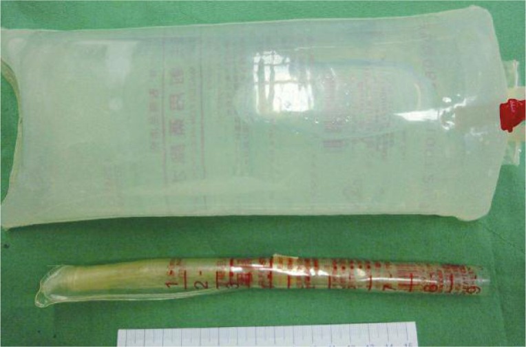
The homemade retrieval bag is made from a commercial sterile intravenous infusion container (Baxter Healthcare, USA), which is a foldable, flexible and waterproof PVC (polyvinyl chloride) bag (Photo 1). The price of this bag sold in this country is about one to two US dollars per piece. This bag has a capacity of 1000 ml in volume, measuring 26 cm × 15 cm in size. It is extremely tough and resistant to perforation while morcellating the spleen. The side with the infusion port of the container is then cut off to create a bag mouth with a short upper lip and a long lower lip. The storage capacity of the bag is large enough to contain a massive spleen up to 900 g in weight. A bigger bag of 2000 ml in volume can also be adopted in this technique.
Photo 1.
The rolled and unrolled versions of the commercial bag are shown
Retrieval of the specimen
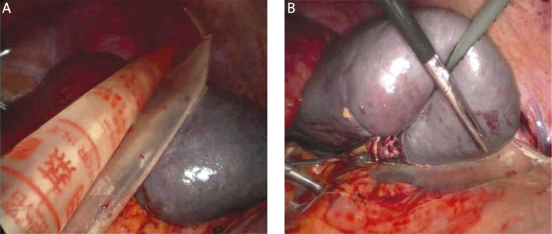
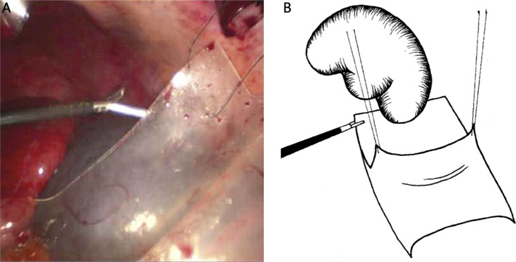
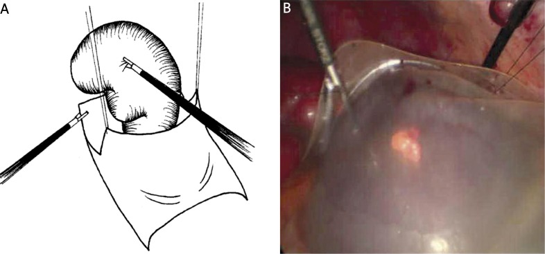
After freeing the spleen, the retrieval bag is rolled into a cigar shape and inserted through the port site into the abdominal cavity (Photo 2A). The bag is deployed and laid in the left abdominal cavity, with the mouth orientated to the splenic fossa where the resected spleen is placed (Photo 2B). Two transabdominal suture sites are selected below the level of the lateral two port sites. A straight needle with suture is inserted into the abdominal cavity to penetrate the lateral side of the upper lip of the bag mouth (Figure 1A). The suture is then pulled through the abdominal wall by a specific hook-pin [14] and tied on the abdominal skin. Another similar suture is made on the medial side of the upper lip. Finally, the whole upper lip of the bag mouth is thus anchored and fixed to the peritoneum of the anterior abdominal wall (Figure 1B). At this time, the laparoscope was changed to the middle one of the three subcostal trocars. By this way, the operative field would not be obscured by the large retrieval bag and the bag can easily be opened by holding and keeping the lower lip of the bag mouth tucked under the spleen with a 5-mm grasper (Figure 2A). Another grasper is inserted into the abdominal cavity to pull the spleen into the bag (Figure 2B). After the spleen is already placed in the bag, the two anchoring sutures are loosened and the edges of the bag are brought out of the abdomen via the lateral port. A ring forceps is used to morcellate the splenic tissue to facilitate extraction. A drainage tube is routinely placed in the splenic area in patients with portal hypertension or intraoperative blood loss > 200 ml. The tube was then removed if the daily amount of the discharge decreased to 50 ml and the color became clear.
Photo 2.
The rolled retrieval bag is inserted through the port site into the abdominal cavity (A). The bag is deployed and laid in the left abdominal cavity and the resected spleen is placed on the lower lip of the bag (B)
Figure 1.
A transabdominal suture is made on the lateral side of the upper lip of the bag mouth (A). Another similar suture is made on the medial side of the upper lip. The bag can easily be opened by holding and keeping the lower lip of the bag mouth tucked under the spleen with a 5-mm grasper (B)
Figure 2.
Another grasper is inserted into the abdominal cavity to pull the spleen into the bag, while the upper lip of the bag is suspended by two transabdominal sutures (A). The major portion of the spleen has been placed in the re trieval bag (B)
Results
Patient characteristics and indications for surgery are shown in Table I. Laparoscopic splenectomy was successfully completed in 49 of these patients. There was one conversion to an open procedure due to massive intraoperative bleeding. Overall, mean operative time was 149 ±31 min (range: 100–252 min). Estimated blood loss was minimal in the majority of patients with only 3 patients experiencing blood loss greater than 500 ml. Median estimated blood loss was 189 ±155 cc (range: 50–920 cc). The drainage tube could be removed within 4 days postoperatively in the great majority of patients. There were no other major complications and no mortality. Out of the 50 patients with LS, 2 (4.0%) presented with trocar site infection and 5 had postoperative mild fever without significant leukocytosis for a short period. Minor complications and the period of hospital are shown in Table II. In all patients the aggregated weights of morcellated splenic tissues were more than 600 g (mean: 729 ±74 g; range: 632–930 g). Time to discharge after surgery ranged from 3 to 9 (mean: 4.7 ±1.7 days).
Table I.
Patient characteristics and diagnosis of the disorders
| Age [years] | 20–73 (mean, 56 ±11) |
| Sex (male: female) | 31: 19 |
| Diagnosis | n |
| Liver cirrhosis | 40 |
| Hereditary spherocytosis | 5 |
| β-Thalassemia | 3 |
| Splenic abscess | 1 |
| Splenic cyst | 1 |
| Child-Pugh classification for liver cirrhosis | |
| A | 25 |
| B | 13 |
| C | 2 |
Table II.
Perioperative data in patients with laparoscopic splenectomy (n = 50)
| Operative time [min] | 100–252 (mean 149 ±31) |
| Blood loss [ml] | 50–920 (median 189 ±155) |
| Postoperative complications | n |
| Trocar site infection | 2 |
| Fever | 5 |
| Flank hematoma | 5 |
| Hospital stay [days] | 3–9 (mean 4.7 ±1.7) |
| Splenic weight [g] | 610–930 (mean 729 ±74) |
Discussion
Successful LS with normal-sized and mildly enlarged spleens has prompted many surgeons to increase the use of this procedure in patients with massive splenomegaly [4, 8–10] since it was first described in 1991 [1]. In patients with severe liver disease, thrombocytopenia secondary to hypersplenism is a clinical feature that may represent an obstacle to chemotherapy, and anti-viral treatment. Previously patients presenting portal hypertension were not indicated for LS, most likely due to the risk of life-threatening hemorrhage and difficulties in achieving hemostasis in such patients [15, 16]. Recently it has been reported that laparoscopic splenectomy in patients with hepatitis C cirrhosis can be done safely to allow application of antiviral treatment and potentially avoid transplantation [17]. Moreover, it has been reported that an enlarged spleen can be removed successfully by laparoscopy with improvement of the instrument and refinement of the techniques of LS, although massive splenomegaly was once considered as a relative contraindication to LS, especially hypersplenism secondary to liver cirrhosis [8–10, 15, 16]. In this series, we also confirmed that LS is feasible and safe in cirrhotic patients with hypersplenism. Rarely, total or partial splenectomy via the laparoscopic approach may be employed as a surgical alternative for splenic infarction due to sleeve gastrectomy or sclerosing angiomatoid nodular transformation of the spleen [18, 19].
A few potential difficulties have been encountered in the laparoscopic approach to massive splenomegaly. During LS for splenomegaly the operative field in the abdominal cavity is diminished by an enlarged spleen, and intra-abdominal manipulation of the bulky organ becomes more technically difficult. To solve the difficulty of retraction and prevent the risk of massive hemorrhage, we suggest early ligation of the splenic artery before mobilization of the spleen to shrink the size of the spleen by facilitating autotransfusion of the sinusoid blood. Laparoscopic splenectomy can be more secure to manipulate through this procedure. Several reports have indicated that early ligation of the splenic artery increases the safety of the surgery and reduces blood loss [17, 20]. The use of preoperative splenic embolization has been reported [21, 22] but does not appear necessary if the splenic blood supply is controlled early. Retrieval of the massively enlarged spleen is another obstacle to be overcome and may be the most challenging step of LS. There are several kinds of methods and various devices used for this purpose [23–25]. Usually a commercial endoscopic bag is employed for spleen removal. However, it can be very difficult, and even impossible, to introduce the enlarged organ into a small endoscopic bag. It has been found that the process of deploying and opening the bag for specimen retrieval, using conventional laparoscopic instruments, may be annoying and time-consuming [23–25]. Sometimes, a separate abdominal incision [26] or the hand port technique [23, 27] is used when attempting to remove massive spleens from the abdomen laparoscopically. However, extending the wound of the hand port site or making another large secondary abdominal incision defeats the advantages associated with laparoscopic surgery. It may not only add operative time but also increase the expense of laparoscopic surgery. Poulin and Thibault [4] proposed the intraperitoneal morcellation of the spleen and its retrieval in several pieces, followed by intraperitoneal irrigation to avoid splenic tissue implantation. However, the risks of subsequent splenosis due to splenic tissue deposition and injury of surrounding structures during morcellation should be avoided.
In this study two patients (4%) developed surgical site infection which could be controlled after intensive and adequate wound care. It is well known that potentially life-threatening infection is a major long-term risk in post-splenectomized or hyposplenic patients. Early and local surgical site infection also occurs much more often than could be expected [28]. However, the benefits of local or systemic antibiotic prophylaxis against local infection remain questionable [28]. Contrarily, it is generally accepted to prevent overwhelming post-splenectomy infection (OPSI) by appropriate vaccination and life-long antibiotic chemoprophylaxis [29]. There is a consensus among the various guidelines regarding immunization prior to elective splenectomy and post-trauma. Asplenic individuals should be educated and repeatedly reminded about the risks and health precautions which are required to avoid infectious hazards [30]. Regarding the necessity of routine drainage after laparoscopic splenectomy, several studies confirmed that the length of hospital stay is shorter when drainage is abandoned [31]. Moreover, “drain fever syndrome”, which is a febrile symptom without accompanying signs of infection, may disappear and patients with postoperative abdominal pain also noticed considerable improvement after the drain's removal [31].
We have described a simple technique, using a self-designed retrieval bag, to facilitate the removal of massively enlarged spleens laparoscopically. The ideal bag for specimen retrieval should be easy to use and keep wide open during the entrapping procedure. It must also be strong enough to allow for the safe morcellation of the spleen and to prevent causing damage to the surrounding organs and spillage of the splenic tissue in the peritoneal cavity. Our technique facilitates removal of the massively enlarged spleen from the abdomen, and can be accomplished with low morbidity and low risk of splenosis. No additional incision of the abdominal wall or intraperitoneal fragmentation of the spleen is required. Compared to other techniques, our proposed method provides distinct advantages over other methods of controlling the retrieval bag. The bag is also readily available and inexpensive and the technique is not difficult. This technique significantly shortens this phase of spleen retrieval and thus the operative time. Our preliminary experiences are consistent with the conclusion that laparoscopic-assisted surgery for massive spleens is both feasible and effective, and may be a well-tolerated alternative to open splenectomy.
References
- 1.Delaitre B, Maignien B. Laparoscopic splenectomy: one case. Presse Med. 1991;44:2263. [PubMed] [Google Scholar]
- 2.Carroll BJ, Phillips EH, Semel CJ, et al. Laparoscopic splenectomy. Surg Endosc. 1992;6:183–5. doi: 10.1007/BF02210877. [DOI] [PubMed] [Google Scholar]
- 3.Kathkouda N, Hurtwitz MB, Rivera RT, et al. Laparoscopic splenectomy. Outcome and efficacy in 103 consecutive cases. Ann Surg. 1998;228:568–78. doi: 10.1097/00000658-199810000-00013. [DOI] [PMC free article] [PubMed] [Google Scholar]
- 4.Poulin EC, Thibault C. Laparoscopic splenectomy for massive splenomegaly: operative technique and case report. Can J Surg. 1995;38:69–72. [PubMed] [Google Scholar]
- 5.Park A, Marcaccio M, Sternbach M, et al. Laparoscopic versus open splenectomy. Arch Surg. 1999;134:1263–9. doi: 10.1001/archsurg.134.11.1263. [DOI] [PubMed] [Google Scholar]
- 6.Rege RV, Joehl RJ. A learning curve for laparoscopic splenectomy at an academic institution. J Surg Res. 1999;81:27–32. doi: 10.1006/jsre.1998.5485. [DOI] [PubMed] [Google Scholar]
- 7.Trías M, Targarona EM, Espert JJ, et al. Impact of hematological diagnosis on short and long term follow up after laparoscopic splenectomy. Surg Endosc. 2000;14:556–60. doi: 10.1007/s004640000149. [DOI] [PubMed] [Google Scholar]
- 8.Yee JC, Akpata MO. Laparoscopic splenectomy for congenital spherocytosis with splenomegaly: a case report. Can J Surg. 1995;38:73–6. [PubMed] [Google Scholar]
- 9.Terrosu G, Donini A, Baccarani U, et al. Laparoscopic versus open splenectomy in the management of splenomegaly: our preliminary experience. Surgery. 1998;24:839–43. [PubMed] [Google Scholar]
- 10.Targarona EM, Espert JJ, Balague C, et al. Splenomegaly should not be considered a contraindication for laparoscopic splenectomy. Ann Surg. 1998;228:35–9. doi: 10.1097/00000658-199807000-00006. [DOI] [PMC free article] [PubMed] [Google Scholar]
- 11.Grahn SW, Alvarez J, 3rd, Kirkwood K. Trends in laparoscopic splenectomy for massive splenomegaly. Arch Surg. 2006;141:755–62. doi: 10.1001/archsurg.141.8.755. [DOI] [PubMed] [Google Scholar]
- 12.Patel AG, Parker JE, Wallwork B. Massive splenomegaly is associated with significant morbidity after laparoscopic splenectomy. Ann Surg. 2003;238:235–40. doi: 10.1097/01.sla.0000080826.97026.d8. [DOI] [PMC free article] [PubMed] [Google Scholar]
- 13.Targarona EM, Espert JJ, Cerdan G, et al. Effect of spleen size on splenectomy outcome. Surg Endosc. 1999;13:559–62. doi: 10.1007/s004649901040. [DOI] [PubMed] [Google Scholar]
- 14.Hsieh JS, Wu CF, Chen FM, et al. Simple laparoscopic gastrostomy using a homemade hook-pin. Endoscopy. 2007;39(Suppl 1):E226. doi: 10.1055/s-2006-945160. [DOI] [PubMed] [Google Scholar]
- 15.Hashizume M, Tanoue K, Morita M, et al. Laparoscopic gastric devascularization and splenectomy for sclerotherapy- resistant esophagogastric varices with hypersplenism. J Am Coll Surg. 1998;187:263–70. doi: 10.1016/s1072-7515(98)00181-1. [DOI] [PubMed] [Google Scholar]
- 16.Kercher KW, Carbonell AM, Heniford BT, et al. Laparoscopic splenectomy reverses thrombocytopenia in patients with hepatitis C cirrhosis and portal hypertension. J Gastrointest Surg. 2004;8:120–6. doi: 10.1016/j.gassur.2003.10.009. [DOI] [PubMed] [Google Scholar]
- 17.Nicholson IA, Falk GL, Mulligan SC. Laparoscopically assisted massive splenomegaly: a preliminary report of the technique of early hilar devascularization. Surg Endosc. 1998;12:73–5. doi: 10.1007/s004649900598. [DOI] [PubMed] [Google Scholar]
- 18.Budzyński A, Demczuk S, Kumiega B, et al. Sclerosing angiomatoid nodular transformation of the spleen treated by laparoscopic partial splenectomy. Videosurgery Miniinv. 2011;6:249–55. doi: 10.5114/wiitm.2011.26261. [DOI] [PMC free article] [PubMed] [Google Scholar]
- 19.Michalik M, Budziński R, Orlowski M, et al. Splenic infarction as a complication of laparoscopic sleeve gastrectomy. Videosurgery Miniinv. 2011;6:92–8. [Google Scholar]
- 20.Palanivelu C, Jani K, Malladi V, et al. Early ligation of the splenic artery in the leaning spleen approach to laparoscopic splenectomy. J Laparoendosc Adv Surg Tech A. 2006;16:339–44. doi: 10.1089/lap.2006.16.339. [DOI] [PubMed] [Google Scholar]
- 21.Iwase K, Higaki J, Yoon HE. Splenic artery embolization using contour emboli before laparoscopic or laparoscopically assisted splenectomy. Surg Laparosc Endosc Percutan Tech. 2002;12:331–6. doi: 10.1097/00129689-200210000-00005. [DOI] [PubMed] [Google Scholar]
- 22.Poulin EC, Mamazza J, Schlachta CM. Splenic artery embolization before laparoscopic splenectomy. An update. Surg Endosc. 1998;12:870–5. doi: 10.1007/s004649900732. [DOI] [PubMed] [Google Scholar]
- 23.Targarona EM, Balague C, Cerdan G, et al. Hand-assisted laparoscopic splenectomy (HALS) in cases of splenomegaly: a comparison analysis with conventional laparoscopic splenectomy. Surg Endosc. 2002;16:426–30. doi: 10.1007/s00464-001-8104-z. [DOI] [PubMed] [Google Scholar]
- 24.Zacharoulis D, O'Boyle C, Royston CM, et al. Splenic retrieval after laparoscopic splenectomy: a new bag. J Laparoendosc Adv Surg Tech A. 2006;16:128–32. doi: 10.1089/lap.2006.16.128. [DOI] [PubMed] [Google Scholar]
- 25.Greene AK, Hodin RA. Laparoscopic splenectomy for massive splenomegaly using a Lahey bag. Am J Surg. 2001;181:543–6. doi: 10.1016/s0002-9610(01)00632-8. [DOI] [PubMed] [Google Scholar]
- 26.Choy C, Cacchione R, Moon V, et al. Experience with seven cases of massive splenomegaly. J Laparoendosc Adv Surg Tech A. 2004;14:197–200. doi: 10.1089/lap.2004.14.197. [DOI] [PubMed] [Google Scholar]
- 27.Demetrius EM, Litwin MD, Darzi A. Hand-assisted laparoscopic surgery (HALS) with the handport system: initial experience with 68 patients. Ann Surg. 2000;231:715–23. doi: 10.1097/00000658-200005000-00012. [DOI] [PMC free article] [PubMed] [Google Scholar]
- 28.Migaczewski M, Zub-Pokrowiecka A, Budzyński P, et al. Prevention of early infective complications after laparoscopic splenectomy with the Garamycin sponge. Videosurgery Miniinv. 2012;7:105–10. doi: 10.5114/wiitm.2011.27151. [DOI] [PMC free article] [PubMed] [Google Scholar]
- 29.Brigden ML, Pattullo AL. Prevention and management of overwhelming postsplenectomy infection – an update. Crit Care Med. 1999;27:836–42. doi: 10.1097/00003246-199904000-00050. [DOI] [PubMed] [Google Scholar]
- 30.El-Alfy MS, El-Sayed MH. Overwhelming postsplenectomy infection: is quality of patients knowledge enough for prevention? Haematol J. 2004;5:77–80. doi: 10.1038/sj.thj.6200328. [DOI] [PubMed] [Google Scholar]
- 31.Major P, Matłok M, Pędziwiatr M, et al. Do we really need routine drainage after laparoscopic adrenalectomy and splenectomy? Videosurgery Miniinv. 2012;7:33–9. doi: 10.5114/wiitm.2011.25610. [DOI] [PMC free article] [PubMed] [Google Scholar]


