Abstract
During fluid phase endocytosis (FPE) in plant storage cells, the vacuole receives a considerable amount of membrane and fluid contents. If allowed to accumulate over a period of time, the enlarging tonoplast and increase in fluids would invariably disrupt the structural equilibrium of the mature cells. Therefore, a membrane retrieval process must exist that will guarantee membrane homeostasis in light of tonoplast expansion by membrane addition during FPE. We examined the morphological changes to the vacuolar structure during endocytosis in red beet hypocotyl tissue using scanning laser confocal microscopy and immunohistochemistry. The heavily pigmented storage vacuole allowed us to visualize all architectural transformations during treatment. When red beet tissue was incubated in 200 mM sucrose, a portion of the sucrose accumulated entered the cell by means of FPE. The accumulation process was accompanied by the development of vacuole-derived vesicles which transiently counterbalanced the addition of surplus endocytic membrane during rapid rates of endocytosis. Topographic fluorescent confocal micrographs showed an ensuing reduction in the size of the vacuole-derived vesicles and further suggest their reincorporation into the vacuole to maintain vacuolar unity and solute concentration.
Keywords: exocytosis, retrograde vesicles, sucrose transport, tonoplast
Introduction
Plant storage cells accumulate a portion of their photoassimilates in the vacuole via fluid phase endocytosis (FPE),1-6 a process characterized by the nonspecific bulk uptake of extracellular solutes in plasma membrane-bound vesicles.7 The portion of assimilates taken up by FPE varies according to the apoplastic solute concentration,8 the remaining difference being transported by membrane-bound carriers. Although firmly established that all types of endocytic systems (i.e., clathrin-mediated, caveolin-mediated, lipid raft-dependent, flotillin-dependent and macropinocytosis)9 carry a fluid-phase component,10,11 FPE is distinguished by their relatively larger vesicle size11,12 and cargo capacity. Solutes trapped by FPE eventually reach the vacuole;13,14 however,some systematic cargo distribution within the endosome has been observed using a combination of soluble membrane-impermeable endocytic markers5,15 and fluorescent nano-particles.16
In plants, FPE has been documented primarily for storage cells.3-6,8,17-19 During accumulation of photoassimilates at low external concentrations, membrane-bound solute transporters support the cellular energy and metabolic demands, in addition to transporting surplus solutes to the vacuole.20-22 This constitutes the hyperbolic, high-affinity phase of the characteristic curvilinear photoassimilate uptake mechanism.23 At high extracellular sucrose, mannitol or glucose concentrations, FPE is triggered and functions largely to carry surplus photoassimilates into the vacuole bypassing the cytosol,5,8 constituting the linear phase of the uptake curve.23 The simultaneous action of these multiple transport mechanisms was demonstrated using fluorescent deoxy-glucose, evacuolated cytoplasts and inhibitors of both transport systems24 and their coordinated action detailed by Pozueta-Romero et al.8 Similar conclusions were obtained by Bandmann and Homann5 with tobacco BY-2 cells. The results provided evidence that FPE and membrane transporters can work in parallel and are under strict control of the external photoassimilate supply. A number of studies have revealed the formation of endocytic vesicles of different sizes ranging from 0.5–6 µm,6,25 a size more in accordance with macropinocytosis.26 However, membrane capacitance measurements by Bandmann and Homann5 and Gall et al.27 suggested an endocytic vesicle diameter of 80–220 nm. Whether or not the larger vesicles carrying endocytic markers observed during FPE result from the fusion of smaller vesicles28,29 prior to merging with the vacuole has not been determined.
During the vesicle delivery process, the vacuole receives a considerable amount of membrane and fluid content which, if allowed to accumulate over a period of time, would invariably disrupt the structural equilibrium of the mature cells. Furthermore, given the disproportional membrane surface-area/volume added by endocytic vesicles, membrane influx in the absence of a compensatory efflux process would result in increased dilution of the vacuolar contents. Therefore a retrieval process must exist that would guarantee membrane homeostasis in light of tonoplast expansion by membrane addition during FPE.30 Despite the volumes of literature on endocytosis in both animal and plant cells, the structural integrity of the tonoplast/lysosome during endocytosis remains unexplained, largely overlooked by the difficulty in visualizing, measuring and quantifying such changes. In an attempt to explain the lack of observed variations in the lysosome volume during macropinocytosis in macrophages, Swanson26 suggested a system of retrograde vesicles of much reduced in size compared with the endocytic vesicles, a way to minimize solute loss while disproportionally removing excessive membrane surface area. If the diameter of the efflux vesicles were considerably smaller than that of the macropinosome (endocytic vesicle) the cell could balance incoming and outgoing membrane, and at the same time, restrict the retro-efflux of fluid. Nevertheless, regardless of the size of the exocytic vesicles, some portion of the vacuolar fluids would be returned to the exterior, creating an ancillary futile cycle. The remaining water and other permeable solutes would cross the lysosome membrane leaving membrane impermeable solutes to concentrate in the lysosome. Such a system could explain how solutes are concentrated in the storage lysosome/vacuole thereby maintaining the membrane surface area without significant loss of trapped solutes.31 In support of this proposed system, clathrin coated retrieval vesicles have been observed at the surface of lysosomes,32 whereas retrograde membrane traffic systems from the vacuole have been identified as part of the membrane recycling process in Saccharomyces cerevisiae,33,34 in tobacco BY-2 cells35 and from the pre-vacuolar compartment in Arabidopsis.36
Throughout our investigations into FPE in plant cells with a variety of membrane-impermeable probes,2,15,16,25 we consistently observed their eventual deposition in the vacuole. For such mature cells to endocytose constitutively, a stable tonoplast surface area/volume ratio must be preserved by an unknown accompanying process of membrane turnover. Based on reports of retrograde vesicle budding,32 the exceptional plasticity of the tonoplast37 and our unpublished observations, we hypothesize the existence of one or several cellular, morphological and/or metabolic events that will assist in vacuolar stability during endocytosis. As part of our continued investigation on FPE in plant cells, we examined the morphological changes to the vacuolar structure during rapid rates of endocytosis. To follow these essential modifications, we used red beet hypocotyl tissue. The heavily pigmented vacuole of the storage parenchyma cells allowed us to microscopically visualize all architectural transformations during treatment and distinguish these from any possible plasma membrane-derived osmocytotic structures as those reported by Goodwin et al.38 and Oparka et al.39 The present study revealed that, under conditions of rapid FPE proliferation, the cell transiently counterbalances the addition of surplus membrane by vacuole fragmentation with a system of vacuole-derived vesicles. Topographic fluorescent confocal micrographs showed an ensuing reduction in the size of the vacuole-derived vesicles and further suggested their reincorporation into the vacuole.
Results
Endoytic sucrose uptake
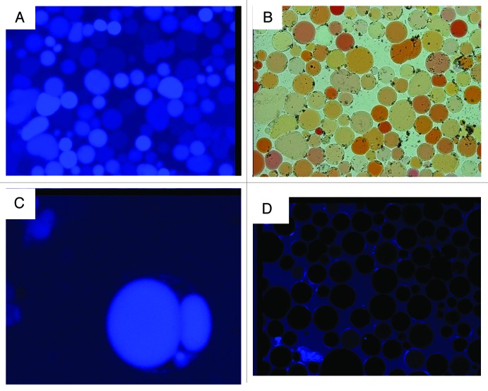
Protoplasts prepared from red beet storage tissue after incubation in 200 mM sucrose and the blue-fluorescent membrane-impermeable endocytic marker Alexa-350 for 24 h exhibited a pronounced blue fluorescence in the vacuole (Fig. 1A and C). The blue fluorescence was not due to autofluorescence by the vacuolar pigment betacyanin. Although betacyanin has a wide fluorescent spectrum (560–750 nm)40 it does not overlap with that of Alexa-350 (400–550 nm). Furthermore, and most notably, protoplasts prepared from control tissue incubated in Alexa-350 and betaine instead of sucrose showed no fluorescence (Fig. 1D). Sucrose induced FPE has been reported for other tissues including turnip storage parenchyma,8 citrus juice cells3 and sycamore cultured cells.2,16
Figure 1. Red beet protoplasts isolated from tissue incubated in the presence and absence of the membrane-impermeable, endocytic-marker blue-fluorescent Alexa-350.(A) Fluorescent micrograph of protoplasts isolated from tissue incubated in 200 mM sucrose and 100 μM Alexa-350. (B) Light micrograph of (A). (C) High magnification fluorescent micrograph of a protoplast demonstrating Alexa-350 blue fluorescence in the vacuole and vacuole derived vesicles. (D) Fluorescent micrograph of protoplasts prepared from control tissue incubated in betaine.
Consistent with the participation of FPE in the process of sucrose uptake, determinations of membrane flux indicated a significant movement of plasma membrane elements to the tonoplast. A 20% increase in vanadate-sensitive ATPase activity (plasma membrane H+-ATPase marker) was measured in purified tonoplast fractions from tissue incubated in sucrose when compared with control samples incubated in equal concentrations of betaine (Table 1). The above observations are in agreement with other storage tissues1-6 and collectively indicate that transport of Alexa-350 to the vacuole (Fig. 1) was mediated in part by sucrose-induced FPE.
Table 1. Changes of plasma membrane marker vanadate-sensitive ATPase activity in red beet cells incubated in sucrose or betaine for 24 h.
| 24 h | ||||
|---|---|---|---|---|
| Time 0 | Betaine | Sucrose | % increase | |
|
Vanadate sensitive P-ATPase (*Units/ total ATPase in tonoplast fraction) |
0.17 ± 0.03 | 0.23 ± 0.01 | 0.31 ± 0.02 | 20% |
One unit defined as 1 nkat. Purified tonoplast fractions were isolated from fresh tissue and from tissue incubated in sucrose or betaine for 24 h. V-ATPase activity was measured in the presence and absence of vanadate.
Vacuole restructuring during endocytosis
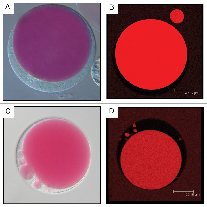
During dormant or resting stage, red beet hypocotyl storage cells contained predominantly one large central vacuole (Fig. 2A and B). However, after tissue incubation in 200 mM sucrose for 24 h, a series of vacuole-derived organelles become evident (Fig. 2C and D), a condition scarcely observed when betaine was used as osmotic control. These vesicles are unquestionably of vacuolar origin given their red betacyanin content (Fig. 2C and D). The vacuole-derived vesicles not only varied in size and number between cells from different beet hypocotyl preparations, but also between cells from the same hypocotyl. Lack of synchrony is not unusual since not all cells participate equally in the endocytic process.26,30
Figure 2. Light Nomarski (Aand C) and fluorescent laser scanning confocal micrographs (BandD) of protoplasts from red beet storage tissue. Protoplasts were prepared from hypocotyls at dormant state (AandB) and after 24 h incubation in 200 mM sucrose (Cand D).
The absence of vacuole-derived vesicles in protoplasts obtained from control tissue cultured in betaine is in clear contrast to the prominent tonoplast vesiculation observed by Diekmann et al.41 using guard cells protoplasts subjected to osmotic excursions. However, it is noteworthy that guard cells are highly differentiated cells primarily specialized to undergo rapid volume adjustments based on vacuole fragmentation and reformation in response to osmotic changes42 as opposed to sucrose accumulating beet storage parenchyma cells.
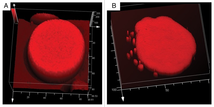
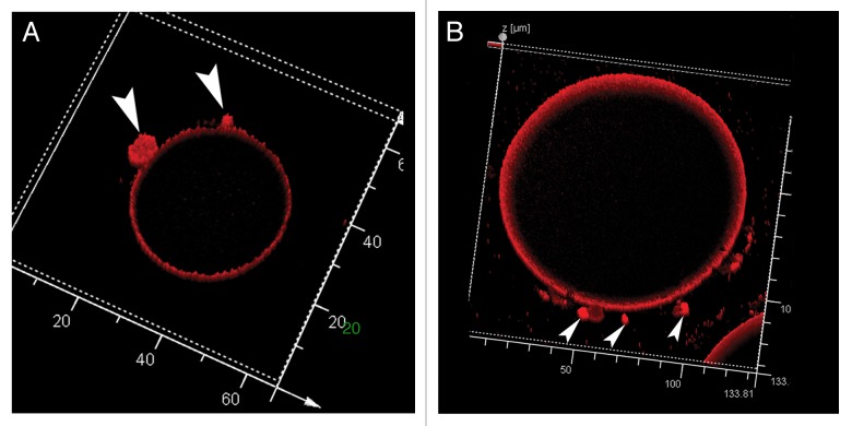
A Z-stack 3D scanning laser confocal analysis revealed the abundance of vesicles and their large variation in size (Fig. 3A and B). Whether the larger vacuole-derived vesicles resulted from the fusion of smaller ones cannot be determined from the data generated by these static images. Nevertheless, merger of vesicles would not offer the cell any organizational advantage during photoassimilate accumulation since fusion of two spherical compartments would generate a larger surface area/volume structure resulting in solute dilution.
Figure 3. 3D reconstruction of Z-stack fluorescent images of red beet protoplasts isolated from tissue incubated 24 h in 200 mM sucrose. Given the wide fluorescence spectrum of betacyanin, samples were examined under an emission range of 620–650 nm. (A) Entire protoplast showing vacuole and vacuole-derived vesicles of a wide range of sizes. (B) Close up of a section of a protoplast. The dark region represents the cytosol, whereas the red structures are the vacuole, vacuole-derived vesicles and external medium still fluorescing from residual dye.
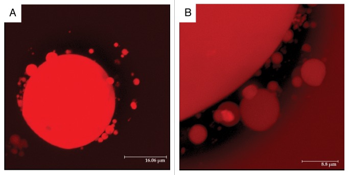
Fluorescence comparison of vacuole and vacuole-derived vesicles revealed comparable fluorescence intensity (Fig. 4). Since signal strength is a function of the concentration of fluorescent molecules,27 the concentration of betacyanin (and hence all solutes) within the vacuole-derived vesicles must, therefore, be reasonably similar in all compartments (Fig. 4B). This apparent uniformity in fluorescence remained despite some expected pigment dilution generated by incoming endocytic fluids.
Figure 4. Topographic fluorescence analysis of red beet protoplasts isolated from tissue incubated 24 h in 200 mM sucrose at two different stages. (A) Protoplast from dormant beet hypocotyls with a large central vacuole. (B) Protoplast from tissue incubated in 200 mM sucrose for 24 h. Fluorescence intensity of vacuole-derived vesicles is similar to that emanating from the vacuole.
Apparent volume contraction in vacuole-derived vesicles
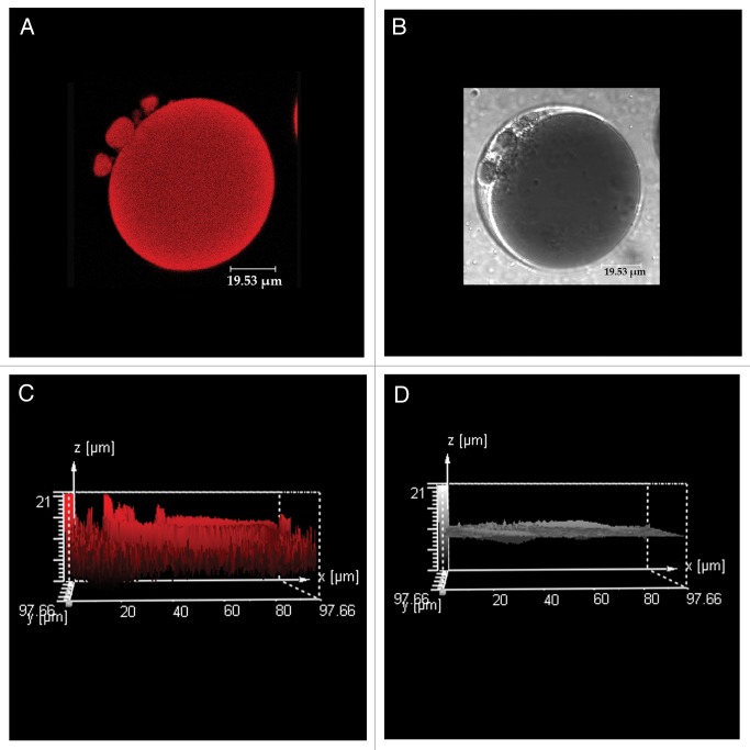
The vacuole-derived vesicles formed during incubation in sucrose underwent visible changes when cells were further incubated for an additional 24 h in the absence of sucrose. At the end of the additional incubation period, there were no observable vesicles, or when present, their numbers were substantially reduced suggesting a re-fusion process with the large central vacuole. The likelihood that the pigmented vacuole-derived vesicles merged with the plasma membrane in an exocytic manner was disregarded since there was no betacyanin bleeding into the media during incubation. During a more rigorous examination, the enduring vesicles appeared darker in color under light microscopy, although not so apparent in photomicrographs (Fig. 5A and B). However, the perceived increase in color intensity was confirmed by a landscape view of a topographic fluorescence analysis (Fig. 5C). The vesicle fluorescence peaks were an average 20–30% taller than those emanating from the vacuole indicating higher betacyanin concentration.
Figure 5. Red beet protoplast isolated from tissue incubated 24 h in 200 mM sucrose followed by a 24 h period of sucrose starvation. (AandB) represent fluorescent and light laser scanning confocal micrographs, respectively. (Cand D) are topographic analyses of (A andB), respectively. Fluorescent peaks on (C) correspond to the vacuole-derived vesicles in (A).
In a distinct type of image analysis performed on Z-stacks obtained from separate cells, topographic surface reconstruction of maximum fluorescence intensity analysis of protoplasts only revealed the outlining tonoplast (Fig. 6A and B). At this setting, fluorescence of the central vacuole was eliminated whereas the vacuole-derived vesicle(s) remained intensely fluorescent indicating that the concentration of betacyanin (and thus all membrane impermeable solutes) within the vacuole-derived vesicles was considerably higher than in the large central vacuole (arrows). The increase in fluorescence intensity strongly suggests that vacuole-derived vesicles underwent a process of size reduction and their disappearance denotes their eventual fusion with the vacuole.
Figure 6. Topographic surface reconstruction of maximum fluorescence intensity of two red beet protoplasts prepared from tissue after a 24 h incubation in 200 mM sucrose followed by a 24 h period of sucrose starvation. Vacuole-derived vesicles (arrows) remain highly fluorescent.
Discussion
In eukaryotic cells, the dynamics of membrane maintenance requires a constant turnover of endosome-intrinsic constituents mediated by vesicle transport and the processes of exocytosis and endocytosis.43 In each of these processes, membrane vesicles composed of lipid bilayers and various membrane proteins are pinched off from one organelle and delivered to other subcellular or extracellular compartments that are undergoing expansion or reconstruction.43 During cell growth, much of this trafficking is directed outwards toward the plasma membrane and, in plants, toward the enlargement and assembling of the vacuole. However, in non-expanding cells, membrane degrading and transport pathways must be kept in balance to maintain the proper functioning size of cellular constituents.44 As part of internal trafficking between different organelles, several inter-endosomal retrograde systems have been hypothesized that undoubtedly contribute to membrane homeostasis.32,33,45
In mature plant storage cells, sugar accumulation takes place in the confined space of a non-expanding vacuole. Aside from sugar transport by tonoplast-bound transporters,20-22 the partial delivery of sugars in cargo-filed endocytic vesicles to the vacuole imposes a significant structural and spatial constraint difficult to overcome by the reported clathrin-dependent vacuolar retrograde systems.32-35 Merging of the relatively large FPE vesicles with the vacuole generates a dual imbalance of membrane and vacuolar fluids. The data we present in this communication confirm the endocytic uptake of extracellular fluids induced by sucrose (Fig. 1) and reveal the transitional re-arrangement of the tonoplast/vacuole architecture during this process (Figs. 2–6). Vacuole fragmentation allows for the temporary adjustment of membrane surplus and, at the same time, assuring the eventual concentration of solutes. This conclusion is supported by three interrelated pieces of evidence. First, incorporation of the fluorescent endocytic marker Alexa-350 into the vacuole in the presence of sucrose indicated the involvement of FPE. Second, a substantial and consistent fragmentation of the vacuole was observed upon sucrose accumulation by FPE (Figs. 2–4). Third, the increase intensity of pigment fluorescence denoted volume shrinkage of the vacuole-derived vesicles (Figs. 5 and 6). Although speculative at this moment, disappearance of the vacuole-derived vesicles suggests their merging with the vacuole. A similar conclusion was reached for Saccharomyces at times of membrane expansion where vesicle trafficking and nuclear membrane deformations maintain nuclear/cell volume ratio.46 In plant cells, fragmentation of the vacuole is commonly observed in glandular cells of carnivorous plants during endocytosis.6 Another type of tonoplast deformation (termed “bulbs”) has been implicated in the rearrangement of the vacuole.47 Bulbs are tonoplast- delimited spherical protrusions of 1–3 μm and formed from cytoplasmic folds protruding into the vacuole. These are believed to serve as reservoir of tonoplast to prepare for quick volume changes.48
The process of vacuole fragmentation described here is fundamentally similar in concept to that conjectured by Traub et al.32 and Swanson26 in that excess membrane from the vacuole/lysosome is removed in a retrograde manner. However, our findings differ in that the “retrograde” vesicles generated during FPE are unequally sized and are not recycled within the endosome as would be those using a clathrin-assisted vesicle system. Fundamental to the reduction in size of the vacuole-derived vesicles (Figs. 5 and 6) is the participation of an additional, yet unknown, ancillary mechanism(s) involved in the removal of membrane components and volume regulation. Much of this permanent reduction can be performed by vacuolar lipases, which have been shown to be essential for membrane digestion during the macroautophagocytic process in yeast.49 In fact, mutation of cvt17, which encodes for a putative lipase in yeast, disrupted the proper turnover of endocytic membrane.49 In our experiments, it is possible that tonoplast digestion also took place in the large vacuole; however, since its volume was considerably larger than that of vacuole-derived vesicles, any reduction in size would have been proportionally smaller than that of vesicles. This disproportional reduction in size would allow the vacuole-derived vesicles to outshine the vacuole (Figs. 5 and 6).
Involvement of other proposed mechanisms that could conceivably participate in reducing or preventing expansion of the tonoplast during FPE are not supported by our data. For example, macrophagocytosis, the engulfing of protoplasm into the vacuole by tonoplast foldings,50 if operative, would have appeared as discolored areas within the vacuole, a situation never observed in our 3-D reconstructions of Z-stacks. The same absence of membrane inclusions makes microphagocytosis of vacuolar tubules an unlikely event. Finally, “kiss-and-run,” the fast release of contents by the endocytic vesicle into the targeted organelle and its immediate retrieval,51 can be disregarded by the observed increase presence of plasma membrane P-ATPase in vacuolar fractions (Table 1).
The tonoplast’s inherent elasticity is often overlooked as an essential factor in vacuolar maintenance.36,52,53 Capable of withstanding substantial deformations, the tonoplast can absorb extensive changes in surface area without the need for permanent removal. In fact this elasticity is likely the initial buffer mechanism during FPE before the formation of vacuole-derived vesicles.
Studies of membrane rearrangement during endocytosis have been precluded by the absence of an indisputable recycling cargo,44,54 a limitation overcame in this study by the use of red beet hypocotyl cells. These data indicate that tonoplast homeostasis during FPE is a coordinated event that may involve one or many processes. Here, we present evidence for the architectural remodeling of the tonoplast during rapid rates of FPE and suggests the participation of an axillary membrane recycling mechanism where vacuole-derived vesicles diminish their size before re-incorporation to the vacuole and maintaining vacuolar unity.
Materials and Methods
Plant material
Fresh red beet hypocotyls were purchased at a local grocery store and maintained refrigerated until use. Beets were brought to room temperature 24 h before experimentation.
Tissue incubation
Tissue disks of approximately 0.5 mm in thickness were obtained from cylinders excised using a #5 cork borer. The disks were thoroughly rinsed in deionized water until bleeding stopped. After blotting dry, 1 g of tissue was incubated in a 25 mL Erlenmeyer flask containing 5 mL of a solution of 10 mM MES (pH 5.6), 200 mM sucrose, 1 mM CaCl2, 1 mM MgCl2 and 2 mM DTT, incubated for 24 h at 30°C while being agitated at 80 rpm. Treatment solutions were changed every 24 h as required by individual experiments. When indicated, inhibitors of endocytosis (10 µM Latrunculin-B) and membrane-bound sucrose carriers (1 µM pCMBS) were added to pre-incubation and incubation solutions. The pre-incubation solution was similar to the incubation solution with 200 mM betaine added instead of sucrose.
At the end of the incubation time, tissue was rinsed three times with 5 mL of pre-incubation media for 10 min each. Samples were immediately frozen in 5 mL of 25 mM Tris/MES (pH 5.6) until further analysis. Sucrose was determined following the procedure of Van Handel55 after thawing the tissue in a rotary shaker for 1 h at 40 rpm and 30°C.
Protoplast preparation
Protoplasts were prepared from tissue incubated according to individual experiments by replacing the incubation medium with 10 mL of 100 mM MES (pH 5.6), 2% Cellulase from Trichoderma viride (Onozuka-RS; SERVA Cat no. 16420), 2% Pectinase from Aspergillus niger (CalBiochem Cat no. 515883), 1% Macerozyme R-10 from Rhizopus sp (SERVA cat no. 28302), 1% BSA, 125 mM CaCl2, 125 mM KCl and incubated overnight in a rotary shaker at 30°C and 40 rpm. Prior to microscopic observation, the protoplasts were washed with a similar solution without hydrolyzing enzymes.
Tonoplast and plasma membrane vesicle isolation
Highly purified tonoplast and plasma membrane samples were isolated using a discontinuous sucrose gradient as described by Bennett et al.56 and Etxeberria and Gonzalez.57 The purities of the membrane fractions were corroborated measuring the total ATPase activities with and without vanadate and nitrate inhibitors. Individual membrane fractions were brought to 1.0 mL with storage buffer containing 250 mM sucrose, 10 mM MES pH 5.6 and 2 mM DTT and stored at −80°C.
ATPase activity
V-ATPase activity in tonoplast fractions was determined by coupling the production of ADP to the oxidation of NADH at 340 nm using a pyruvic kinase/lactate dehydrogenase system (PK-LDH). ATPase reaction solution contained 50 mM BTP/MES (pH 7.5), 4 mM dithiothreitol (DTT), 0.4 mg mL−1 bovine serum albumin (BSA), 250 mM sorbitol, 50 mM KCl, 10 mM gramicidin, 4 mM ATP, 4 mM, MgSO4, 5 mL PK:LDH enzymes (Sigma P-0294), 0.25 mg mL−1 NADH, 1 mM phosphoenolpyruvate and approximately 100 mg vesicle protein in a total volume of 1.0 mL.58 ADP generated was calculated from the extinction coefficient of NAD (6.22) at 340 nm. All reactions were carried with and without 50 mM vandate at 30°C in a Shimadzu UV-160 .
Microscopy
Light microscopy was performed using an Olympus BH-2 microscope equipped with a Canon Power Shot S3-15 digital camera connected to a Dell monitor. Fluorescent images were obtained using a Leica TCS-SL confocal laser scanning microscopy.
Given the wide range of fluorescence emission by the vacuolar pigment betacyanin, for experiments containing the red fluorescent membrane marker FM 4-64 FX, we used ex/em = 488/490-510. We captured FM 4-64 fluorescence at ex/em = 543/650-750. For all other experiments not containing FM 4-64, betacyanin was observed at ex/em = 543/590-650. Images were analyzed and created using Leica imaging system. To observe protoplast loaded with Alexa 350 we used a Zeiss Scope A-1 fluorescent microscope equipped with Zeiss Filter No. 49 (DAPI; Ex: G365,Em:BP445/50) and Zeiss Axio Cam ICc1.
Glossary
Abbreviations:
- ADP
adenosine-5′-diphosphate
- ATP
adenosine-5′-triphosphate
- BSA
bovine serum albumin
- BTP
bis-tris-propane
- DTT
dithiothreitol
- FPE
fluid phase endocitosis
- MES
2-(N-morpholino)ethanesulfonic acid
- NAD
nicotinamide adenine dinucleotide
- NADH
nicotinamide adenine dinucleotide-H
- pCMBS
p-Chloromercuribenzene sulfonate
- Tris
tris(hydroxymethyl)aminomethane
- V-ATPase
vacuolar-type H+-ATPase
Disclosure of Potential Conflicts of Interest
No potential conflicts of interest were disclosed.
Footnotes
Previously published online: www.landesbioscience.com/journals/psb/article/24793
References
- 1.Baluška F, Baroja-Fernandez E, Pozueta-Romero J, Hlavacka J, Etxeberria E, Samaj J. Endocytic uptake of nutrients, cell wall molecules, and fluidized cell wall portions into heterotrophic plant cells. In: Šamaj J, Baluška F, Menzel D, ed(s) Plant Endocytosis, Plant Cell Monograph, Springer Verlag, Berlin. 2005:1-17. [Google Scholar]
- 2.Etxeberria E, Baroja-Fernandez E, Muñoz FJ, Pozueta-Romero J. Sucrose-inducible endocytosis as a mechanism for nutrient uptake in heterotrophic plant cells. Plant Cell Physiol. 2005;46:474–81. doi: 10.1093/pcp/pci044. a. [DOI] [PubMed] [Google Scholar]
- 3.Etxeberria E, Gonzalez P, Pozueta-Romero J. Sucrose transport into Citrus juice cells: evidence for an endocytic transport system. J Am Soc Hortic Sci. 2005;130:269–74. b. [Google Scholar]
- 4.Etxeberria E, Gonzalez P, Pozueta-Romero J. Mannitol-enhanced, fluid-phase endocytosis in storage parenchyma cells of celery (Apium graveolens; Apiaceae) petioles. Am J Bot. 2007;94:1041–5. doi: 10.3732/ajb.94.6.1041. a. [DOI] [PubMed] [Google Scholar]
- 5.Bandmann V, Homann U. Clathrin-independent endocytosis contributes to uptake of glucose into BY-2 protoplasts. Plant J. 2012;70:578–84. doi: 10.1111/j.1365-313X.2011.04892.x. [DOI] [PubMed] [Google Scholar]
- 6.Adlassnig W, Koller-Peroutka M, Bauer S, Koshkin E, Lendl T, Lichtscheidl IK. Endocytotic uptake of nutrients in carnivorous plants. Plant J. 2012;71:303–13. doi: 10.1111/j.1365-313X.2012.04997.x. [DOI] [PubMed] [Google Scholar]
- 7.Khalil IA, Kogure K, Akita H, Harashima H. Uptake pathways and subsequent intracellular trafficking in nonviral gene delivery. Pharmacol Rev. 2006;58:32–45. doi: 10.1124/pr.58.1.8. [DOI] [PubMed] [Google Scholar]
- 8.Pozueta-Romero D, Gonzalez P, Pozueta-Romero J, Etxeberria E. The hyperbolic and linear phases of the sucrose accumulation curve in turnip (Brassica campestris) storage cells denote carrier-mediated and fluid-phase endocytic transport, respectively. J Am Soc Hortic Sci. 2008;133:612–8. [Google Scholar]
- 9.Lin AE-J, Guttman JA. Hijacking the endocytic machinery by microbial pathogens. Protoplasma. 2010;244:75–90. doi: 10.1007/s00709-010-0164-2. [DOI] [PubMed] [Google Scholar]
- 10.Holstein SE. Clathrin and plant endocytosis. Traffic. 2002;3:614–20. doi: 10.1034/j.1600-0854.2002.30903.x. [DOI] [PubMed] [Google Scholar]
- 11.Kruth HS, Jones NL, Huang W, Zhao B, Ishii I, Chang J, et al. Macropinocytosis is the endocytic pathway that mediates macrophage foam cell formation with native low density lipoprotein. J Biol Chem. 2004;280:2352–60. doi: 10.1074/jbc.M407167200. [DOI] [PubMed] [Google Scholar]
- 12.Cao H, Chen J, Awoniyi M, Henley JR, McNiven MA. Dynamin 2 mediates fluid-phase micropinocytosis in epithelial cells. J Cell Sci. 2007;120:4167–77. doi: 10.1242/jcs.010686. [DOI] [PubMed] [Google Scholar]
- 13.Lazzaro MD, Thompson WW. Endocytosis of lanthanum nitrate in organic acid-secreting trichomes of chickpea (Cicer arietinum) Am J Bot. 1992;79:1113–8. doi: 10.2307/2445210. [DOI] [PubMed] [Google Scholar]
- 14.Šamaj J, Šamajova O, Peters M, Baluška F, Lichtscheidl I, Knox JP, et al. Immunolocalization of LM2 arabinogalactan protein epitope associated with endomembranes of plant cells. Protoplasma. 2000;212:186–96. doi: 10.1007/BF01282919. [DOI] [Google Scholar]
- 15.Etxeberria E, Gonzalez P, Pozueta-Romero J. Evidence for two endocytic transport pathways in plant cells. Plant Sci. 2009;177:341–8. doi: 10.1016/j.plantsci.2009.06.014. [DOI] [Google Scholar]
- 16.Etxeberria E, Gonzalez P, Baroja-Fernandez E, Pozueta-Romero J. Fluid phase uptake of artificial nano-spheres and fluorescent quantum-dots by sycamore cultured cells. Plant Signal Behav. 2006;1:196–200. doi: 10.4161/psb.1.4.3142. [DOI] [PMC free article] [PubMed] [Google Scholar]
- 17.Paramonova NV. Structural bases of interrelationships between the symplast and apoplast in the root of Beta vulgaris during the period of assimilate influx from the leaves. Физиол раст. 1974;21:578–88. [Google Scholar]
- 18.Baroja-Fernandez E, Etxeberria E, Muñoz FJ, Morán-Zorzano MT, Alonso-Casajús N, Gonzalez P, et al. An important pool of sucrose linked to starch biosynthesis is taken up by endocytosis in heterotrophic cells. Plant Cell Physiol. 2006;47:447–56. doi: 10.1093/pcp/pcj011. [DOI] [PubMed] [Google Scholar]
- 19.Onelli E, Prescianotto-Baschong C, Caccianiga M, Moscatelli A. Clathrin-dependent and independent endocytic pathways in tobacco protoplasts revealed by labelling with charged nanogold. J Exp Bot. 2008;59:3051–68. doi: 10.1093/jxb/ern154. [DOI] [PMC free article] [PubMed] [Google Scholar]
- 20.Briskin DP, Thornley WR, Wyse RE. Membrane transport in isolated vesicles from sugarbeet taproot. Plant Physiol. 1985;78:871–5. doi: 10.1104/pp.78.4.871. [DOI] [PMC free article] [PubMed] [Google Scholar]
- 21.Getz HP. Sucrose transport in tonoplast vesicles of beet roots is linked to ATP hydrolysis. Planta. 1991;185:261–8. doi: 10.1007/BF00194069. [DOI] [PubMed] [Google Scholar]
- 22.Schulz A, Beyhl D, Marten I, Wormit A, Neuhaus E, Poschet G, et al. Proton-driven sucrose symport and antiport are provided by the vacuolar transporters SUC4 and TMT1/2. Plant J. 2011;68:129–36. doi: 10.1111/j.1365-313X.2011.04672.x. [DOI] [PubMed] [Google Scholar]
- 23.Ayre BG. Membrane-transport systems for sucrose in relation to whole-plant carbon partitioning. Mol Plant. 2011;4:377–94. doi: 10.1093/mp/ssr014. [DOI] [PubMed] [Google Scholar]
- 24.Etxeberria E, González PC, Tomlinson P, Pozueta-Romero J. Existence of two parallel mechanisms for glucose uptake in heterotrophic plant cells. J Exp Bot. 2005;56:1905–12. doi: 10.1093/jxb/eri185. c. [DOI] [PubMed] [Google Scholar]
- 25.Etxeberria E, Gonzalez P, Pozueta-Romero J. Fluid phase endocytosis in Citrus juice cells is independent from vacuolar pH and inhibited by chlorpromazine, a PI-3 kinase and clathrin-mediated endocytosis inhibitor. J Hortic Sci Biotechnol. 2007;82:900–7. b. [Google Scholar]
- 26.Swanson JA. Phorbol esters stimulate macropinocytosis and solute flow through macrophages. J Cell Sci. 1989;94:135–42. doi: 10.1242/jcs.94.1.135. [DOI] [PubMed] [Google Scholar]
- 27.Gall L, Stan RC, Kress A, Hertel B, Thiel G, Meckel T. Fluorescent detection of GFP allows for the in vivo estimation of endocytic vesicle sizes in plant cells with sub-diffraction accuracy. Traffic. 2010;11:548–59. doi: 10.1111/j.1600-0854.2010.01037.x. [DOI] [PubMed] [Google Scholar]
- 28.Murphy RF. Analysis and isolation of endocytic vesicles by flow cytometry and sorting: demonstration of three kinetically distinct compartments involved in fluid-phase endocytosis. Proc Natl Acad Sci USA. 1985;82:8523–6. doi: 10.1073/pnas.82.24.8523. [DOI] [PMC free article] [PubMed] [Google Scholar]
- 29.Cupers P, Veithen A, Kiss A, Baudhuin P, Courtoy PJ. Clathrin polymerization is not required for bulk-phase endocytosis in rat fetal fibroblasts. J Cell Biol. 1994;127:725–35. doi: 10.1083/jcb.127.3.725. [DOI] [PMC free article] [PubMed] [Google Scholar]
- 30.Müller O, Sattler T, Flötenmeyer M, Schwarz H, Plattner H, Mayer A. Autophagic tubes: vacuolar invaginations involved in lateral membrane sorting and inverse vesicle budding. J Cell Biol. 2000;151:519–28. doi: 10.1083/jcb.151.3.519. [DOI] [PMC free article] [PubMed] [Google Scholar]
- 31.Swanson J, Yirinec B, Burke E, Bushnell A, Silverstein SC. Effect of alterations in the size of the vacuolar compartment on pinocytosis in J774.2 macrophages. J Cell Physiol. 1986;128:195–201. doi: 10.1002/jcp.1041280209. [DOI] [PubMed] [Google Scholar]
- 32.Traub LM, Bannykh SI, Rodel JE, Aridor M, Balch WE, Kornfeld S. AP-2-containing clathrin coats assemble on mature lysosomes. J Cell Biol. 1996;135:1801–14. doi: 10.1083/jcb.135.6.1801. [DOI] [PMC free article] [PubMed] [Google Scholar]
- 33.Bryant NJ, Piper RC, Weisman LS, Stevens TH. Retrograde traffic out of the yeast vacuole to the TGN occurs via the prevacuolar/endosomal compartment. J Cell Biol. 1998;142:651–63. doi: 10.1083/jcb.142.3.651. [DOI] [PMC free article] [PubMed] [Google Scholar]
- 34.Brown CR, Hung G-C, Dunton D, Chiang H-L. The TOR complex 1 is distributed in endosomes and in retrograde vesicles that form from the vacuole membrane and plays an important role in the vacuole import and degradation pathway. J Biol Chem. 2010;285:23359–70. doi: 10.1074/jbc.M109.075143. [DOI] [PMC free article] [PubMed] [Google Scholar]
- 35.Yano K, Matsui S, Tsuchiya T, Maeshima M, Kutsuna N, Hasezawa S, et al. Contribution of the plasma membrane and central vacuole in the formation of autolysosomes in cultured tobacco cells. Plant Cell Physiol. 2004;45:951–7. doi: 10.1093/pcp/pch105. [DOI] [PubMed] [Google Scholar]
- 36.Otegui MS, Spitzer C. Endosomal functions in plants. Traffic. 2008;9:1589–98. doi: 10.1111/j.1600-0854.2008.00787.x. [DOI] [PubMed] [Google Scholar]
- 37.Makarenko SP, Konenkina TA, Salyaev RK. Characteristics of fatty acid composition of lipids in higher plant vacuolar membranes. Membr Cell Biol. 2000;13:687–95. [PubMed] [Google Scholar]
- 38.Goodwin PB, Shepherd V, Erwee MG. Compartmentation of florescent tracers injected into the epidermal cells of Egeria densa leaves. Planta. 1990;181:129–36. doi: 10.1007/BF00202335. [DOI] [PubMed] [Google Scholar]
- 39.Oparka KJ, Prior DAM, Harris N. Osmotic induction of fluid-phase endocytosis in onion epidermal cells. Planta. 1990;180:555–61. doi: 10.1007/BF02411454. [DOI] [PubMed] [Google Scholar]
- 40.Rabasovic MS, Sevic D, Terzic M, Savic-Sevic S, Muric B, Pantelic D, et al. Measurement of beet root extract flurescence using TR-LIF technique. Acta Physiol Pol. 2009;116:570–2. [Google Scholar]
- 41.Diekman W, Hedrich R, Raschke K, Robinson DG. Osmocytosis and vacuolar fragmentation in guard cell protoplasts: their relevance to osmotically-induced volume changes in guard cells. J Exp Bot. 1993;44:1569–77. doi: 10.1093/jxb/44.10.1569. [DOI] [Google Scholar]
- 42.Gao X-Q, Li C-G, Wei P-C, Zhang X-Y, Chen J, Wang X-C. The dynamic changes of tonoplasts in guard cells are important for stomatal movement in Vicia faba. Plant Physiol. 2005;139:1207–16. doi: 10.1104/pp.105.067520. [DOI] [PMC free article] [PubMed] [Google Scholar]
- 43.Verma DPS, Hong Z. The ins and outs in membrane dynamics: tabulation and vesiculation. Trends Pharmacol Sci. 2005;10:159–65. doi: 10.1016/j.tplants.2005.02.004. [DOI] [PubMed] [Google Scholar]
- 44.Efe JA, Botelho RJ, Emr SD. The Fab1 phosphatidylinositol kinase pathway in the regulation of vacuole morphology. Curr Opin Cell Biol. 2005;17:402–8. doi: 10.1016/j.ceb.2005.06.002. [DOI] [PubMed] [Google Scholar]
- 45.Hawes C. The ER/Golgi interface – is there anything in-between? Frontiers in Plant Sci. 2012 doi: 10.3389/fpls.2012.00073. [DOI] [PMC free article] [PubMed] [Google Scholar]
- 46.Webster MT, McCaffery JM, Cohen-Fix O. Vesicle trafficking maintains nuclear shape in Saccharomyces cerevisiae during membrane proliferation. J Cell Biol. 2010;191:1079–88. doi: 10.1083/jcb.201006083. [DOI] [PMC free article] [PubMed] [Google Scholar]
- 47.Saito C, Uemura T, Awai C, Tominaga M, Ebine K, Ito J, et al. The occurrence of ‘bulbs’, a complex configuration of the vacuolar membrane, is affected by mutations of vacuolar SNARE and phospholipase in Arabidopsis. Plant J. 2011;68:64–73. doi: 10.1111/j.1365-313X.2011.04665.x. [DOI] [PubMed] [Google Scholar]
- 48.Saito C, Ueda T, Abe H, Wada Y, Kuroiwa T, Hisada A, et al. A complex and mobile structure forms a distinct subregion within the continuous vacuolar membrane in young cotyledons of Arabidopsis. Plant J. 2002;29:245–55. doi: 10.1046/j.0960-7412.2001.01189.x. [DOI] [PubMed] [Google Scholar]
- 49.Teter SA, Eggerton KP, Scott SV, Kim J, Fischer AM, Klionsky DJ. Degradation of lipid vesicles in the yeast vacuole requires function of Cvt17, a putative lipase. J Biol Chem. 2001;276:2083–7. doi: 10.1074/jbc.C000739200. [DOI] [PMC free article] [PubMed] [Google Scholar]
- 50.Toyooka K, Moriyasu Y, Goto Y, Takeuchi M, Fukuda H, Matsuoka K. Protein aggregates are transported to vacuoles by a macroautophagic mechanism in nutrient-starved plant cells. Autophagy. 2006;2:96–106. doi: 10.4161/auto.2.2.2366. [DOI] [PubMed] [Google Scholar]
- 51.Taraska JW, Almers W. Bilayers merge even when exocytosis is transient. Proc Natl Acad Sci USA. 2004;101:8780–5. doi: 10.1073/pnas.0401316101. [DOI] [PMC free article] [PubMed] [Google Scholar]
- 52.Makarenko SP, Salyaev RK. The structure of plant vacuolar membranes according to infrared spectroscopy. Membr Cell Biol. 1998;12:385–400. [PubMed] [Google Scholar]
- 53.Reisen D, Marty F, Leborgne-Castel N. New insights into the tonoplast architecture of plant vacuoles and vacuolar dynamics during osmotic stress. BMC Plant Biol. 2005;5:13. doi: 10.1186/1471-2229-5-13. [DOI] [PMC free article] [PubMed] [Google Scholar]
- 54.Floyd FE. 1942. The carnivorous plants. Ronald Press, New York [Google Scholar]
- 55.Van Handel E. Direct microdetermination of sucrose. Anal Biochem. 1968;22:280–3. doi: 10.1016/0003-2697(68)90317-5. [DOI] [PubMed] [Google Scholar]
- 56.Bennett AB, O’neill SD, Spanswick RM. M. H+-ATPase activity from storage tissue of Beta vulgaris. I. Identification and characterization of an anion sensitive H+-ATPase. Plant Physiol. 1984;74:538–44. doi: 10.1104/pp.74.3.538. [DOI] [PMC free article] [PubMed] [Google Scholar]
- 57.Etxeberria E, Gonzalez P. Simultaneous isolation of tonoplast and plasma membrane from Citrus juice cells: combination of sucrose gradient and two phase partitioning. HortScience. 2004;39:174–6. [Google Scholar]
- 58.Brune A, Muller M, Taiz L, Gonzalez PC, Etxeberria E. Vacuolar acidification in citrus fruit: Comparison between acid lime (Citrus aurantifolia) and sweet lime (Citrus limettioides) juice cells. J Am Soc Hortic Sci. 2002;127:171–7. [Google Scholar]


