

Abstract
Cysticercosis and Taeniasis continue to be a major public health burden in the developing countries like India. Humans are the only definitive hosts infected by ingestion of eggs of Taenia species. Appendicular Taeniasis is rare with few isolated case reports during the past 30 years. Here, we report the case of a 38-year-old female patient from Nepal who presented with acute pain abdomen, was operated and diagnosed as suffering from gangrenous appendicitis caused by eggs of Taenia species.
Keywords: Pain abdomen, Gangrenous appendicitis, Taeniasis
Introduction
India, a developing country is still fighting against the various communicable diseases. Among them, parasitic infection is rampant particularly in the underprivileged society. Cysticercosis and Taeniasis, both caused by the Taenia solium and Taenia saginata is a major public health problem in India. Humans serve as the only definitive host of Taenia with adult tapeworms residing in the intestine (also known as Taeniasis). Cattle and pigs are the usual intermediate host while humans also may be an accidental intermediate host with larva of Taenia species affecting various organs of body (also known as Cysticercosis) (Prasad et al. 2008). Thus, cattle, pigs and sometimes humans get affected by ingestion of Taenia eggs excreted through the faeces of an infected human T. solium carrier. Appendicular Taeniasis is rare with few isolated case reports during the past 30 years (Lejbkwicz et al. 2002). Here, we report the case of a 38-year-old female from Nepal who presented with acute pain abdomen, was operated and diagnosed as suffering from gangrenous appendicitis caused by eggs of Taenia species.
Case report
A 38-year-old female presented to the Emergency Department with complaint of severe pain in the abdomen for the last 4 h. The patient also vomited once during this period. On examination, the patient was having a toxic appearance and had a temperature of 101.5 °F. The pulse rate was 120 beats per minute and was in sinus rhythm. There was severe pain, tenderness and guarding in the periumbilical region and right iliac fossa. The Mc Burney’s point was severely tender. The patient was clinically diagnosed as suffering from acute appendicitis. The routine blood tests were done which showed a neutrophilic leucocytosis and a high erythrocyte sedimentation rate. The patient had an emergency appendicectomy and the sample was submitted for histopathological opinion. The gross examination revealed an 8-cm long appendix with attached mesoappendix. The outer surface was congested and blackish in color. The diameter was 0.7 cm and the tip was filled with dirty granular material. The sections stained with Haematoxylin and Eosin stain showed histopathological features of a gangrenous appendicitis with lumen filled with eggs of Taenia species (Figs. 1 and 2). The stool samples of the patient also showed presence of eggs of Taenia. Subsequent enquiry revealed that the patient who originally belonged to a hill tribe of Nepal used to regularly consume pork meat but did not take beef. She was treated post-operatively with Albendazole (400 mg for 3 days) and the dose was repeated after 2 weeks. Following that, stool examination for three consecutive days was negative for Taenia eggs.
Fig. 1.
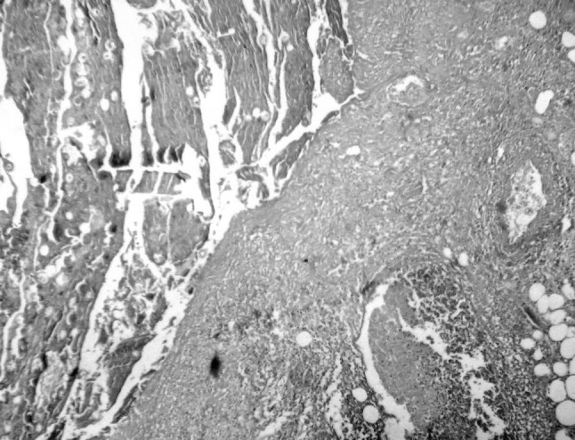
Microphotograph showing histopathological features of gangrenous appendicitis (right side) with mucosal ulceration. The exudate in the lumen (left side) show eggs of Taenia species. (Hematoxylin and Eosin stain, ×100 magnification)
Fig. 2.
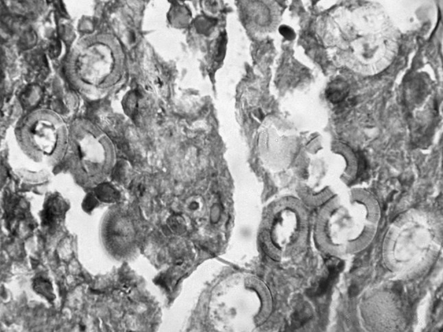
Microphotograph showing a closer look at the eggs of Taenia species. (Hematoxylin and Eosin stain, ×400 magnification)
Discussion
Taenia saginata and Taenia solium are cyclophyllidean tape worms whose adult forms are harbored in human intestines, who act as definitive hosts. Cattle and pigs serve as the intermediate hosts for T. saginata and T. solium, respectively (Kia et al. 2004). Human acquires Taeniasis following ingestion of raw or insufficiently cooked pork or beef containing the larval forms. Following ingestion, the scolex attaches itself to the intestine and subsequently starts to develop proglottids which ultimately develop into a long strobila (Lejbkwicz et al. 2002). The gravid proglottids containing Taenia eggs become detached and are excreted in human faeces, thus contaminating soil and water. The Taenia eggs are spherical and measure 30–40 μm in diameter but cannot be morphologically distinguished whether they belong to T. solium or T. saginata. These eggs can then be ingested by cattle, swine or man. Once in the digestive tract, the eggs lose their coat by the action of gastric and pancreatic enzymes and liberate oncospheres which, aided by their hooklets cross the intestinal wall, local venules, enter systemic circulation. They are then subsequently carried to different organs of the host (skeletal muscles, CNS, subcutaneous tissue, eye, etc.) After getting lodged to the host organ, the oncospheres lose their hooklets and gradually evolve into cysticerci. The life-cycle gets completed when undercooked pork or beef infested with cysticerci is again eaten by human beings (Prasad et al. 2008). Thus, it is evident that incidence of Taeniasis and Cysticercosis will be high in areas with poor hygiene, overcrowding, in communities which live in close association with cattle and swine or in those who take undercooked pork or beef. So much so, Cysticercosis has been designated as a “biological marker” of the social and economic development of a community (Carpio et al. 1998). In humans, gravid proglottids or their eggs may get lodged in the appendiceal lumen and result in appendicitis. However, appendicular Taeniasis is uncommon. Kia et al. (2004) in their study of 879 appendicectomy specimens also found a single case of Taeniasis. Gupta et al. (1989) in their review of 2,921 appendicectomies reported an incidence of 0.05 % Taeniasis. Silva et al. (2008) in their retrospective study of 1,600 appendectomies performed during a 10-year period found a single case of appendicular Taeniasis. 2 cases were reported by Lejbkwicz et al. (2002) and 3 cases by Khodjet El Khil et al. (1980) while 1 case each were reported by Sartorelli et al. (2005) and Payne et al. (1970). Barring these, reports of appendicular Taeniasis have been sparse. Recognition of this entity by meticulous histopathological examination of all appendicectomy specimens is thus important particularly in developing countries like ours for further treatment and life-style modification of patients to prevent further complications.
References
- Carpio A, Escobar A, Hauser WA. Cysticercosis and epilepsy: a critical review. Epilepsia. 1998;39:1025–1040. doi: 10.1111/j.1528-1157.1998.tb01287.x. [DOI] [PubMed] [Google Scholar]
- Gupta SC, Gupta AK, Keswani NK, Singh PA, Tripathi AK, Krishna V. Pathology of tropical appendicitis. J Clin Pathol. 1989;42(11):1169–1172. doi: 10.1136/jcp.42.11.1169. [DOI] [PMC free article] [PubMed] [Google Scholar]
- Khodjet El Khil A, Zitouna MM, Chadli A, Maamouri MT, Kennou MF, Ben Rachid MS. Appendicular Taeniasis. Report of 3 cases. Arch Inst Pasteur Tunis. 1980;57(4):349–354. [PubMed] [Google Scholar]
- Kia EB, Afshar-Moghadam N, Kazemzade H. Appendicular taeniasis: an association with acute gangrene appendicitis in Isfahan, Iran. Southeast Asian. J Trop Med Public Health. 2004;35:259–261. [Google Scholar]
- Lejbkwicz F, Abel AB, Tsilman B, Cohen HI. Taenia infestation in the appendix: a report of two cases. J Med Microbiol. 2002;51:90–91. doi: 10.1099/0022-1317-51-1-90. [DOI] [PubMed] [Google Scholar]
- Payne JE. Taenia infestation of the vermiform appendix. Med J Aust. 1970;1:768–769. doi: 10.5694/j.1326-5377.1970.tb116844.x. [DOI] [PubMed] [Google Scholar]
- Prasad KN, Prasad A, Verma A, Singh AK. Human cysticercosis and Indian scenario: a review 2008. J Biosci. 2008;33:571–582. doi: 10.1007/s12038-008-0075-y. [DOI] [PubMed] [Google Scholar]
- Sartorelli AC, da Silva MG, Rodrigues MA, da Silva RJ. Appendiceal taeniasis presenting like acute appendicitis. Parasitol Res. 2005;97(2):171–172. doi: 10.1007/s00436-005-1408-5. [DOI] [PubMed] [Google Scholar]
- Silva DF, Silva RJ, Silva MG, Sartorelli AC, Takegawa BK, Rodrigues MA. Parasitic infection of the appendix and its possible relationship to acute appendicitis. Arq Gastroenterol. 2008;45(2):166–168. doi: 10.1590/s0004-28032008000200015. [DOI] [PubMed] [Google Scholar]