Abstract
Gonadotropin-releasing hormone (GnRH) and the GnRH receptor (GnRHR) play an important role in vertebrate reproduction. Although many GnRHR genes have been identified in a large variety of vertebrate species, the evolutionary history of GnRHR in vertebrates is unclear. To trace the evolutionary origin of GnRHR we examined the conserved synteny of chromosomes harboring GnRHR genes and matched the genes to linkage groups of reconstructed vertebrate ancestor chromosomes. Consistent with the phylogenetic tree, three pairs of GnRHR subtypes were identified in three paralogous linkage groups, indicating that an ancestral pair emerged through local duplication before two rounds of whole genome duplication (2R). The 2R then led to the generation of six subtypes of GnRHR. Some subtypes were lost during vertebrate evolution after the divergence of teleosts and tetrapods. One subtype includes mammalian GnRHR and a coelacanth GnRHR that showed the greatest response to GnRH1 among the three types of GnRH. This study provides new insight into the evolutionary relationship of vertebrate GnRHRs.
Introduction
Families of neuropeptides and receptors have emerged through evolutionary processes such as gene/chromosome duplications. In particular, two rounds (2R) of whole genome duplication (WGD) during early vertebrate evolution contributed to the expansion of family members [1]–[8]. Moreover, teleost fish underwent a teleost-specific third round (3R) of genome duplication [6]. In addition to WGD, local tandem duplication of some genes and loss of redundant genes occurred before and after 2R [9]–[14], therefore the evolutionary scheme of a gene family can be highly complicated. Genome duplication events have produced paralogous chromosomal regions, also called paralogons, that provide a basis for exploring orthologous and paralogous relationships among gene family members as well as predicting gene loss in the paralogons [1], [2], [3], [14], [15]. In addition, tracing the family of genes on reconstructed pre-2R vertebrate ancestral chromosomes (VAC) is a fast and relatively accurate way to explore relationships among members of a family that contains a large number of paralogous genes [14], [16]. In particular, because this method provides a WGD scheme for each ancestral linkage group, gene duplication and/or loss during each step of genome duplication can be traced [16].
The neuropeptide gonadotropin-releasing hormone (GnRH) and its G protein-coupled receptor GnRHR play a pivotal role in the control of reproduction in vertebrates [17], [18]. To date, many GnRH and GnRHR genes have been identified in invertebrate and vertebrate species using conventional biochemical and bioinformatics tools [13], [19]–[28]. Most vertebrate species have two or three forms of GnRH in the brain (GnRH1, GnRH2, and GnRH3) [13], [19], [20], [29]. The corresponding genes are likely to have emerged through 2R because the three subtypes of the GnRH gene are on three different paralogons that share common neighbor gene families [13]. Most vertebrate species also possess multiple forms of GnRHR. For example, teleost fish possess four or five isoforms of GnRHR [25], [29], whereas amphibian and reptilian species have three forms of GnRHR [23], [30]. In our previous study using synteny analysis, we proposed four vertebrate GnRHR clades: non-mammalian GnRHR n1, n2, and n3, and mammalian GnRHR m1. GnRHR n1 and n2 were suggested to have emerged through local duplication before 2R because these genes are on the same linkage group of vertebrate chromosomes [13].
In an extension of our previous study, the present study examined more diverse vertebrate and invertebrate species including lamprey (Petromyzon marinus), coelacanth (Latimeria chalumnae), spotted gar (Lepisosteus oculatus), Ciona intestinalis, amphioxus (Brnachiostoma floridae), Caenorhabditis elegans, and Drosophila melanogaster. Synteny analysis and relocation of GnRHR genes and their neighboring genes on vertebrate ancestral linkage groups revealed that three pairs of GnRHRs reside on three paralogous chromosomal regions. When combined with phylogenetic analysis, this observation suggests the presence of six subtypes of vertebrate GnRHRs that arose through local duplication on the same chromosome followed by 2R. The fourth pair of GnRHR was probably lost during 2R. In addition, our study suggests that a coelacanth GnRHR is an ortholog of mammalian GnRHR because the proteins share a high degree of amino acid sequence identity and both receptors show the highest activity in response to GnRH1 among three GnRH types, thus resolving an enduring question regarding the origin of mammalian GnRHR.
Results
Phylogenetic analysis of GnRHR and evolutionary history of the vertebrate GnRHR genes
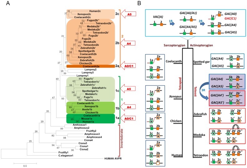
The amino acid sequences of GnRHR from various vertebrate taxa including 13 species (human, mouse, anole lizard, chicken, Xenopus, coelacanth, spotted gar, zebrafish, medaka, fugu, stickleback, tetraodon, and lamprey) (Table 1) and invertebrate taxa including 4 species (C. elegans, Drosophila, Ciona, and amphioxus) (Table S1) were retrieved from ENSENBL and NCBI databases. Phylogenetic analysis reveals that vertebrate GnRHRs are grouped monophylogenetically excluding invertebrate GnRHR-like receptors (Figure 1A). To explore the evolutionary relationship among the vertebrate GnRHR lineage, we examined the location of each GnRHR lineage on reconstructed linkage groups of vertebrate ancestors proposed by Nakatani et al. [6]. The Nakatani model suggests the presence of 10–13 pre-2R vertebrate ancestral chromosomes (VAC), defined as the A-J linkage groups. These VACs then underwent 2R to generate approximately 40 post-2R gnathostome ancestor chromosomes (GACs A0-J1). Because chromosomal segments of medaka, chicken, mouse, and human have been matched to these reconstructed linkage groups, the location of a current gene can be traced back to the linkage groups [14], [16]. Our data showed that three pairs of GnRHR subtypes (1a–2a, 1b–2b, and 1c–2c) are found in three related GAC linkage groups, GAC A0/C1, A4, and A5 (Figure 1A). Based on phylogenetic analysis and the gene location on GACs, we propose that vertebrate GnRHRs originated from two ancestral GnRHR genes designated types 1 and 2 that emerged through a local duplication on the same chromosome before 2R. During 2R each ancestor gene gave rise to subtypes ‘a’ on GAC A0/C1, ‘b’ on GAC A4, and ‘c’ on GAC A5, resulting in six subtypes (1a,b,c and 2a,b,c) of GnRHR in vertebrates. The subtype 1a includes mammalian GnRHR and a coelacanth GnRHR. No ray-finned fish (actinopterygii including spotted gar) GnRHRs belong to the 1a subtype. The subtypes 1b and 1c are included in the same branch. The 1b subtype includes tetrapod and lobe-finned fish (sarcopterygii) receptors whereas the 1c subtype contains only ray-finned fish receptors. This observation may lead to misunderstanding of the origin of these subtypes. For example, 1b and 1c subtype receptors might be considered to be orthologous. However, as 1b and 1c subtype receptors are on different GACs (GAC A4 and A5, respectively) they are more likely to be paralogs generated through a second round of WGD. Therefore, it seems likely that the 1b subtype was lost in ray-finned fish and the 1c subtype was lost in lobe-finned fish after 2R. The teleost-specific 3R may have generated an additional form, the 1c′ subtype. The 2a subtype includes only a coelacanth receptor therefore this subtype was lost in other vertebrates. The 2b subtype includes both ray-finned and lobe-finned fish receptors. The evolution of the 2b subtype in teleosts is highly complicated. The teleost-specific 3R might have contributed to generation of the 2b′ subtype, which was followed by local duplication for emergence of the 2b″ subtype. The 2c subtype is composed of human, Xenopus, and coelacanth receptors, but no ray-finned fish receptors (Figure 1A).
Table 1. Classification of vertebrate GnRHR based on phylogenetic and synteny analysis.
| Vertebrate species | Subtype (GAC) | |||||
| 1a (A0/C1) | 1b (A4) | 1c (A5) | 2a (A0/C1) | 2b (A4) | 2c (A5) | |
| Human (H. sapiens) | GnRHR (m1) AAI13547 (Ch4) | GnRHR (n3/m2) NR_002328 (Ch1) | ||||
| Mouse (M. musculus) | GnRHR (m1) NP_034453 (Ch7) | |||||
| Anole lizard (A. carolinensis) | GnRHR (n2) ENSACAP00000004352 (Scaffold:GL343392) | |||||
| Chicken (G. gallus) | GnRHR (n2) NP_989984 (Ch10) | GnRHR (n1) NP_001012627 (Ch10) | ||||
| Western clawed frog (X. tropicalis) | GnRHR (n2) NP_001107548 (Scaffold:GL172999) | GnRHR (n1) NP_001107547 (Scaffold: GL172658) | GnRHR (n3/m2) NP_001107549 (Scaffold:GL176594) | |||
| Coelacanth (L.chalumnae) | GnRHR1a ENSLACG00000002185 (Scaffold:JH130998) | GnRHR1b ENSLACG00000016584 (Scaffold:JH126619) | GnRHR2a ENSLACG00000013490 (Scaffold:JH127011) | GnRHR2b ENSLACG00000018198 (Scaffold:JH126611) | GnRHR2c ENSLACG00000014236 (Scaffold:JH126664) | |
| Spotted gar (L.oculatus) | GnRHR ENSLOCG00000008760 (Ch LG24) | GnRHR ENSLOCG00000014360 (Ch LG3) | ||||
| Zebrafish (D. renio) | GnRHR (n3) NP_001170921 (Ch16) | GnRHR (n2) NP_001138451.1 (Ch7) | ||||
| GnRHR′ (n3b) NP_001138452 (Scaffold:Zv-NA22) | GnRHR′ (n1) NP_001091663 (Ch18) | |||||
| Medaka (O. latipes) | GnRHR (n3) NP_001098392 (Scaffold:2457) | GnRHR (n1) NP_001098393 (Ch6) | ||||
| GnRHR′ (n2) ENSORLP00000015859 (Scaffold:1670) | ||||||
| GnRHR″ (n1b) NP_001098352 (Ch3) | ||||||
| Green pufferfish (T. nigroviridis) | GnRHR (n3) BAE45702 (Ch_Unrandom) | GnRHR (n1) BAE45698 (Ch13) | ||||
| GnRHR′ (n3b) BAE45700 (Ch17) | GnRHR′ (n2) ENSORLP00000015859 (Scaffold:1670) | |||||
| GnRHR″ (n1b) BAE45694 (ChUnrandom) | ||||||
| Stickleback (G.aculeatus) | GnRHR (n3) ENSGACP00000004101 (Ch20) | GnRHR (n1) ENSGACP00000014249 (Ch19) | ||||
| GnRHR′ (n3b) ENSGACP00000000651 (Scaffold:101) | GnRHR′ (n2) ENSGACP00000021774 (Ch2) | |||||
| GnRHR″ (n1b) ENSGACP00000019583 (Ch2) | ||||||
| Fugu (T. rubripes) | GnRHR(n3) ENSTRUP00000014399 (Scaffold:270) | GnRHR (n1) ENSTRUP00000019152 (Scaffold:217) | ||||
| GnRHR′ (n3b) ENSTRUP00000005457 (Scaffold:291) | GnRHR′ (n2) ENSTRUP00000014430 (Scaffold:174) | |||||
| GnRHR″ (n1b) ENSTRUP00000018665 (Scaffold:92) | ||||||
| Lamprey (P. marinus) | GnRHR1 ENSPMAP00000010120 (Scaffold:GL493405) | GnRHR2 ENSPMAP00000003609 (Scaffold:GL483393) | GnRHR3 ENSPMAP00000011312 (Scaffold:GL483841) |
The subtype annotations were based on phylogeny and location on gnathostome ancestor gene linkage blocks GAC A0/C1, GAC A4, and GAC A5. The accession numbers for NCBI and ENSEMBL and gene location are given below the names of the receptors. The receptor names according to our previous classification [13] are shown in parentheses. The five new coelacanth GnRHRs follow current subtype annotation. ′, product of 3R; ″, product of a local duplication after 3R.
Figure 1. Phylogenetic tree construction for GnRHR-like receptors and evolutionary relationship of vertebrate GnRHRs genes.
A, Amino acid sequences of vertebrate species (human, mouse, chicken, anole lizard, Xenopus, coelacanth, spotted gar, zebrafish, medaka, fugu, stickleback, tetraodon, and lamprey) and invertebrate species (C. elegans, Drosophila, Ciona, and amphioxus) were aligned using MUSCLE and the phylogeny tree was generated by MEGA 5.05. The human arginine vasopressin receptor 1a protein sequence was used as an outgroup. The vertebrate GnRHRs are grouped monophylogetically. The vertebrate gene types 1 and 2 are illustrated by green and orange, respectively. The arrows indicate the location of each vertebrate gene type on the ancestral linkage group. B, Prior to 2R, GnRHR types 1 and 2 emerged as a product of local duplication on the VAC A. Following 2R, a, b, and c subtypes of each ancestral type 1 and 2 were produced on GAC A0, GAC A4, and, GAC A5 respectively and GnRHR type 1a was translocated to GAC C1. During the evolution of vertebrate species some GnRHR subtypes have been lost or/and duplicated. The empty box indicates a pseudogene in a human chromosome. An empty box with a cross indicates loss of the gene.
Based on these results, we propose an evolutionary history of vertebrate GnRHRs. Type 1 and 2 receptors may have emerged through a local duplication on VAC A before 2R. During the first round (1R) of WGD, two genes were duplicated on GAC A0/A1 and A4/5 linkage groups. According to the Nakatani model, GAC A2/3 separated from the GAC A4/5 linkage group by fission after 1R [6] therefore GAC A2/3 is not discussed here. The GAC A4/5 linkage group doubled during the second round (2R) of WGD to produce pairs of GnRHR on GACs A4 and A5. The second round of WGD may not have generated the fourth GnRHR pair on GAC A1. Alternatively, the fourth GnRHR pair on GAC A1 might have been lost after 2R. After 2R, the 1a subtype gene appears to have moved to the GAC C1 linkage group, probably through chromosomal rearrangement or a single gene translocation (Figure 1B). Thus, gnathostome ancestors may have six subtypes of GnRHR. Some subtypes of GnRHR have been lost after the divergence of ray-finned and lobe-finned fish. For example, coelacanth lost the 1c subtype and Xenopus lacks 1a, 2a, and 1c subtypes. Chicken has only two subtypes, 1b and 2b, and humans possess one functional receptor of the 1a subtype and one nonfunctional pseudogene of the 2c subtype [24]. In early actinopterygians, GnRHRs on GAC A0/C1 are likely to have disappeared. Thus, spotted gar contains only two GnRHRs (2b and 1c) on GAC A4 and A5. However, GnRHRs on GACs A4 and A5 were duplicated through the teleost-specific 3R, followed by loss of some subtypes. Zebrafish contains four subtypes of receptors, 2b, 2b′, 1c, and 1c′. In medaka and tetraodon, the 2c′ subtype was lost but a 2b″ subtype emerged, probably through a local duplication (Figure 1B). Although we propose a scenario for a doublet GnRHR before 2R, a triplet GnRHR before 2R is also possible because the 1a subtype is phylogenetically distinct from 1b and 1c subtypes. Such a scenario involves an additional local duplication, generating three types (1a, 1b/c, and 2a/b/c) before 2R.
Synteny of vertebrate chromosomes containing GnRHRs
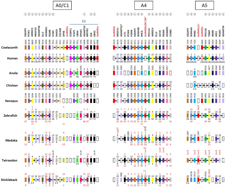
To corroborate the gene/genome duplication scheme of the vertebrate GnRHR family, we performed synteny analysis of chromosomal fragments containing GnRHRs. The coelacanth scaffold JH127011 contains the GnRHR 2a subtype and its neighboring genes, which are also found in regions of human chromosomes 5 and 4 that fall into GAC A0 and C1, respectively. It is noteworthy that this region of chromosome 4 contains the GnRHR 1a subtype (Figure 2). Unfortunately, 1a and 2a subtypes are not found in other vertebrate species. The presence of neighbor genes belonging to either GAC A0 or C1 in the coelacanth scaffold JH127011 suggests the possibility that the linkage groups including GAC A0 and C1 were previously linked. This possibility is partly supported by observations that the paralogous genes ADAMTS6 and ADAMTS3 are on either GAC A0 or C1 and that some genes in fish, including ADAMTS3, EPHA5, and NPFFR2 on GAC C1, are on the same chromosomal regions as genes that map on GAC A0 (Figure 2). An alternative scenario is that GnRHR 1a and some neighbors were translocated to the GAC C1 linkage group.
Figure 2. Synteny analysis for chromosome fragments harboring vertebrate GnRHRs.
GnRHR and neighboring genes in human, coelacanth, anole, chicken, Xenopus, zebrafish, medaka, tetraodon, and stickleback were matched on ancestral linkage groups. The GnRHRs are illustrated in red. Orthologous genes are aligned in the same column and indicated by the same color. The chromosome number and gene location (in megabases) are indicated above and below the indicated genes respectively. For genes located on scaffolds, the gene positions are indicated vertically with only the last four digits of the scaffold number (e.g., JH127011 in coelacanth is indicated as 7011). For teleost species, the duplicated genes are represented by two lines of numbers because of the third round of whole genome duplication. The broken boxes indicate loss or absence of genes. The common paralogs across GAC A0, GAC A4, GAC A5, and GAC C1 are indicated by open circles containing numbers as follows:  indicates the gene families found on GACs A0, A4, and A5;
  indicates the gene families observed in GACs A0 and A4;
  indicates the genes in GACs A4 and A5. In the case of stickleback and medaka, (*) indicates that 2b″ was generated from local gene duplication of 2b′.
The GnRHR 1b and 2b subtypes are on coelacanth scaffolds JH126619 and JH126611, respectively. Neighbor genes on these two scaffolds are found on the same chromosomes in human (chromosome 15) and chicken (chromosome 10). The 1b subtype is absent in teleosts. The teleost-specific 3R might have contributed to doubling of the 2b subtype to produce 2b and 2b′ subtypes as these subtypes are found in chromosomal regions that seem to have been duplicated through 3R [6]. In addition, medaka, tetraodon, and stickleback have an additional subtype 2b″ that is likely to have emerged through a local duplication after 3R. In stickleback, the 2b′ and 2b″ subtypes are on the same chromosome (Figure 2). This observation is consistent with the phylogenetic tree (Figure 1A).
Chromosomal regions with GnRHR 1c or 2c subtypes share neighbors in vertebrate chromosomal regions. The 2c subtype is present in coelacanth, human, and Xenopus, but not in ray-finned fish. In contrast, the 1c subtype is present in ray-finned fish but absent in lobe-finned fish and tetrapods. There are two 1c subtype genes in teleost fish. As one gene is on a short scaffold with few neighbors, it is unclear whether these genes were generated by the teleost 3R or local duplication (Figure 2).
Many paralogous genes were shared between these linkage groups (Figure 2). For example, IQGAP, ADAMTS, MEGF/PEAR, RGM/HFE, and ARRDC/TXNIP family genes are commonly present in the three linkage groups. Paralogous genes for PCSK1, ELL2, HOMER1, and CCNO on GAC A0 are present in GAC A4 and paralogous genes for SEMA4B, RHCG, PWX11A, and IGF1R on GAC A4 are seen in GAC A5. This result is consistent with the Nakatani model proposing that the three linkage groups shown in Figure 2 were generated through the 2R.
Subtype-specific motifs in vertebrate GnRHRs
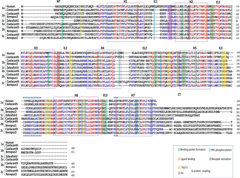
In a search for new GnRHR genes we identified five new genes in coelacanth. Our synteny and phylogenetic analysis revealed that one of these coelacanth GnRHR genes corresponds to mammalian type I GnRHR (the 1a subtype in this study) and three genes correspond to non-mammalian GnRHRs GnRHRn1 (2b), GnRHRn2 (1b), and GnRHRn3 (2c) according to our previous classification [13]. Surprisingly, one coelacanth GnRHR (2a) is unique and cannot readily be compared with other vertebrate GnRHRs. The five coelacanth GnRHRs were aligned with human 1a, Xenopus 1b, 2a, 2b, 2c, and zebrafish 1c GnRHRs (Figure 3). Alignment for all amino acid sequences of GnRHR examined in this study is shown in Figure S1. Sequence alignment analysis revealed that all subtypes of GnRHRs share many critical residues involved in ligand binding, binding pocket formation, G protein coupling, and receptor activation (Figure 3). The major difference between types 1 and 2 is found in intracellular loop 3 (IL3). The type 2 receptors have a longer IL3 that contains a highly conserved SKExxLRr/cS motif. The type 1 receptors have subtype-specific residues in IL3: the 1a subtype has a Tr/qVLh/rQDP motif, the 1b subtype contains KQm/lK motif, and the 1c subtype possesses a slightly longer IL3 without pronounced sequence conservation (Figure S1). Because IL3 is involved in G-protein coupling, receptor desensitization, and internalization [31], [32], this subtype specificity may result in differences in signaling cascades and down-regulation among receptors. Extracellular loop 3 (EL3) also contains subtype-specific motifs: Sd/eP for 1a, PEY for 1b and 1c, Pe/pS for 2a and 1c, and Sh/qS for 2b. These motifs are known to be involved in ligand specificity [33]-[36]. Major differences between subtype 1a mammalian GnRHR and other subtype receptors are the absence/presence of the cytoplasmic tail and residue Asn2.50/Asp2.50 in transmembrane helix 2 (TMH2) [37]-[40]. Coelacanth GnRHR1a possesses specific characteristics present in mammalian type I GnRHR (the 1a subtype) – it lacks the cytoplasmic tail and contains Asn2.50 in TMH2. Thus, coelacanth GnRHR 1a may be an ortholog of mammalian type I GnRHR.
Figure 3. Sequence alignment of coelacanth GnRHRs with vertebrate GnRHRs that represent each subtype.
The five coelacanth GnRHRs were aligned with human GnRHR1a, Xenopus GnRHR1b, zebrafish GnRHR1c, Xenopus GnRHR2b, and Xenopus GnRHR2c. Residues with 50% and 90% similarity are indicated in blue and red, respectively. Predicted N terminal (NT), transmembrane helix (H), intracellular loop (IL), extracellular loop (EL) and C-terminal (CT) domains are indicated. The putative residues involved in ligand binding, binding pocket formation, receptor activity, PKC phosphorylation, and G protein coupling are illustrated in different colors. ▾ indicates the subtype-specific motifs.
Ligand selectivity of coelacanth GnRHR 1a
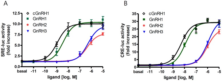
To further examine functional orthology between coelacanth GnRHR 1a and mammalian type I GnRHR, we examined the ligand selectivity of coelacanth GnRHR 1a. HEK293 cells were transfected with plasmid containing coelacanth GnRHR 1a and the reporter vector SRE-luc or CRE-luc. Cells were then treated with human GnRH1, coelacanth GnRH1, chicken GnRH2, or salmon GnRH3. In response ligand stimulation, cells transfected with coelacanth GnRHR 1a showed an increase in both SRE-luc and CRE-luc activities, indicating that coelacanth GnRHR 1a may be coupled to both Gq/11- and Gs-mediated signaling pathways (Figure 4). Coelacanth GnRHR 1a responded best to human and coelacanth GnRH1 but poorly to GnRH2 and GnRH3, indicating that coelacanth GnRHR 1a has highest affinity for GnRH1, as does the mammalian type I receptor [33], [34].
Figure 4. Activity of GnRH peptides with coelacanth GnRHR 1a.

Plasmid containing coelacanth GnRHR 1a sequence was co-transfected into HEK293T cells with SRE-luc (A) or CRE-luc (B) vector in 48-well plates. Forty-eight hours after transfection, cells were treated with the indicated concentrations of peptides (◊ coelacanth (c) GnRH1; ▪ GnRH1; • GnRH2; ▾ GnRH3) for 6 h, and luciferase activity was examined.
Discussion
The evolutionary relationship and history of a gene family can be deciphered by conventional phylogenetic analysis and synteny analysis of chromosomal regions containing members of the gene family [5], [8],[13]. The accumulation of genome sequence information for various invertebrate and vertebrate species has facilitated entire genome comparisons of evolutionarily distant taxa, allowing reconstruction of hypothetical chromosomes of vertebrate or chordate ancestors [6], [7]. Recently, relocating gene family members on these reconstructed vertebrate ancestral linkage groups was found to be a complementary tool to examine the evolutionary history of a gene family [14], [16]. This method is particularly useful for a gene family with a large number of paralogs that arose before WGD [14]. Because conventional small-scale synteny covers relatively short chromosomal regions, neighbor genes located far from the gene of interest on the chromosome or genes on other chromosomes are often excluded from the analyses. In contrast, the size of the reconstructed ancestral linkage group is much greater than that used for small-scale synteny and the ancestral linkage group also includes genes clusters that translocated to other chromosomes [14], [16]. However, this method suffers from poor resolution in some ancestral linkage groups due to either inaccurate reconstruction because of massive chromosome rearrangements, or a single gene translocation that caused it to move from its authentic chromosomal fragments during or after 2R [14], [16]. Thus, combining phylogenic and syntenic analyses is necessary for a better understanding of the evolutionary relationship and history of a gene family [14], [16].
By applying the above approaches, we were able to propose an evolutionary history of the GnRHR family in vertebrates. In our previous study, we classified vertebrate GnRHRs into four subtypes, one mammalian 1 subtype (m1) and three non-mammalian subtypes (n1, n2, and n3) [13]. In the present study, through identification of a novel subtype of GnRHR and improved synteny methods, we propose the presence of six subtypes (1a–2c) of GnRHRs in vertebrates. The m1 subtype is equivalent to the 1a subtype that includes a coelacanth GnRHR. The n1 and n2 subtypes are largely shuffled in the present study (Table 1). The tetrapod n2 matches with 1b, whereas teleost n2 belongs to 2b. The tetrapod n1 and teleost n1 are included in the 2b subtype. The n3 group splits into 1c (teleost receptors) and 2c (tetrapod receptors). This confusion is largely due to multiple local duplications of GnRHR before 2R (and after 3R in teleosts) followed by either lobed-finned fish- or ray-finned fish-specific loss of the subtype gene. For example, the n1 and n2 subtypes (1b and 2b in this study) emerged via a local duplication on the same chromosome before 2R, and additional subtypes 2b′ and 2b″ in teleosts arose through 3R and a subsequent local duplication. Thus, our previous synteny failed to explore the fine mechanisms of the emergence of GnRHR subtypes through genome duplications and local duplications [13]. A new GnRHR found in coelacanth is likely to belong to a new subtype 2a.
The phylogenetic trees constructed in our study and by others reveal that the 1a subtype receptors are phylogenetically closer to invertebrate receptors than to other receptors [27] suggesting that, among vertebrate receptors, the 1a subtype receptors are probably closer to an ancestral form of GnRHR. Interestingly, amphioxus has four GnRHRs, two of which are similar to the protostomian GnRHR found in Drosophila and C. elegans whereas the other two are similar to vertebrate GnRHR [27], [41]. Indeed, a recent study showed that the vertebrate-like amphioxus GnRHRs exhibited a high affinity to vertebrate GnRHs but not to protostomian GnRHs, whereas protostomian-like amphioxus GnRHRs had poor affinity to vertebrate GnRHs [27]. Thus, amphioxus contains two GnRHR lineages, vertebrate-like GnRHR and protostomian-like GnRHR. The protostomian GnRHRs were not found in the basal vertebrate lamprey. Instead, lamprey contains three receptors which are likely to belong to type 2 receptors. As the scaffolds containing lamprey GnRHRs are short and do not have any neighbors, syntenic analysis to explore the evolutionary position of these genes is impossible.
Our chromosome analysis showed that three pairs of the subtypes (1a–2a, 1b–2b, and 1c–2c) are present on three paralogous chromosomal regions. Although pairs of the subtype are on the same chromosomes, members in each pair are phylogenetically distant; instead, the phylogeny tree shows a close relationship among type 1 subtypes (1a, 1b, and 1c) and type 2 subtypes (2a, 2b, and 2c). This indicates that a local duplication of GnRHR occurred before 2R and that consecutive chromosome duplications have contributed to the generation of three pairs. The fourth pair was probably lost after 2R. There is accumulating evidence for local duplication gene families before 2R. In the case of somatostatin and its paralogous genes cortistatin, urotensin II, and urotensin II-like peptide, two pairs of the genes are on two paralogous chromosomes [11]. The neuropeptide Y receptor (NPYR) triplet is proposed to have existed before 2R [9]. In addition, prolactin-releasing hormone receptors, which are phylogenetically very close paralogous receptors for NPYR, are also in the near vicinity of NPYR on the same chromosomes [10]. The secretin-like receptors and their peptides genes are also clustered on paralogous chromosomes [14], showing that paralogous genes that arose through local duplications reside in the near vicinity on the same chromosomes or on reconstructed ancestral chromosomes.
The origin of type 1 mammalian GnRHR has been questioned because orthologs of the mammalian receptor have not been found in other vertebrates and the structure of the mammalian receptor is quite different from that of other receptor subtypes [13], [27], [42]. In our study, we found that a coelacanth receptor is similar to the mammalian receptor based on amino acid sequence. Like mammalian GnRHR, this coelacanth receptor lacks the C-terminal cytoplasmic tail, has conserved Asn2.50/Asp7.49 residues in TMH2 and THM7, and harbors an Sd/eP motif in ECL3. The presence or absence of the C-terminal cytoplasmic tail is important for receptor down-regulation and internalization [37]. For example, addition of the C-terminal cytoplasmic tail to mammalian GnRHR greatly enhances the internalization and desensitization rates [37]. Furthermore, the C-terminal cytoplasmic tail is likely to be involved in signaling pathway. Deletion of the C-terminal tail from bullfrog GnRHR-1 remarkably decreased its ability to induce Gs-mediated adenylyl cyclase (AC)/protein-kinase A (PKA) signaling. In contrast, addition of this motif to mammalian GnRHR improved its ability to induce the AC/PKA-linked signaling pathway [38]. Interaction of the Asn2.50 residue in TMH2 with the Asp7.49 residue in TMH7 of mammalian GnRHR is essential for receptor conformation and activation whereas non-mammalian receptors have Asp2.50/Asp7.49 in both TMH2 and TMH7 [39], [40]. The GnRH binding residues (i.e. Asp98, Asn102, Lys121, Asn212, Trp280, Trp289, and Tyr290) of mammalian GnRHR-1 are highly conserved among vertebrate GnRHRs [43]–[47]. Thus, a mammalian GnRHR-specific motif, the Sd/eP motif in ECL3, was considered to be important for high affinity toward GnRH1 [33]–[35]. Indeed, our study shows that the coelacanth receptor with the SDP motif responded best to human and coelacanth GnRH1 but poorly to GnRH2 and GnRH3. In particular, differences in the specific domains (SD/EP, PEY, and PPS) at the ECL3 have been used for classification of GnRHR [36], [42]. Taken together, these findings indicate that the coelacanth receptor is a functional ortholog of the mammalian type 1 GnRHR, thus the 1a subtype receptor is no longer unique to mammals.
In summary, the evolutionary history of the GnRHR represents repetitions of continuous duplications and losses within a gene family during metazoan evolution. In particular, during emergence of early vertebrates, at least two GnRHRs appear to have been generated through local duplication. This doublet was amplified as three pairs through WGD, followed by loss of redundant members before the divergence of ray-finned and lobed-finned fish. Evolution of GnRHR in teleosts is more complicated than that of tetrapods due to teleost-specific 3R, local duplication after 3R, and loss of genes before or after 3R. Because of the complexity of GnRHR evolutionary history, a combination of phylogenetic analysis, small-scale synteny, and relocating gene family on the reconstructed ancestor chromosome is required for better understanding of the evolutionary history of the gene family. This issue will be further clarified when improved genome data of other vertebrate and invertebrate species are available.
Materials and Methods
Phylogeny of the GnRHR family
ENSEMBE (http://www.ensembel.org) and GenBank (http://www.ncbi.nlm.nih.gov/Entrez/) databases were used to retrieve all annotated amino acid sequences for GnRHR of vertebrates including lamprey, spotted gar, zebrafish, medaka, fugu, stickleback, tetraodon, coelacanth, anole lizard, Xenopus, chicken, mouse, and human, and of invertebrates including C. elegans, Drosophila, Ciona, and amphioxus [26], [27], [30], [41], [48]–[51]. Orthologous or paralogous genes from vertebrate and invertebrate species were identified through research tools provided by the ENSEMBEL database. To obtain additional sequences other than the well-annotated query sequences, a TBLASTN algorithm was performed against a genome database of selected species [14]. The phylogenetic analysis of GnRHR sequences was accomplished as previously described [14]. The MUSCLE algorithm with default settings was used to align the sequences as implemented in MEGA 5.05. A maximum likelihood phylogenetic tree was created following the Jones-Taylor-Thornton model with a confidence level of 100 replicate bootstrap samples.
Synteny analysis and location of GnRHR genes on reconstructed ancestral linkage group
For synteny analysis, contig views of genome regions surrounding the GnRHR loci were compared. The exact map location of orthologs/paralogs of neighboring genes was obtained from the ENSEMBLE database [14]. Next, the chromosomal segments of three taxa (medaka, chicken, and human) with a reliable synteny were matched with vertebrate ancestral linkage groups according to the Nakatani model [6] as described previously [14], [16]. The Nakatani model provides vertebrate ancestral linkage blocks displayed on individual human chromosomes compared with the corresponding chromosome locations of medaka, chicken, and mouse [6]. Comparison of results from each taxon resolved the locations of the gene blocks at consecutives stages of vertebrate genome evolution.
Chemicals and hormones
All chemicals were obtained from Sigma-Aldrich (St. Louis, MO, USA) unless otherwise stated. Restriction enzymes were obtained from New England Biolabs (Ipswich, MA, USA). Human (hu) GnRH1, coelacanth GnRH1 (pyroEYWSYDLRPG-NH2), chicken GnRH2, and salmon GnRH3 were synthesized by AnyGen (Gwangju, Korea). The purities of the synthesized peptides were greater than 98% based on high-performance liquid chromatography. All peptides were dissolved in distilled water and then diluted in media to the desired working concentrations.
Plasmid constructs
The pcDNA3.1 expression vector was purchased from Invitrogen Corp. (San Diego, CA, USA). The CRE-luc vector containing four copies of the cyclic AMP-responsive element (CRE; TGACGTCA), and the SRE-luc vector containing a single copy of the serum response element (SRE, CCATATTAGG) were purchased from Stratagene (La Jolla, CA, USA). The cDNA for coelacanth GnRHR 1a was synthesized by GenScript (Piscataway, NJ, USA). The cDNA was incorporated into the EcoRI and XhoI sites of pcDNA3.1 and the resulting construct was verified by sequencing.
Cell transfection and luciferase assay
HEK293T cells (American Type Culture Collection, Manassas, VA, USA) were maintained in Dulbecco's modified Eagle's medium (DMEM) supplemented with 10% fetal bovine serum, 100 U/ml penicillin G, and 100 µg/ml streptomycin (Invitrogen). Cells were seeded in 48-well plates at a density of 1.4×10⁴ cells per well 1 day before transfection. A mixture containing 75 ng of CRE-luc (or SRE-luc), 75 ng of the coelacanth GnRHR 1a plasmid, and 2 µl of Effectene reagent (Qiagen, Chatsworth, CA, USA) was prepared and added to each well according to the manufacturer's instructions. Approximately 48 h after transfection, cells were treated with the respective ligands for 6 h. For SRE-luc analysis, cells were maintained in serum-free DMEM for at least 16 h before ligand treatment. Cells were lysed by addition of 100 µl lysis buffer and the luciferase activity in 40 µl of cell extract was determined on a luciferase assay system according to the standard protocol for the Synergy 2 Multi-Mode Microplate Reader (BioTek, Winooski, VT, USA).
Data analysis
Data analysis was performed by nonlinear regression with a sigmoidal dose-response. The agonist concentrations that induced half-maximal stimulation (EC50) were calculated with GraphPad PRISM4 software (GraphPad Software Inc., San Diego, CA, USA). All data were presented as mean ± S.E. of at least three independent experiments.
Supporting Information
Sequence alignment of vertebrate GnRHRs. GnRHRs from human, coelacanth, anole, chicken, Xenopus, zebrafish, medaka, tetraodon, fugu, and stickleback are shown. Residues with 50% and 90% similarity are indicated in blue and red respectively. Predicted N terminal (NT), transmembrane helix (H), intracellular loop (IL), extracellular loop (EL), and C terminal (CT) domains are indicated.
(PDF)
The accession numbers for NCBI and ENSEMBL of C. elegans, Drosophila, Ciona, and amphioxus GnRHRs are given beneath receptor's name as well as gene location in brackets.
(PDF)
Funding Statement
This work was supported by grants from the Brain Research Program (2011-0019205) and Basic Science Research Program (2013R1A2A2A01068295) of the National Research Foundation of Korea. The funders had no role in study design, data collection and analysis, decision to publish, or preparation of the manuscript.
References
- 1. Lundin LG (1993) Evolution of the vertebrate genome as reflected in paralogous chromosomal regions in man and the house mouse. Genomics 16: 1–19. [DOI] [PubMed] [Google Scholar]
- 2. Fredriksson R, Lagerström MC, Lundin L-G, Schiöth HB (2003) The G-protein-coupled receptors in the human genome form five main families. Phylogenetic analysis, paralogon groups, and fingerprints. Molecular pharmacology 63: 1256–1272. [DOI] [PubMed] [Google Scholar]
- 3. Larhammar D, Lundin L-G, Hallböök F (2002) The human Hox-bearing chromosome regions did arise by block or chromosome (or even genome) duplications. Genome research 12: 1910–1920. [DOI] [PMC free article] [PubMed] [Google Scholar]
- 4. Lee LT, Siu FK, Tam JK, Lau IT, Wong AO, et al. (2007) Discovery of growth hormone-releasing hormones and receptors in nonmammalian vertebrates. Proceedings of the National Academy of Sciences 104: 2133–2138. [DOI] [PMC free article] [PubMed] [Google Scholar]
- 5. Lee YR, Tsunekawa K, Moon MJ, Um HN, Hwang J-I, et al. (2009) Molecular evolution of multiple forms of kisspeptins and GPR54 receptors in vertebrates. Endocrinology 150: 2837–2846. [DOI] [PubMed] [Google Scholar]
- 6. Nakatani Y, Takeda H, Kohara Y, Morishita S (2007) Reconstruction of the vertebrate ancestral genome reveals dynamic genome reorganization in early vertebrates. Genome research 17: 1254–1265. [DOI] [PMC free article] [PubMed] [Google Scholar]
- 7. Putnam NH, Butts T, Ferrier DE, Furlong RF, Hellsten U, et al. (2008) The amphioxus genome and the evolution of the chordate karyotype. Nature 453: 1064–1071. [DOI] [PubMed] [Google Scholar]
- 8. Larsson T, Olsson F, Sundstrom G, Lundin L-G, Brenner S, et al. (2008) Early vertebrate chromosome duplications and the evolution of the neuropeptide Y receptor gene regions. BMC evolutionary biology 8: 184. [DOI] [PMC free article] [PubMed] [Google Scholar]
- 9. Tostivint H, Joly L, Lihrmann I, Parmentier C, Lebon A, et al. (2006) Comparative genomics provides evidence for close evolutionary relationships between the urotensin II and somatostatin gene families. Proceedings of the National Academy of Sciences of the United States of America 103: 2237–2242. [DOI] [PMC free article] [PubMed] [Google Scholar]
- 10. Wraith A, Törnsten A, Chardon P, Harbitz I, Chowdhary BP, et al. (2000) Evolution of the neuropeptide Y receptor family: gene and chromosome duplications deduced from the cloning and mapping of the five receptor subtype genes in pig. Genome research 10: 302–310. [DOI] [PMC free article] [PubMed] [Google Scholar]
- 11. Lagerström MC, Fredriksson R, Bjarnadóttir TK, Fridmanis D, Holmquist T, et al. (2005) Origin of the prolactin-releasing hormone (PRLH) receptors: evidence of coevolution between PRLH and a redundant neuropeptide Y receptor during vertebrate evolution. Genomics 85: 688–703. [DOI] [PubMed] [Google Scholar]
- 12. Daza DO, Sundström G, Bergqvist CA, Larhammar D (2012) The evolution of vertebrate somatostatin receptors and their gene regions involves extensive chromosomal rearrangements. BMC evolutionary biology 12: 231. [DOI] [PMC free article] [PubMed] [Google Scholar]
- 13. Kim D-K, Cho EB, Moon MJ, Park S, Hwang J-I, et al. (2011) Revisiting the evolution of gonadotropin-releasing hormones and their receptors in vertebrates: secrets hidden in genomes. General and comparative endocrinology 170: 68–78. [DOI] [PubMed] [Google Scholar]
- 14. Hwang J-I, Moon MJ, Park S, Kim D-K, Cho EB, et al. (2013) Expansion of Secretin-Like G Protein-Coupled Receptors and Their Peptide Ligands via Local Duplications Before and After Two Rounds of Whole-Genome Duplication. Molecular biology and evolution 30: 1119–1130. [DOI] [PubMed] [Google Scholar]
- 15. Um HN, Han JM, Hwang JI, Hong SI, Vaudry H, et al. (2010) Molecular coevolution of kisspeptins and their receptors from fish to mammals. Annals of the New York Academy of Sciences 1200: 67–74. [DOI] [PubMed] [Google Scholar]
- 16. Yegorov S, Good S (2012) Using paleogenomics to study the evolution of gene families: origin and duplication history of the relaxin family hormones and their receptors. PloS one 7: e32923. [DOI] [PMC free article] [PubMed] [Google Scholar]
- 17. Matsuo H, Baba Y, Nair R, Arimura A, Schally A (1971) Structure of the porcine LH-and FSH-releasing hormone. I. The proposed amino acid sequence. Biochemical and biophysical research communications 43: 1334–1339. [DOI] [PubMed] [Google Scholar]
- 18. Reinhart J, Mertz L, Catt K (1992) Molecular cloning and expression of cDNA encoding the murine gonadotropin-releasing hormone receptor. The Journal of biological chemistry 267: 21281–21284. [PubMed] [Google Scholar]
- 19. Powell J, Zohar Y, Elizur A, Park M, Fischer W, et al. (1994) Three forms of gonadotropin-releasing hormone characterized from brains of one species. Proceedings of the National Academy of Sciences 91: 12081–12085. [DOI] [PMC free article] [PubMed] [Google Scholar]
- 20. White RB, Eisen JA, Kasten TL, Fernald RD (1998) Second gene for gonadotropin-releasing hormone in humans. Proceedings of the National Academy of Sciences 95: 305–309. [DOI] [PMC free article] [PubMed] [Google Scholar]
- 21. Fernald RD, White RB (1999) Gonadotropin-releasing hormone genes: phylogeny, structure, and functions. Frontiers in neuroendocrinology 20: 224–240. [DOI] [PubMed] [Google Scholar]
- 22. Millar R, Lowe S, Conklin D, Pawson A, Maudsley S, et al. (2001) A novel mammalian receptor for the evolutionarily conserved type II GnRH. Proceedings of the National Academy of Sciences 98: 9636–9641. [DOI] [PMC free article] [PubMed] [Google Scholar]
- 23. Wang L, Bogerd J, Choi HS, Seong JY, Soh JM, et al. (2001) Three distinct types of GnRH receptor characterized in the bullfrog. Proceedings of the National Academy of Sciences 98: 361–366. [DOI] [PMC free article] [PubMed] [Google Scholar]
- 24. Morgan K, Conklin D, Pawson AJ, Sellar R, Ott TR, et al. (2003) A transcriptionally active human type II gonadotropin-releasing hormone receptor gene homolog overlaps two genes in the antisense orientation on chromosome 1q. 12. Endocrinology 144: 423–436. [DOI] [PubMed] [Google Scholar]
- 25. Moncaut N, Somoza G, Power DM, Canário AV (2005) Five gonadotrophin-releasing hormone receptors in a teleost fish: isolation, tissue distribution and phylogenetic relationships. Journal of molecular endocrinology 34: 767–779. [DOI] [PubMed] [Google Scholar]
- 26. Tello JA, Rivier JE, Sherwood NM (2005) Tunicate gonadotropin-releasing hormone (GnRH) peptides selectively activate Ciona intestinalis GnRH receptors and the green monkey type II GnRH receptor. Endocrinology 146: 4061–4073. [DOI] [PubMed] [Google Scholar]
- 27. Tello JA, Sherwood NM (2009) Amphioxus: beginning of vertebrate and end of invertebrate type GnRH receptor lineage. Endocrinology 150: 2847–2856. [DOI] [PubMed] [Google Scholar]
- 28. Silver MR, Kawauchi H, Nozaki M, Sower SA (2004) Cloning and analysis of the lamprey GnRH-III cDNA from eight species of lamprey representing the three families of Petromyzoniformes. General and comparative endocrinology 139: 85–94. [DOI] [PubMed] [Google Scholar]
- 29. Kah O, Lethimonier C, Somoza G, Guilgur L, Vaillant C, et al. (2007) GnRH and GnRH receptors in metazoa: a historical, comparative, and evolutive perspective. General and comparative endocrinology 153: 346–364. [DOI] [PubMed] [Google Scholar]
- 30. Ikemoto T, Park M (2005) Identification and molecular characterization of three GnRH ligands and five GnRH receptors in the spotted green pufferfish. Molecular and cellular endocrinology 242: 67–79. [DOI] [PubMed] [Google Scholar]
- 31. Karges B, Karges W, de Roux N (2003) Clinical and molecular genetics of the human GnRH receptor. Human reproduction update 9: 523–530. [DOI] [PubMed] [Google Scholar]
- 32.Naor Z, Huhtaniemi I (2012) Interactions of the GnRH receptor with heterotrimeric G proteins. Frontiers in neuroendocrinology. [DOI] [PubMed]
- 33. Wang C, Yun O, Maiti K, Kim KK, Chae CH, et al. (2004) Position of Pro and Ser near Glu7. 32 in the extracellular loop 3 of mammalian and nonmammalian gonadotropin-releasing hormone (GnRH) receptors is a critical determinant for differential ligand selectivity for mammalian GnRH and chicken GnRH-II. Molecular Endocrinology 18: 105–116. [DOI] [PubMed] [Google Scholar]
- 34. Li JH, Choe H, Wang AF, Maiti K, Wang C, et al. (2005) Extracellular loop 3 (EL3) and EL3-proximal transmembrane helix 7 of the mammalian type I and type II gonadotropin-releasing hormone (GnRH) receptors determine differential ligand selectivity to GnRH-I and GnRH-II. Molecular pharmacology 67: 1099–1110. [DOI] [PubMed] [Google Scholar]
- 35. Flanagan CA, Becker II, Davidson JS, Wakefield IK, Zhou W, et al. (1994) Glutamate 301 of the mouse gonadotropin-releasing hormone receptor confers specificity for arginine 8 of mammalian gonadotropin-releasing hormone. Journal of Biological Chemistry 269: 22636–22641. [PubMed] [Google Scholar]
- 36. Flanagan CA, Chen C-C, Coetsee M, Mamputha S, Whitlock KE, et al. (2007) Expression, structure, function, and evolution of gonadotropin-releasing hormone (GnRH) receptors GnRH-R1SHS and GnRH-R2PEY in the teleost, Astatotilapia burtoni. Endocrinology 148: 5060–5071. [DOI] [PubMed] [Google Scholar]
- 37. Heding A, Vrecl M, Bogerd J, McGregor A, Sellar R, et al. (1998) Gonadotropin-releasing hormone receptors with intracellular carboxyl-terminal tails undergo acute desensitization of total inositol phosphate production and exhibit accelerated internalization kinetics. Journal of Biological Chemistry 273: 11472–11477. [DOI] [PubMed] [Google Scholar]
- 38. Oh DY, Song JA, Moon JS, Moon MJ, Kim JI, et al. (2005) Membrane-proximal region of the carboxyl terminus of the gonadotropin-releasing hormone receptor (GnRHR) confers differential signal transduction between mammalian and nonmammalian GnRHRs. Molecular Endocrinology 19: 722–731. [DOI] [PubMed] [Google Scholar]
- 39. Zhou W, Flanagan C, Ballesteros JA, Konvicka K, Davidson JS, et al. (1994) A reciprocal mutation supports helix 2 and helix 7 proximity in the gonadotropin-releasing hormone receptor. Molecular pharmacology 45: 165–170. [PubMed] [Google Scholar]
- 40. Flanagan CA, Zhou W, Chi L, Yuen T, Rodic V, et al. (1999) The functional microdomain in transmembrane helices 2 and 7 regulates expression, activation, and coupling pathways of the gonadotropin-releasing hormone receptor. Journal of Biological Chemistry 274: 28880–28886. [DOI] [PubMed] [Google Scholar]
- 41. Lindemans M, Liu F, Janssen T, Husson SJ, Mertens I, et al. (2009) Adipokinetic hormone signaling through the gonadotropin-releasing hormone receptor modulates egg-laying in Caenorhabditis elegans. Proceedings of the National Academy of Sciences 106: 1642–1647. [DOI] [PMC free article] [PubMed] [Google Scholar]
- 42. Morgan K, Millar RP (2004) Evolution of GnRH ligand precursors and GnRH receptors in protochordate and vertebrate species. General and comparative endocrinology 139: 191–197. [DOI] [PubMed] [Google Scholar]
- 43. Davidson JS, McArdle CA, Davies P, Elario R, Flanagan CA, et al. (1996) Asn102 of the gonadotropin-releasing hormone receptor is a critical determinant of potency for agonists containing C-terminal glycinamide. Journal of Biological Chemistry 271: 15510–15514. [DOI] [PubMed] [Google Scholar]
- 44. Flanagan CA, Rodic V, Konvicka K, Yuen T, Chi L, et al. (2000) Multiple interactions of the Asp2. 61 (98) side chain of the gonadotropin-releasing hormone receptor contribute differentially to ligand interaction. Biochemistry 39: 8133–8141. [DOI] [PubMed] [Google Scholar]
- 45. Hövelmann S, Hoffmann SH, Kühne R, ter Laak T, Reiländer H, et al. (2002) Impact of aromatic residues within transmembrane helix 6 of the human gonadotropin-releasing hormone receptor upon agonist and antagonist binding. Biochemistry 41: 1129–1136. [DOI] [PubMed] [Google Scholar]
- 46. Millar RP, Lu Z-L, Pawson AJ, Flanagan CA, Morgan K, et al. (2004) Gonadotropin-releasing hormone receptors. Endocrine Reviews 25: 235–275. [DOI] [PubMed] [Google Scholar]
- 47. Seong JY, Wang L, Yun O, Maiti K, Li JH, et al. (2003) Ala/Thr201 in extracellular loop 2 and Leu/Phe290 in transmembrane domain 6 of type 1 frog gonadotropin-releasing hormone receptor confer differential ligand sensitivity and signal transduction. Endocrinology 144: 454–466. [DOI] [PubMed] [Google Scholar]
- 48. Hauser F, Søndergaard L, Grimmelikhuijzen CJ (1998) Molecular cloning, genomic organization and developmental regulation of a novel receptor from drosophila melanogaster structurally related to gonadotropin-releasing hormone receptors from vertebrates. Biochem Bioph Res Co 249: 822–828. [DOI] [PubMed] [Google Scholar]
- 49. Kusakabe T, Mishima S, Shimada I, Kitajima Y, Tsuda M (2003) Structure, expression, and cluster organization of genes encoding gonadotropin-releasing hormone receptors found in the neural complex of the ascidian Ciona intestinalis. Gene 322: 77–84. [DOI] [PubMed] [Google Scholar]
- 50. Kakar SS, Musgrove LC, Devor DC, Sellers JC, Neill JD (1992) Cloning, sequencing, and expression of human gonadotropin releasing hormone (GnRH) receptor. Biochem Bioph Res Co 189: 289–295. [DOI] [PubMed] [Google Scholar]
- 51. Joseph NT, Aquilina-Beck A, MacDonald C, Decatur WA, Hall JA, et al. (2012) Molecular cloning and pharmacological characterization of two novel GnRH receptors in the lamprey (Petromyzon marinus). Endocrinology 153: 3345–3356. [DOI] [PMC free article] [PubMed] [Google Scholar]
Associated Data
This section collects any data citations, data availability statements, or supplementary materials included in this article.
Supplementary Materials
Sequence alignment of vertebrate GnRHRs. GnRHRs from human, coelacanth, anole, chicken, Xenopus, zebrafish, medaka, tetraodon, fugu, and stickleback are shown. Residues with 50% and 90% similarity are indicated in blue and red respectively. Predicted N terminal (NT), transmembrane helix (H), intracellular loop (IL), extracellular loop (EL), and C terminal (CT) domains are indicated.
(PDF)
The accession numbers for NCBI and ENSEMBL of C. elegans, Drosophila, Ciona, and amphioxus GnRHRs are given beneath receptor's name as well as gene location in brackets.
(PDF)


