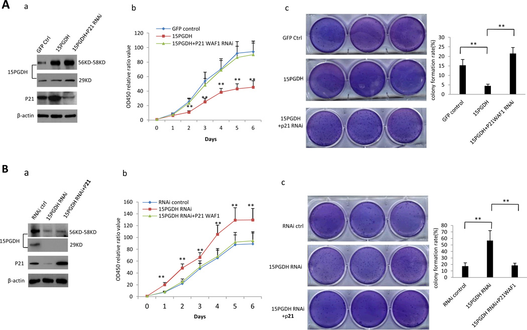
Figure 8. 15-PGDH inhibits HCC cell growth through p21WAF1/Cip1.
A. Huh7 cells transfected with 15-PGDH overexpression vector with or without co-transfection of p21 RNAi. a Western blotting for 15-PGDH and p21 (β-actin was used as loading control). b WST cell proliferation assay. Each sample was assayed in triplicate for 6 consecutive days. Data are means±SEM from three independent experiments (**p < 0.01). c Soft-agar colony formation assay. The results are representative of three independent experiments (**p < 0.01).
B. Huh7 stable cell transfected with 15-PGDH RNAi vector with or without co-transfection of p21 expression vector. a Western blotting for 15-PGDH and p21 (β-actin was used as loading control). b WST cell proliferation assay. Each sample was assayed in triplicate for 6 days consecutively. Data are mean±SEM from three independent experiments (**p < 0.01). c Soft-agar colony formation assay. The results are representative of three independent experiments (**p < 0.01).

