Fig. 2.

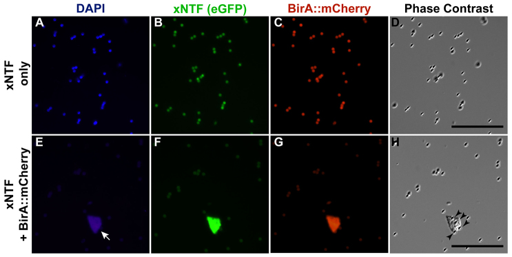
Affinity isolation of xNTF-tagged nuclei. Streptavidin-coated magnetic beads incubated with nuclei from embryos injected with mRNA encoding xNTF with or without BirA::mCherry. (A-D) Fluorescent DAPI (A), xNTF (eGFP; B), BirA::mCherry (Texas Red; C) and phase-contrast (D) images of magnetic beads after nuclear purification of xNTF in the absence of BirA. Autofluorescing beads are detectable throughout the field, but no nuclei. (E-H) Fluorescent DAPI (E), eGFP (F), Texas Red (G) and phase-contrast (H) images of magnetic beads after nuclear purification of xNTF. Note the presence of eGFP and BirA::mCherry-positive nucleus (arrow) and presence of magnetic beads coating nucleus (arrowheads). Scale bars: 50 μm.
