

Abstract
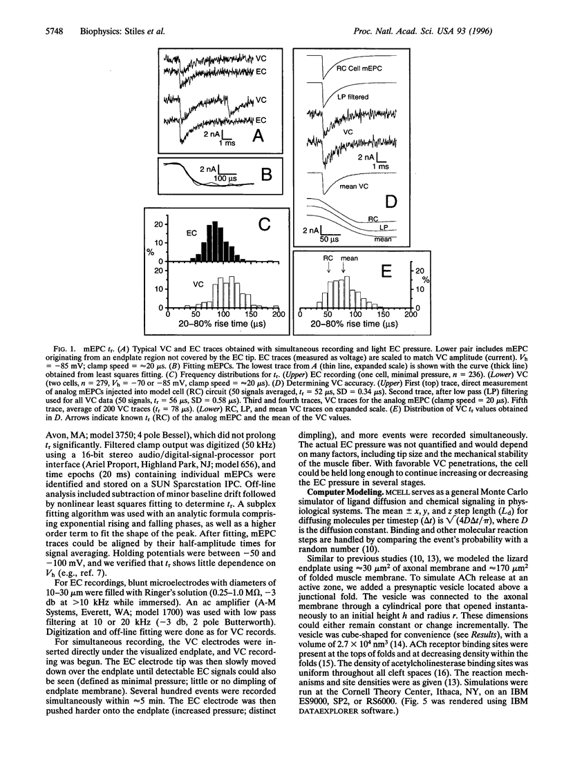
We recorded miniature endplate currents (mEPCs) using simultaneous voltage clamp and extracellular methods, allowing correction for time course measurement errors. We obtained a 20-80% rise time (tr) of approximately 80 micros at 22 degrees C, shorter than any previously reported values, and tr variability (SD) with an upper limit of 25-30 micros. Extracellular electrode pressure can increase tr and its variability by 2- to 3-fold. Using Monte Carlo simulations, we modeled passive acetylcholine diffusion through a vesicle fusion pore expanding radially at 25 nm x ms(-1) (rapid, from endplate omega figure appearance) or 0.275 nm x ms(-1) (slow, from mast cell exocytosis). Simulated mEPCs obtained with rapid expansion reproduced tr and the overall shape of our experimental mEPCs, and were similar to simulated mEPCs obtained with instant acetylcholine release. We conclude that passive transmitter diffusion, coupled with rapid expansion of the fusion pore, is sufficient to explain the time course of experimentally measured synaptic currents with trs of less than 100 micros.
Full text
PDF


Images in this article


Selected References
These references are in PubMed. This may not be the complete list of references from this article.
- Almers W., Tse F. W. Transmitter release from synapses: does a preassembled fusion pore initiate exocytosis? Neuron. 1990 Jun;4(6):813–818. doi: 10.1016/0896-6273(90)90134-2. [DOI] [PubMed] [Google Scholar]
- Anglister L., Stiles J. R., Salpeter M. M. Acetylcholinesterase density and turnover number at frog neuromuscular junctions, with modeling of their role in synaptic function. Neuron. 1994 Apr;12(4):783–794. doi: 10.1016/0896-6273(94)90331-x. [DOI] [PubMed] [Google Scholar]
- Bartol T. M., Jr, Land B. R., Salpeter E. E., Salpeter M. M. Monte Carlo simulation of miniature endplate current generation in the vertebrate neuromuscular junction. Biophys J. 1991 Jun;59(6):1290–1307. doi: 10.1016/S0006-3495(91)82344-X. [DOI] [PMC free article] [PubMed] [Google Scholar]
- Dionne V. E. Characterization of drug iontophoresis with a fast microassay technique. Biophys J. 1976 Jul;16(7):705–717. doi: 10.1016/S0006-3495(76)85723-2. [DOI] [PMC free article] [PubMed] [Google Scholar]
- Gage P. W., McBurney R. N. Effects of membrane potential, temperature and neostigmine on the conductance change caused by a quantum or acetylcholine at the toad neuromuscular junction. J Physiol. 1975 Jan;244(2):385–407. doi: 10.1113/jphysiol.1975.sp010805. [DOI] [PMC free article] [PubMed] [Google Scholar]
- Katz B., Miledi R. The binding of acetylcholine to receptors and its removal from the synaptic cleft. J Physiol. 1973 Jun;231(3):549–574. doi: 10.1113/jphysiol.1973.sp010248. [DOI] [PMC free article] [PubMed] [Google Scholar]
- Khanin R., Parnas H., Segel L. Diffusion cannot govern the discharge of neurotransmitter in fast synapses. Biophys J. 1994 Sep;67(3):966–972. doi: 10.1016/S0006-3495(94)80562-4. [DOI] [PMC free article] [PubMed] [Google Scholar]
- Land B. R., Harris W. V., Salpeter E. E., Salpeter M. M. Diffusion and binding constants for acetylcholine derived from the falling phase of miniature endplate currents. Proc Natl Acad Sci U S A. 1984 Mar;81(5):1594–1598. doi: 10.1073/pnas.81.5.1594. [DOI] [PMC free article] [PubMed] [Google Scholar]
- Land B. R., Salpeter E. E., Salpeter M. M. Acetylcholine receptor site density affects the rising phase of miniature endplate currents. Proc Natl Acad Sci U S A. 1980 Jun;77(6):3736–3740. doi: 10.1073/pnas.77.6.3736. [DOI] [PMC free article] [PubMed] [Google Scholar]
- Meyhöfer E., Howard J. The force generated by a single kinesin molecule against an elastic load. Proc Natl Acad Sci U S A. 1995 Jan 17;92(2):574–578. doi: 10.1073/pnas.92.2.574. [DOI] [PMC free article] [PubMed] [Google Scholar]
- Monck J. R., Fernandez J. M. The exocytotic fusion pore and neurotransmitter release. Neuron. 1994 Apr;12(4):707–716. doi: 10.1016/0896-6273(94)90325-5. [DOI] [PubMed] [Google Scholar]
- Salpeter M. M. Electron microscope radioautography as a quantitative tool in enzyme cytochemistry. II. The distribution of DFP-reactive sties at motor endplates of a vertebrate twitch muscle. J Cell Biol. 1969 Jul;42(1):122–134. doi: 10.1083/jcb.42.1.122. [DOI] [PMC free article] [PubMed] [Google Scholar]
- Spruce A. E., Breckenridge L. J., Lee A. K., Almers W. Properties of the fusion pore that forms during exocytosis of a mast cell secretory vesicle. Neuron. 1990 May;4(5):643–654. doi: 10.1016/0896-6273(90)90192-i. [DOI] [PubMed] [Google Scholar]
- Torri-Tarelli F., Grohovaz F., Fesce R., Ceccarelli B. Temporal coincidence between synaptic vesicle fusion and quantal secretion of acetylcholine. J Cell Biol. 1985 Oct;101(4):1386–1399. doi: 10.1083/jcb.101.4.1386. [DOI] [PMC free article] [PubMed] [Google Scholar]
- Van der Kloot W. The rise times of miniature endplate currents suggest that acetylcholine may be released over a period of time. Biophys J. 1995 Jul;69(1):148–154. doi: 10.1016/S0006-3495(95)79884-8. [DOI] [PMC free article] [PubMed] [Google Scholar]