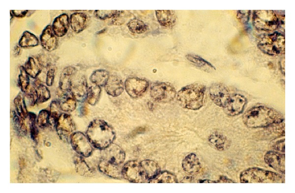
Figure 5.

Unusual expression and cell compartmentalization of p53 protein in breast carcinoma cells. By immunohistochemical staining, p53 appears as discrete dot-shaped regions within the nucleus of the cell. p53 cancer-associated mutants localize to distinct domains in the nucleoli and at and around the nuclear membrane. The antibody reacted with a distinct perinuclear structure in almost all of the cells. Cytoplasmic p53 was observed as faint staining. Smaller tumor cell nuclei showed ball-like shape (frozen section, immunoperoxidase with antibody DO-7, nuclear counterstain with haematoxylin, ×250).
