Abstract
Background:
Endodontic obturating materials should form monoblocks, reinforcing the treated teeth against fracture.
Aim:
To evaluate and compare the effect of two resin sealers and a MTA sealer on the fracture resistance of endodontically treated teeth.
Materials and Methods:
Fifty single-rooted mandibular premolars, decoronated at cemento-enamel junction, were divided into 5 groups (n = 10 each). Group 1 and group 2 served as negative and positive controls. Cleaning and shaping of root canals was done using ProTaper rotary files and 3% sodium hypochlorite irrigation. Obturation was done using AH plus (Dentsply, Germany) (group 3), MetaSEAL (Parkell, USA) (group 4), MTA Fillapex (Angeles, Brazil) (group 5) sealers and gutta-percha. Teeth were subjected to vertical loading using a universal testing machine and the point at which fracture of the roots occurred was recorded. The data was subjected to statistical analysis using one way analysis of variance (ANOVA) followed by pair-wise comparison using Tukey's Post-hoc test.
Results:
AH Plus showed better fracture resistance among the sealer groups. Statistically, no significant difference was found between MetaSEAL and Fillapex groups.
Conclusion:
MTA Fillapex as a root canal sealer was not able to reinforce the tooth against fracture.
Keywords: Endodontic monoblocks, MTA, root canal sealers, universal testing machine, vertical root fracture
INTRODUCTION
Endodontically treated teeth are structurally different from unrestored vital teeth and require specialized restorative treatment. It is generally accepted that the strength of an endodontically treated tooth is directly related to the amount of remaining sound tooth structure.[1,2] Vertical root fracture (VRF) is a serious clinical concern, with an unfavorable prognosis, resulting almost inevitably in extraction of the tooth or resection of the affected root, thus leading to endodontic failure.
As the loss of dentin creates an increased susceptibility to fracture, the use of obturating materials that strengthen the root and compensate for the weakening effect is mandatory. Resin-based dental materials have been proposed as a means to reinforce endodontically treated teeth through the use of adhesive sealers in the root canal system. However, for a root canal sealer to reinforce the root, it must bond to dentin. Bondable root canal sealers have a desirable property of creating monoblocks within the root canal space with their sealing ability to wet and infiltrate dentin.[3,4]
Numerous studies have shown that the bond strength of epoxy resin-based sealer, AH plus was significantly higher than zinc-oxide eugenol, calcium hydroxide and glass ionomer-based sealers.[5,6] MetaSEAL, a fourth generation self-etching, dual - cured methacrylate resin-based sealer was suggested to form a hybrid layer that creates a strong bond to the dentinal walls.[7] Recently introduced mineral trioxide aggregate sealer, MTA Fillapex is biocompatible, seals the canal better and has good mechanical properties like having the modulus of elasticity similar to that of dentin.[8] But, the ability of these MTA sealers in enhancing the fracture resistance of endodontically treated teeth was not addressed much till now.
The purpose of this in vitro study was to assess the influence of AH plus (Dentsply, Germany), MetaSEAL (Parkell, USA) and MTA Fillapex (Angeles, Brazil) sealers on the fracture resistance of endodontically treated teeth and further, to determine whether MTA-based sealer has the ability to reinforce the root.
MATERIALS AND METHODS
Fifty non-carious, freshly extracted, single-rooted mandibular premolar teeth with approximately similar bucco-lingual and mesio-distal dimensions were used in the study. After extraction, all the collected teeth were immersed in 5% sodium hypochlorite (NaOCl) solution for 2 hours for surface disinfection and dissolution of the superficial soft tissue. Pre-operative radiographs and surgical loupes (Stac Dental Instruments Inc. Canada) at 3.5 X magnification were used to ensure that the collected teeth did not have open apices, multiple canals, calcifications, fractures or craze lines. All teeth were decoronated 1 mm coronal to the cemento-enamel junction (CEJ) using a flexible diamond disk (Novo dental products, India) in a slow speed hand piece under copious amount of water coolant. The root of each tooth was standardized to a length of 12 mm as measured from the apex to the facial CEJ.
The teeth were divided into 3 experimental groups and two control groups using a randomized stratified design. Coronal access for root samples was prepared using #245 bur, and the canal patency was checked. To standardize the working length, a size 10 k file was inserted into the canal until the tip of the instrument was first visualized at the apical foramen. The working length was determined by subtracting 1 mm from this length.
Cleaning and shaping of the root canals were completed with ProTaper rotary Ni-Ti files (Dentsply Maillefer, Switzerland) using a 1:64 gear reduction Anthogyr handpiece (Dentsply) at a speed of 300 rpm. ProTaper shaping and finishing files were used in a sequential order. The canals were irrigated with 2 ml of 3% sodium hypochlorite (NaOCl) after each instrumentation, using 27 gauge side-vented needle and syringe. Then, F 3 master cone guttapercha (Dentsply, Maillefer) matched to the final master apical instrument was placed into the canaland the fit was confirmed radiographically.
Grouping method
Group 1; Received no instrumentation or obturation (negative control)
Group 2; Received instrumentation but no obturation (positive control)
Group 3; Received canal preparation and obturated with AH Plus sealer (Dentsply, Germany)
Group 4; Received canal preparation and were obturated with MetaSEAL sealer (Parkell, USA)
Group 5; Received canal preparation and were obturated with MTA Fillapex sealer (Angeles,Brazil)
Prior to obturation, the root canals in group 3 and 4 root samples were irrigated with 17% EDTA (Canalarge, Ammdent) for 1 minute to remove the smear layer followed by 3% NaOCl irrigation. Each canal was then irrigated with 5 ml of 10% ascorbic acid solution for the anti-oxidative action. A final flush was done with 5 ml of normal saline in all the test samples, and excess moisture from the canals was removed with sterile absorbent points. Sealers were mixed according to the manufacturer instructions. Root canals were coated with sealers using lentulospirals at 300 rpm and obturated using F3 ProTaper guttapercha points.
For all the experimental root samples, post-obturation radiographs were taken in both labio-lingual and mesio-distal directions to ensure homogeneous adequate root filling without voids. The filled roots were stored in an incubator (Nuaire US Autoflow, MN, USA) for 7 days at 37o C and 100% relative humidity to allow the sealer to set completely. For all specimens, the root surface was covered with a paste of silicon-based impression material (Aquasil) up to 2 mm apical to the CEJ to simulate a periodontal ligament and was kept in 100% humidity for 24 hours. Each tooth was then mounted vertically to a depth of 2 mm below the CEJ in polystyrene resin block using ice cube holder moulds.
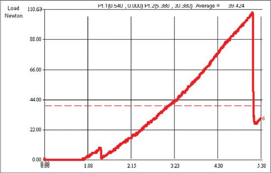
Resistance offered by root samples of all groups against vertical fracture was tested using universal testing machine. A cylindrical hardened steel rod (2.2 mm diameter) with a sharpened conical tip was attached to the upper part of the universal testing machine (Dak system Inc) to apply force to the root-causing VRF. A vertical load was applied at a crosshead speed of 0.5 mm/min until the root fractured. Fracture was defined as the point at which a sharp and instantaneous drop greater than 25% of the applied force was observed [Figure 1]. For most specimens, an audible crack also was heard, and the amount of force required for fracture was recorded in Newtons.
Figure 1.

Tracings obtained from Negative Control Group showing gradual increase in fracture force (N) plotted against penetration depth in millimeters
The load of fracture in Newtons was converted to mega-pascal's by using the following formula

π =3.14 (constant value)
Area of cross.section of plunger = 2.2 (uniform for all specimens)
The fracture load data was subjected to statistical analysis by using SPSS/PC software. A one-way analysis of variance (ANOVA) was used to compare the forces at which fracture of roots occurred. Comparisons among the 5 groups were performed by using Tukey's multiple post-hoc test. The statistical analysis was performed at 95% confidence level.
RESULTS
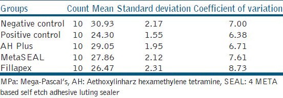
The mean force required to fracture the negative control group was higher (30.93 MPa) compared to any other group [Table 1]. Two resin-based sealers, AH Plus (29.50), MetaSEAL (27.86) and MTA Fillapex group (26.47 MPa), had a mean force to fracture higher than the positive control group (24.30 MPa), but less than the negative control group.
Table 1.
Group statistics:-Mean standard deviation and coefficient of variation of fracture resistance values in MPa for the experimental and control groups

From the results [Table 2], significant difference was observed between 5 groups (F = 15.2686, P < 0.05) at 5% level of significance. The mean fracture resistance values for the negative control group were higher than the AH Plus group, but was not statistically significant. The Positive control group displayed lesser resistance to fracture when compared to AH Plus and MetaSEAL groups, but no significant difference in comparison to Fillapex group.
Table 2.
Comparison of 5 groups with respect to fracture resistance (in MPa) by one way ANOVA test

DISCUSSION
As the endodontic treatment leads to change in the mechanical properties of the tooth, fatigue failures might result from normal functional stresses and from increased functional and para-functional stresses. Finite element analysis study has found that circular canals have lower and more uniform stress distribution than oval canals in which greater stresses are present at the labial and lingual canal extensions and at the cervical and middle thirds.[9] In majority of the premolar samples obtained, the mid-to-apical region had a circular cross-section, which would result in uniform distribution of load and they also simulate the clinical situation where chewing forces are maximum.
The effect of various nickel-titanium rotary files on root dentin is controversial, with some studies reporting an increased risk for craze lines and dentin cracks and reduced root fracture resistance compared to instrumentation with hand files.[10] Apical cracks are more likely to appear when the cleaning and shaping is done upto the root canal length, rather than confining it 1.0 mm short of the root apex.[11] The recommendation has been made that the size of the master apical file should be only 2 to 3 numbers larger than the size of the initial hand file that binds slightly at the cemento-dentinal junction (CDJ). ProTaper rotary instrumentation with the apical enlargement up to F3 was done in this study to avoid thinning of the root dentin.
Stable adhesion to root canal dentin walls and an elastic modulus similar to dentin are the two key factors to improve the fracture resistance of an endodontically treated teeth.[12] Thus, AH Plus and MetaSEAL sealers, both having low solubility and disintegration[13] and good- adhesion,[14] were used. As it is known that MTA does not bond to dentin, the presence of resins in Fillapex sealer increases the flow properties, and the presence of MTA would cause interfacial deposition of hydroxyapatite, which would increase the frictional resistance of the obturating material.
In in vitro fracture resistance tests, the root embedment material should reproduce bone that can absorb masticatory loads and resist the compressive and tangential forces. Periodontal ligament simulation prevents stress concentration in one particular region, and transfers the stresses produced by load application all along the root surface.[15] Thus, artificial periodontal ligament modifies the fracture modes, by fracturing the root at different locations and may have a significant effect on fracture resistance. To simulate the periodontal ligament and alveolar bone, silicone paste and polystyrene resin blocks were used to test the root fracture resistance in the study.
The results displayed that the negative control group has highest fracture resistance values and the positive control group, the lowest. Statistically no significant difference in fracture resistance was found between the negative control group and AH Plus group. Contrary to these results, Jainaen A et al. (2009)[16] showed that fracture resistance of teeth obturated with epoxy resin sealers were not different from sound or instrumented teeth. Results of the present study are in accordance with the study done by Ersev et al. (2012)[17] who stated that AH Plus and Meta SEAL with the matched taper cone technique has significantly higher fracture resistance than the instrumented but not obturated roots.
AH Plus showed the highest root fracture resistance within the sealer groups and displayed statistically higher resistance values compared to Fillapex group. Fillapex sealer by the formation of hydroxyapatite with MTA, having a compressive elastic modulus (14,000-18,600 MPa) similar to dentin, should be able to strengthen the roots. However, this was not shown in the study results, probably due to lack of bonding of MTA to dentin.
Fillapex contains resins as one of its components, and the mechanism of adhesion of resins will be enhanced by removal of smear layer, which will facilitate the penetration of resins into the dentinal tubules. Fillapex contains MTA and on treatment with 17% EDTA would result in decreased hardness, interference in hydration of MTA, poor biocompatibility and poor cell adhesion.[18,19,20] In the present study, irrigation with 17% EDTA to remove the smear layer was not performed for Fillapex samples in order to achieve good sealing of MTA. On the contrary, Assmann et al (2012)[21] used 17% EDTA in all the sealer groups and concluded that, MTA Fillapex and AH Plus has similar bond strengths, pattern of failure and handling characteristics.
On the basis of the present study results and previous studies, MTA Fillapex has the potential to capitalize on the biological and sealing characteristics of MTA, but further investigations are needed to improve the bond strength and other physical properties of the material.
CONCLUSION
AH Plus showed the highest fracture resistance within the sealer groups followed by Meta SEAL and MTA Fillapex
Regardless of the lower bond strength compared to AH Plus, MTA Fillapex presented acceptable fracture resistance values in comparison with MetaSEAL.
Footnotes
Source of Support: Nil
Conflict of Interest: Nil.
REFERENCES
- 1.Gutmann JL. The dentin-root complex: Anatomic and biologic considerations in restoring endodontically treated teeth. J Prosthet Dent. 1992;67:458–67. doi: 10.1016/0022-3913(92)90073-j. [DOI] [PubMed] [Google Scholar]
- 2.Sornkul E, Stennard JG. Strength of roots before and after endodontic treatment and restoration. J Endod. 1992;18:440–3. doi: 10.1016/S0099-2399(06)80845-9. [DOI] [PubMed] [Google Scholar]
- 3.Johnson ME, Stewart GP, Nielson CJ, Halton JF. Evaluation of root reinforcement of endodontically treated teeth. Oral Surg Oral Med Oral Pathol Oral Radiol Endod. 2000;90:360–4. doi: 10.1067/moe.2000.108951. [DOI] [PubMed] [Google Scholar]
- 4.Tay FR, Pashley DH. Monoblocks in root canals: A hypothetical or a tangible goal. J Endod. 2007;33:391–8. doi: 10.1016/j.joen.2006.10.009. [DOI] [PMC free article] [PubMed] [Google Scholar]
- 5.Fisher MA, Berzins DW, Baheall JK. An in-vitro comparison of bond strength of various obturation materials to root canal dentin using a push-out test design. J Endod. 2007;33:856–8. doi: 10.1016/j.joen.2007.02.011. [DOI] [PubMed] [Google Scholar]
- 6.Rahimi M, Jainaen A, Parashas P, Messer HH. Bonding of resin-based sealers to root dentin. J Endod. 2009;35:121–4. doi: 10.1016/j.joen.2008.10.009. [DOI] [PubMed] [Google Scholar]
- 7.Kim YS, Grandini S, Ames JM, Gu L, Kim SK, Pashley DH, et al. Critical review on methacrylate resin-based root canal sealers. J Endod. 2010;36:383–99. doi: 10.1016/j.joen.2009.10.023. [DOI] [PubMed] [Google Scholar]
- 8.Gomes Filho JE, Watanabe S, Bernabé PF, Costa MT. A mineral trioxide aggregate sealer stimulated mineralization. J Endod. 2009;35:256–60. doi: 10.1016/j.joen.2008.11.006. [DOI] [PubMed] [Google Scholar]
- 9.Versluis A, Messer HH, Pintado MR. Changes in compaction stress distributions in roots resulting from canal preparation. Int Endod J. 2006;39:931–9. doi: 10.1111/j.1365-2591.2006.01164.x. [DOI] [PubMed] [Google Scholar]
- 10.Bier CA, Shemesh H, Tanomaru Filho M, Wesselink PR, Wu MK. The ability of different nickel-titanium rotary instruments to induce dentinal damage during canal preparation. J Endod. 2009;35:236–8. doi: 10.1016/j.joen.2008.10.021. [DOI] [PubMed] [Google Scholar]
- 11.Adorno CG, Yoshioka T, Suda H. The effect of root preparation technique and instrumentation length on the development of apical root cracks. J Endod. 2009;35:389–92. doi: 10.1016/j.joen.2008.12.008. [DOI] [PubMed] [Google Scholar]
- 12.Veera L, Araya T, Narold HM. Effects of root canal sealers on vertical root fracture resistance of endodontically treated teeth. J Endod. 2002;28:217–9. doi: 10.1097/00004770-200203000-00018. [DOI] [PubMed] [Google Scholar]
- 13.Versiani MA, Carvalho JR, Junior, Padilha MI, Lacey S, Pascon EA, Sousa Neto MD. A comparative study of physicochemical properties of AH Plus and Epiphany root canal sealants. Int Endod J. 2006;39:464–71. doi: 10.1111/j.1365-2591.2006.01105.x. [DOI] [PubMed] [Google Scholar]
- 14.Sousa Neto MD, Coelho FI, Marchesan MA, Alfredo E, Silva Sousa YT. In vitro study of the adhesion of an epoxy based sealer to human dentine submitted to irradiation with Er: YAG and Nd: YAG lasers. Int Endod J. 2005;38:866–70. doi: 10.1111/j.1365-2591.2005.01027.x. [DOI] [PubMed] [Google Scholar]
- 15.Soares CJ, Gava Pizi EC, Fonseca RB, Martins LR. Influence of root embedment material and periodontal ligament simulation on fracture resistance tests. Braz Oral Res. 2005;19:11–6. doi: 10.1590/s1806-83242005000100003. [DOI] [PubMed] [Google Scholar]
- 16.Jainaen A, Palamara JE, Messer HH. The effect of resin-based sealers on fracture properties of dentine. Int Endod J. 2009;42:136–43. doi: 10.1111/j.1365-2591.2008.01496.x. [DOI] [PubMed] [Google Scholar]
- 17.Ersev H, Yilmaz B, Pehlivanoglu E, Cahskan EO, Erisen FR. Resistance of vertical root fracture of endodontically treated teeth with Meta SEAL. J Endod. 2012;38:653–6. doi: 10.1016/j.joen.2012.02.015. [DOI] [PubMed] [Google Scholar]
- 18.Timpawat S, Vongsavan N, Messer HH. Effect of removal of the smear layer on apical microleakage. J Endod. 2001;27:351–3. doi: 10.1097/00004770-200105000-00011. [DOI] [PubMed] [Google Scholar]
- 19.Yildirim T, Oruçoglu H, Çobankara FK. Long-term evaluation of the influence of smear layer on the apical sealing ability of MTA. J Endod. 2008;34:1537–40. doi: 10.1016/j.joen.2008.08.022. [DOI] [PubMed] [Google Scholar]
- 20.Lee YL, Lin FH, Wang WH, Ritchie HH, Lan WH, Lin CP. Effects of EDTA on the hydration mechanism of mineral trioxide aggregate. J Dent Res. 2007;86:534–8. doi: 10.1177/154405910708600609. [DOI] [PubMed] [Google Scholar]
- 21.Assmann E, Scarparo RK, Bottcher DE, Grecca FS. Dentin bond strength of two Mineral Trioxide Aggregate-based and one Epoxy Resin-based sealers. J Endod. 2012;38:219–21. doi: 10.1016/j.joen.2011.10.018. [DOI] [PubMed] [Google Scholar]


