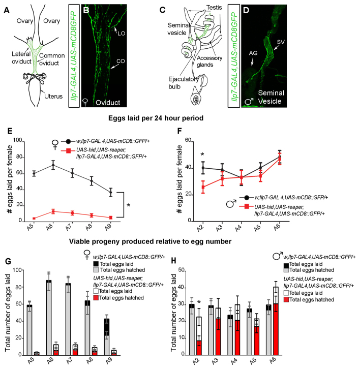
Fig. 3.
Post-embryonic Ilp7 neurons are required only for female fertility. (A-D) Ilp7-GAL4,UAS-mCD8::GFP-expressing neurons project to the female lateral (LO) and common (CO) oviducts and the male seminal vesicle (SV). AG, accessory gland. (E,F) Control and Ilp7-KO females were mated to control males for 24 hours, then males were removed. Thereafter, we counted egg numbers laid per female per 24-hour period, for 5 days (supplementary material Table S1). (E) Ilp7-KO females (red) exhibited severely reduced egg laying compared with controls (black). (F) Control (black) or Ilp7-KO (red) males were mated to control virgin females for 24 hours. Then, mated females were removed and males were provided new virgin females for another 24 hours. This was repeated for 5 days (A2-A5). After females were removed, we counted egg numbers per female over 24 hours. Females mated to Ilp7-KO and control males laid similar egg numbers; only females on assay day 1 had reduced egg numbers. (G,H) Using the mating protocols described for E,F, we counted the total number of viable larvae produced per plate (not per female) within 6-hour assay periods, over 5 days (supplementary material Table S2). (G) Control females produced a high percentage of larvae. The decline in larvae by A9 reflects a lack of mating for 5 days. Ilp7-KO females produced low egg numbers and only ∼40% of eggs produced larvae at each time point (H) Ilp7-KO and control males produced similar percentages of viable larvae at most ages, except for during the first day of the assay. Graphs show mean±s.e.m. for egg number per female. n, number of egg-lay assays.

