Summary:
Hamartoma is a benign tumor-like malformation characterized by a focal mixture of mature cells and tissues normally present in affected area. The hamartoma of the breast is rare. We report a case in an asymptomatic young woman coming to our attention for a left breast lesion detected by ultrasound screening.
Keywords: Hamartoma, Breast, Surgery
Introduction
The hamartoma is a benign tumor-like lesion that can affect various organs of the body, including lungs, kidneys, skin and, more rarely, the breast. Hamartoma is a disorganized focal area of cells and tissues of the organ where it occurs (1–4). Hamartomas represent 4–8% of the benign lesions of the breast in women, with very rare cases in males (5, 6).
As in our case, the hamartoma of the breast is usually asymptomatic.
Case report
A 30 year old, nulliparous woman came to our attention for the occasional detection at breast self-examination of a swelling in the lower quadrants of the left breast. Family history was negative for breast cancer, as well as personal history (no comorbidities or hormonal therapy).
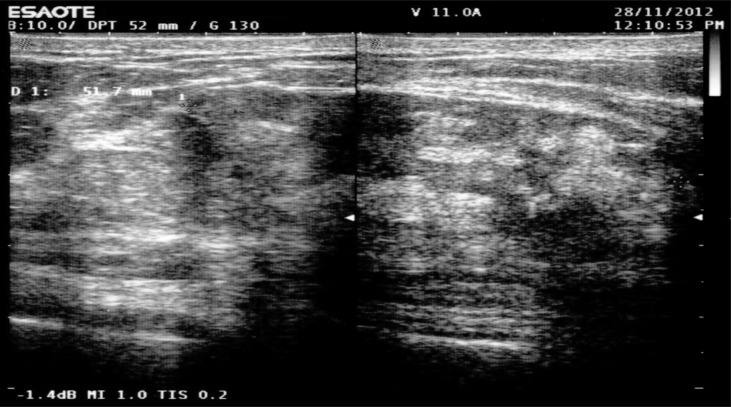
The examination confirmed the presence of the lesion with hard-elastic consistency and margins defined. Ultrasound showed an ovalar, 5.5cm, well capsulated, hypoechogenic lesion with small fluid areas and without suspicious axillary lymph nodes (Fig. 1).
Figure 1.
Ultrasound. The lesion is well-capsulated.
The lesion was resected. The postoperative course was uneventuful.
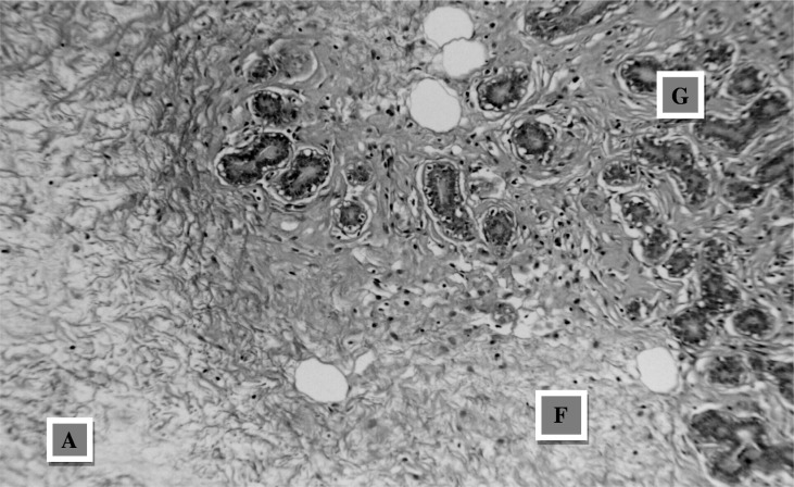
Histologically the lesion appeared with smooth surface and gray-yellow at the cut. Microscopic examination showed a fibrous capsule, normal breast parenchyma with marked stromal fibrosis, cystic dilatation of the ducts, apocrine metaplasia, sclerosing adenosis and typical ductal hyperplasia. The histological findings are indicative of a benign lesion with features of hamartoma (Fig. 2).
Figure 2.
Microscopic examination (H&E, x40). Adipose (A), glandular (G) and fibrous (F) tissue.
Six months after surgery ultrasound follow up is negative for recurrence.
Discussion and conclusion
More often the diagnosis of hamartoma is incidental in women older than 40 years starting mammography screening. Ultrasounds and mammogram show a well-capsulated nodule without calcifications. Imaging features may be similar to those of fibroadenoma (5, 7–13). The definitive diagnosis is only histological (8, 14, 15). Very rarely the hamartoma can turn into malignant tumor; few cases of breast invasive ductal carcinoma from hamartoma are described in literature (16, 17), but the correlation is unproved. However, the resection of the hamartoma of the breast is always recommended (8, 14).
References
- 1.Custureri F, Pasta V, Monti M, Macrì S, Vergine M, Nudo R, Stella S. Possibilità diagnostiche e indicazioni terapeutiche negli amartocondromi. Med Chir Ital. 1987;2 (S.E.R.O.S. Publisher, Roma). [Google Scholar]
- 2.Murugesan JR, Joglekar S, Valerio D, Bradley S, Clark D, Jibril JA. Myoid hamartoma of the breast: case report and review of the literature. Clin Breast Cancer. 2006 Oct;7(4):345–6. doi: 10.3816/CBC.2006.n.050. [DOI] [PubMed] [Google Scholar]
- 3.Stafyla V, Kotsifopoulos N, Grigoriadis K, Bakoyiannis CN, Peros G, Sakorafas GH. Myoid hamartoma of the breast: a case report and review of the literature. [DOI] [PubMed]
- 4.Fischer CJ, Hanby AM, Robinson L, Millis RR. Mammary hamartoma: a review of 35 cases. Histopatology. 1992;20:90–106. doi: 10.1111/j.1365-2559.1992.tb00938.x. [DOI] [PubMed] [Google Scholar]
- 5.Farrokh D, Hashemi J, Ansaripour E. Breast hamartoma: mammographic findings. Iran J Radiol. 2011 Dec;8(4):258–260. doi: 10.5812/iranjradiol.4492. [DOI] [PMC free article] [PubMed] [Google Scholar]
- 6.Ravakhah K, Javadi N, Simms R.Hamartoma of the breast in a man: first case report Breast J 2001July–Aug74266–8. [DOI] [PubMed] [Google Scholar]
- 7.Gomez-Aracil V, Mayayo E, Azua J, Mayayo R, Azua-Romeo J, Arraiza A. Fine needle aspiration cytology of mammary hamartoma: a review of nine cases with histological correlation. Cytopathology. 2003;14:195–200. doi: 10.1046/j.1365-2303.2003.00077.x. [DOI] [PubMed] [Google Scholar]
- 8.Herbert M, Mendlovic S, Liokumovich P, Segal M, Zahavi S, Rath-Wolfson L, Sandbank J. Can hamartoma of the breast be distinguished from fibroadenoma using fine-needle aspiration cytology? Diagn Cytopathol. 2006 May;34(5):326–9. doi: 10.1002/dc.20404. [DOI] [PubMed] [Google Scholar]
- 9.Murat A, Ozdemir H, Yildirim H, Poyraz AK, Ozercan R. Hamartoma of the breast. Australas Radiol. 2007 Oct;51:B37–9. doi: 10.1111/j.1440-1673.2007.01818.x. Spec No. [DOI] [PubMed] [Google Scholar]
- 10.Veneti S, Manek S. Benign phylloides tumors vs fibroadenoma: FNA cytological differentiation. Citopathology. 2001;12:321–328. doi: 10.1046/j.1365-2303.2001.00334.x. [DOI] [PubMed] [Google Scholar]
- 11.Herbert M, Sandbank J, Liokumovich P, et al. Breast hamartomas: clinicopatological and immunoistochemical studies of 24 cases. Histopahology. 2002;41:30–34. doi: 10.1046/j.1365-2559.2002.01429.x. [DOI] [PubMed] [Google Scholar]
- 12.Park SY, Oh KK, Kim EK, Son EJ, Chung WH. Sonographic findings of breast hamartoma: emphasis on compressibility. Yonsei Med J. 2003 Oct 30;44(5):847–54. doi: 10.3349/ymj.2003.44.5.847. [DOI] [PubMed] [Google Scholar]
- 13.Tse GM, Law BK, Ma TK, Chan AB, Pang LM, Chu WC, Cheung HS. Hamartoma of the breast: a clinicopathological review. J Clin Pathol. 2002 Dec;55(12):951–4. doi: 10.1136/jcp.55.12.951. [DOI] [PMC free article] [PubMed] [Google Scholar]
- 14.Arrigoni MG, Dockerty MA, Judd ES. The identification and treatment of mammary hamartomas. Surg Gynecol Obstet. 1971;133:577–82. [PubMed] [Google Scholar]
- 15.Fisher CJ, Hanby AM, Robinson L, et al. Mammary hamartoma-review of 35 cases. Histopathology. 1992;20:99–106. doi: 10.1111/j.1365-2559.1992.tb00938.x. [DOI] [PubMed] [Google Scholar]
- 16.Choi N, Ko ES.Invasive ductal carcinoma in a mammary hamartoma: case report and review of the literature Korean J Radiol 2010November–Dec116687–91. [DOI] [PMC free article] [PubMed] [Google Scholar]
- 17.Daya D, Trus T, D’Souza TJ, et al. Hamartoma of the breast, an underrecognized breast lesion. Am J Clin Pathol. 1995;103:685–9. doi: 10.1093/ajcp/103.6.685. [DOI] [PubMed] [Google Scholar]


