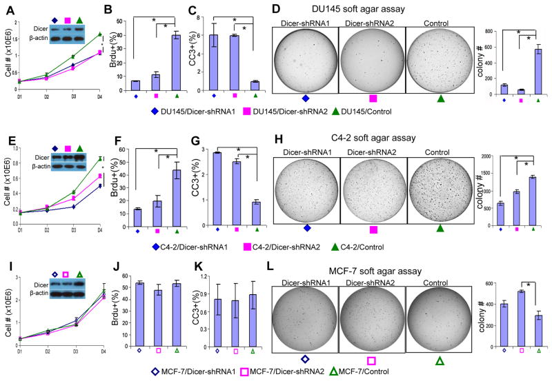
Fig. 5. Suppressing Dicer activity attenuates proliferation and tumorigenesis of the human prostate cancer cells.
(A, E, I) In vitro proliferation curve of control and Dicer shRNA-expressing DU145 cells (A), C4-2 cells (E), and MCF-7 cells (I). Data represent means from three independent experiments. Insets: Western blot assay confirms knockdown of Dicer. (B, F, J) Bar graphs show quantification of BrdU positive proliferating cells in control and Dicer shRNA-expressing DU145 cells (B), C4-2 cells (F), and MCF-7 cells (J). (C, G, K) Bar graphs show quantification of cleaved caspase 3(CC3) positive apoptotic cells in control and Dicer shRNA-expressing DU145 cells (C), C4-2 cells (G), and MCF-7 cells (K). (D, H, L) Soft agar assay using control and Dicer shRNA-expressing DU145 cells (D), C4-2 cells (H), and MCF-7 cells (L). Data represent means ± SD. *: p < 0.05.

