

Abstract
We present here evidence for the existence of a new type of interstitial cell in human myocardial sleeves of pulmonary veins: interstitial Cajal-like cell (ICLC). This cell fulfils the criteria for positive diagnosis of ICLC, including CD 117/c-kit positivity. Transmission electron microscopy revealed typical ICLC with 2 or 3 very long processes (several tens of mm) suddenly emerging from the cellular body. Also, these processes appear moniliform but extremely thin (0.1–0.4 mm) under the resolving power of the usual microscopy. Cell processes establish close spatial relationships between each other, as well as with capillaries and nerve endings. ICLC appear located among the myocardial cells and particularly at the border between the myocardial sleeve and pulmonary vein wall.
Keywords: interstitial Cajal-like cells, arrhythmia, atrium, myocardium, pulmonary veins
A new type of interstitial cell, with ultrastructural features similar to the known pacemaker cells of the gastrointestinal tract, the interstitial cells of Cajal (ICC), has been depicted in a variety of other tissues and organs [1–3]. Although the ICC have been presumed to exist in the heart almost 100 years ago [1], the presence of cells similar to them, the so-called interstitial Cajal-like cells (ICLC), in the human myocardium was demonstrated by transmission electron microscopy 2 years ago [4–7].
It is well accepted that bursts of spontaneous activity in the myocardial sleeves (MS) of the pulmonary veins (PV) can initiate atrial fibrillation [8–12]. Circumferential pulmonary vein ablation provides better recurrence-free survival than antiarrhythmic drug therapy [11] and this substantiates the existence of a structural link between the atrium and PV responsible for atrial fibrillation initiation. In this context, we presumed that the atrial network of ICLC [4, 6] could extend into the MS of PV.
Small tissue specimens of pulmonary veins were obtained during surgery from three patients without atrial fibrillation who were admitted for cardiac surgery. This study was approved by the Institutional Ethics Committee, and written informed consent was obtained from patients. Three other specimens (larger sections) were obtained at autopsy. Immunohistochemistry on human pulmonary veins was performed on 3-mm thick sections from 10% formalin fixed paraffin-embedded specimens using polyclonal CD117 (1:100, DAKO, Glostrup, Denmark) as previously described [6]. Small tissue samples were processed for transmission electron microscopy (TEM) as previously described [4–7]. Digital electron micrographs were recorded with Morada CCD camera and iTEM software (Olympus Soft Imaging Solutions GmbH) on Philips CM12 electron microscope. Computer-based, digitally coloured images were prepared using Adobe Photoshop.
Immunohistochemistry revealed relatively numerous CD117/c-kit positive cells with ICLC morphology and preferentially positioned between atrial myocardial sleeve and pulmonary vein wall (Fig. 1). Isolated ICLC have been observed among myocardial bundles (Fig. 1).
1.
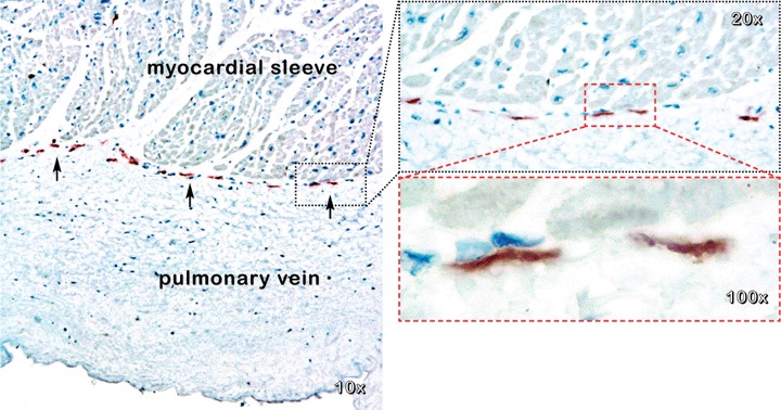
Human pulmonary vein. Immunohistochemistry shows CD117/c-kit positive interstitial cells that create a string between the myocardial sleeve and pulmonary vein wall (arrows). Mayer's haema-toxylin counterstain.
Light microscopy of semithin sections stained with toluidine blue showed that interstitial cells with (very) long and thin processes are located among myocardial cells (Fig. 2A) and in between the MS and PV wall (Fig. 2B). We would like to emphasize that (as far as we know) cell processes with 30–50–70 mm length are to be found only for nerve cells. However, ICLC are not neurons.
2.
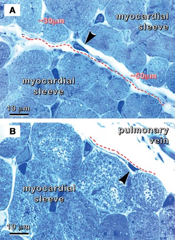
Human pulmonary vein. Specimen processed for transmission electron microscopy (glutaraldehyde/osmium fixation, Epon embedding, ultra-microtomy, semithin sections ∼1μm), but stained with toluidine blue and examined under light microscope. Cross-sections of myocardial cells. (A) Interstitial cell (arrowhead) with long and thin processes (dashed line) located among the myocardial cells. (B) Interstitial cell (arrowhead) between the myocardial sleeve and pulmonary vein wall. This cell with long and thin processes (marked with dashed line) is close to the myocardial cells.
TEM analysis showed that these cells fulfil ultrastructural diagnostic criteria for ICLC [2, 5, 6]: (i) location in the connective interstitium (Figs. 3–6); (ii) characteristic long (several tens of μm), thin and moniliform cell processes (Figs. 3–6); (iii) close vicinity to nerves (Fig. 6) and blood vessels (Figs. 5 and 6); (iv) specialized cell-to-cell junctions; (v) caveolae (Fig. 4 inset); (vi) organelles: mitochondria (about 5% on cytoplasmic volume), relatively well developed smooth and rough endoplasmic reticulum; (vii) intermediate (Fig. 4 inset) and thin filaments, microtubules and undetectable thick filaments.
3.
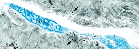
Myocardial sleeve of human pulmonary vein. TEM image corresponding to that shown in Fig. 2A. An interstitial Cajal-like cell (ICLC), computer coloured in blue, is located between myocardial cells. Atrial myocardial cells (M) are easily recognized due to their specific granules (arrows). Note longitudinal (*) and cross-sectioned (**) collagen fibrils in the interstitial space.
6.
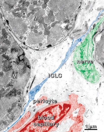
Myocardial sleeve of human pulmonary vein. Digitally coloured electron micrograph emphasizes the relationships between the interstitial Cajal-like cells (ICLC; blue), nerves (green) and blood capillary (brownish). Note an ICLC process running close to myocardial cells (M), nerve fibres and a blood capillary.
5.
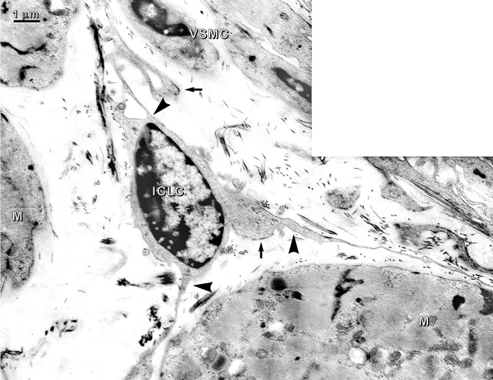
Myocardial sleeve of human pulmonary vein. Transmission electron micrograph shows an interstitial Cajal-like cell (ICLC) with three thin processes that run close to myocardial cells (M) and vascular smooth muscle cells (VSMC). The ICLC processes are narrow at the emergence points (arrowheads) from cellular body. Attachment plaques (arrows) link ICLC with the extracellular matrix.
4.
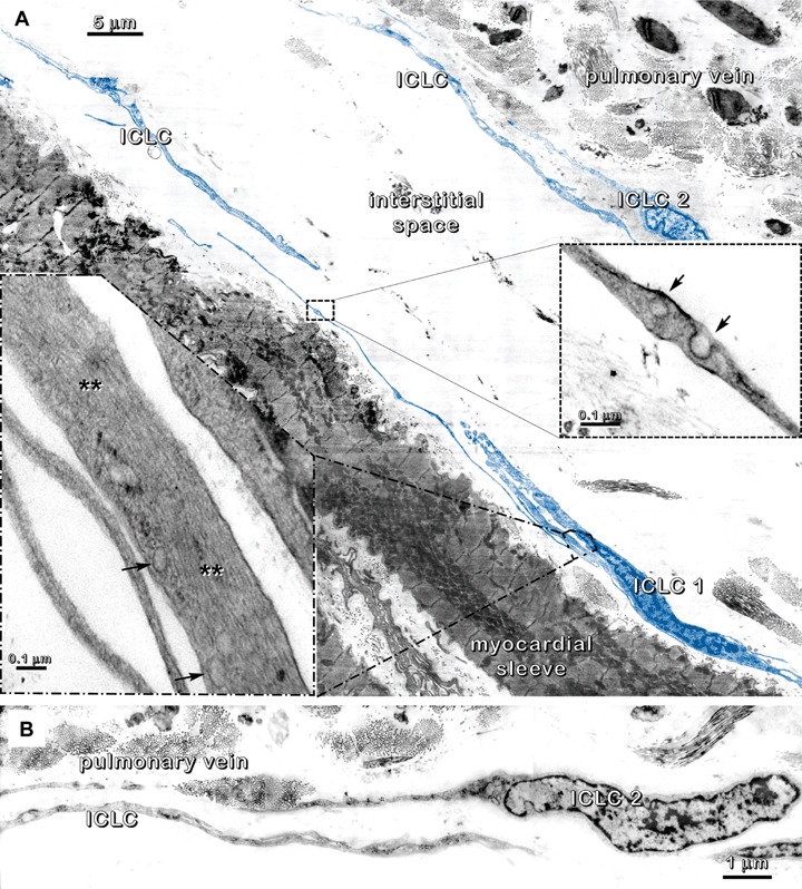
Human pulmonary vein. (A) Digitally coloured TEM micrograph of an area similar to that shown in Fig. 2B. Several interstitial Cajal-like cells (ICLC), highlighted in blue, are located in the interstitium between the pulmonary vein wall and the myocardial sleeve. Insets: ICLC processes have intermediate filaments (**) parallel with the long axis of the cell and caveolae (arrows) along the cell membrane. (B) Details of ICLC 2 from Fig. 4A (above) showing the overlapping of processes in a ‘sheath’ on the pulmonary vein side.
Ultrastructural analysis of the MS showed that, like in the atrium [4, 6], ICLC connect with each other in an interstitial three-dimensional network and run around blood vessels, nerves, and myocardial cells with different orientations (Figs. 3 and 5). One of the most intriguing aspects is that the ICLC were preferentially located at the internal limit of the MS, parallel with the long axis of the PV (Figs. 1 and 4). An incomplete cellular sheath formed by the overlapping ICLC processes seems to border the internal surface of MS and separate it from the PV wall (Fig. 4).
We observed that ICLC have a special relationship with nerve fibres in the atrial sleeves of the PV (Fig. 6). The distance between ICLC and nerves was often less than 100 nm (Fig. 6) and this falls within the molecular interaction range. We also found contact points between ICLC and myocardial cells (without specialized junctional structures) and attachment plaques connecting ICLC to the extracellular matrix (Fig. 5).
Ectopic beats appear to originate from the myocardial sleeves of the pulmonary veins, which are source of arrhythmogenic activity involved in the initiation of atrial fibrillation [11, 12]. In this context, it is essential to point out that interstitial cells identical with ICLC described in atrium [4, 6] or ventriculum [5] are present in the interstitium of the myocardial sleeves of the pulmonary veins. The ICLC seem distinct type of interstitial cells with characteristic long and thin cytoplasmic processes, which form an interstitial cellular network connecting cardiomyocytes, nerves, blood vessels and interstitial immune cells [2, 4–7]. These studies suggest that the ICLC form a tissue-wide network at the level of the myocardium and may have important and so far unsuspected inte-grative functions at the level of the cardiac tissue. It may be speculated that ICLC may be involved in immune surveillance. Also, they may be identified with the so-called ‘stromal mesenchymal stem cells' or could be precursors of several cell types (e.g. ICC, smooth muscle cells and fibroblasts) [13].
This newly described type of cell, ICLC, could be a hidden player in the mechanisms of atrial fibrillation. It is tempting to presume that these ICLC act as mechanoreceptors. ICLC may have a role in tensional integration of the tissue [14], considering their characteristic ultrastructure (extremely long and contorted processes with intermediate filaments and microtubules parallel to the long axis of the cell, attachment plaques connecting it to the extracellular matrix), and their particular distribution in between the MS and PV wall. The pulmonary veins are subjected to stretch from pulsatile blood flow and stretch-induced anionic and cationic currents have been demonstrated that are functionally present in the cardiomyocytes of the main pulmonary veins of rabbits [15]. Therefore, these ICLC could be a key factor in cardiac response to the mechanical stretch induced by the blood flow in the pulmonary veins under normal and/or pathological conditions.
Funding Sources: This work was supported by National Agency for Science (project CEEX 112/2006).
References
- 1.Huizinga JD, Faussone-Pellegrini MS. About the presence of interstitial cells of Cajal outside the musculature of the gastrointestinal tract. J Cell Mol Med. 2005;9:468–73. doi: 10.1111/j.1582-4934.2005.tb00372.x. [DOI] [PMC free article] [PubMed] [Google Scholar]
- 2.Popescu LM, Ciontea SM, Cretoiu D. Interstitial Cajal-like cells in human uterus and fallopian tube. Ann N Y Acad Sci. 2007;1101:139–65. doi: 10.1196/annals.1389.022. [DOI] [PubMed] [Google Scholar]
- 3.Pucovský V, Harhun MI, Povstyan OV, Gordienko DV, Moss RF, Bolton TB. Close relation of arterial ICC-like cells to the contractile phenotype of vascular smooth muscle cell. J Cell Mol Med. 2007;11:764–75. doi: 10.1111/j.1582-4934.2007.00066.x. [DOI] [PMC free article] [PubMed] [Google Scholar]
- 4.Hinescu ME, Popescu LM. Interstitial Cajal-like cells (ICLC) in human atrial myocardium. J Cell Mol Med. 2005;9:972–5. doi: 10.1111/j.1582-4934.2005.tb00394.x. [DOI] [PMC free article] [PubMed] [Google Scholar]
- 5.Popescu LM, Gherghiceanu M, Hinescu ME, Cretoiu D, Ceafalan L, Regalia T, Popescu AC, Ardeleanu C, Mandache E. Insights into the interstitium of ventricular myocardium: interstitial Cajal-like cells (ICLC) J Cell Mol Med. 2006;10:429–58. doi: 10.1111/j.1582-4934.2006.tb00410.x. [DOI] [PMC free article] [PubMed] [Google Scholar]
- 6.Hinescu ME, Gherghiceanu M, Mandache E, Ciontea SM, Popescu LM. Interstitial Cajal-like cells (ICLC) in atrial myocardium: ultrastructural and immunohistochemical characterization. J Cell Mol Med. 2006;10:243–57. doi: 10.1111/j.1582-4934.2006.tb00306.x. [DOI] [PMC free article] [PubMed] [Google Scholar]
- 7.Mandache E, Popescu LM, Gherghiceanu M. Myocardial interstitial Cajal-like cells (ICLC) and their nanostructural relationships with intercalated discs: shed vesicles as intermediates. J Cell Mol Med. 2007;11:1175–84. doi: 10.1111/j.1582-4934.2007.00117.x. [DOI] [PMC free article] [PubMed] [Google Scholar]
- 8.Khan R. Identifying and understanding the role of pulmonary vein activity in atrial fibrillation. Cardiovasc Res. 2004;64:387–94. doi: 10.1016/j.cardiores.2004.07.025. [DOI] [PubMed] [Google Scholar]
- 9.Haissaguerre M, Sanders P, Hocini M, Jais P, Clémenty J. Pulmonary veins in the substrate for atrial fibrillation: the “venous wave’’ hypothesis. J Am Coll Cardiol. 2004;43:2290–2. doi: 10.1016/j.jacc.2004.03.036. [DOI] [PubMed] [Google Scholar]
- 10.Huang CX, Hu CL, Li YB. Atrial fibrillation may be a vascular disease: the role of the pulmonary vein. Med Hypotheses. 2007;68:629–34. doi: 10.1016/j.mehy.2006.07.048. [DOI] [PubMed] [Google Scholar]
- 11.Noheria A, Kumar A, Wylie JV, Jr, Josephson ME. Catheter ablation vs antiarrhythmic drug therapy for atrial fibrillation: a systematic review. Arch Intern Med. 2008;168:581–6. doi: 10.1001/archinte.168.6.581. [DOI] [PubMed] [Google Scholar]
- 12.Rostock T, Steven D, Lutomsky B, Servatius H, Drewitz I, Klemm H, Müllerleile K, Ventura R, Meinertz T, Willems S. Atrial fibrillation begets atrial fibrillation in the pulmonary veins on the impact of atrial fibrillation on the electro-physiological properties of the pulmonary veins in humans. J Am Coll Cardiol. 2008;51:2153–60. doi: 10.1016/j.jacc.2008.02.059. [DOI] [PubMed] [Google Scholar]
- 13.Huizinga JD, White EJ. Progenitor cells of interstitial cells of Cajal: on the road to tissue repair. Gastroenterology. 2008;134:1251–4. doi: 10.1053/j.gastro.2008.02.074. [DOI] [PubMed] [Google Scholar]
- 14.Ingberg DE. Cellular mechanotransduction: putting all the pieces together again. FASEB J. 2006;20:811–27. doi: 10.1096/fj.05-5424rev. [DOI] [PubMed] [Google Scholar]
- 15.Seol CA, Kim WT, Ha JM, Choe H, Jang YJ, Youm JB, Earm YE, Leem CH. Stretch-activated currents in cardiomyocytes isolated from rabbit pulmonary veins. Prog Biophys Mol Biol. 2008;97:217–31. doi: 10.1016/j.pbiomolbio.2008.02.008. [DOI] [PubMed] [Google Scholar]