Abstract
To present a case of impacted artificial denture in esophagus and its removal by flexible endoscope. A 28 year old male presented with history of ingesting his denture 2 h back. It was removed by flexible endoscope and flexible fibre optic forceps. Though rigid endoscopic removal of foreign body is safe and effective, but often requires general anaesthesia. The flexible fibre optic endoscopic removal, which can be done under local anaesthesia in outpatient department is a suitable alternative.
Keywords: Flexible endoscope, Esophagus, Artificial denture
Introduction
Foreign body ingestion is common in children, but frequently seen among adults also. Most common foreign bodies in children are coins but marbles, button, batteries, safety pins and bottle tops are also reported. In adults common foreign bodies are meat bone, fish bone, dentures and metallic wires.
Impaction of dentures in the esophagus is a distressing experience for the patient and can lead to serious consequences such as esophageal perforation. Patients usually present with history of accidental swallowing, often while eating or during sleep or in association with trauma, seizures, or in presence of some degree of psychological dysfunction [1–3]. The common signs and symptoms of an impacted denture are odynophagia, dysphagia, or simply pain and tenderness in the neck or chest. [4–6]
The best method of removing impacted foreign body remains controversial. Rigid endoscopic removal of foreign body was developed by Chevalier Jackson about a century back but it is associated with the hazards of general anaesthesia. Flexible fibre optic endoscopic removal, which can be done under local anaesthesia in outpatient department has gained great popularity over the past decade.
Case Report
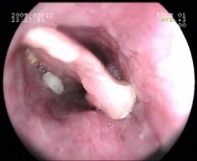
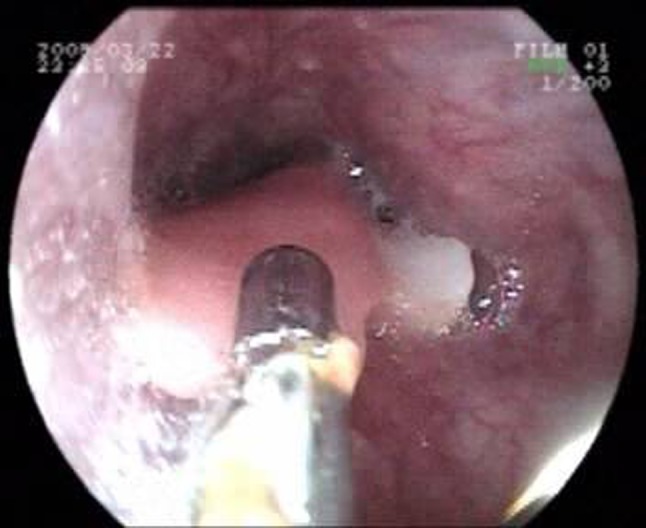
A 28 year old male presented to our emergency department with history of ingesting his denture 2 h back. On flexible endoscopy, we found the denture at about 25 cm from the upper incisors (Fig. 1). The denture was removed with the help of flexible fibreoptic forceps (Fig. 2). No injury to the oesophageal wall was noted post-operatively. The patient was discharged after observation for a few hours.
Fig. 1.

Denture in esophagus
Fig. 2.

Denture removed with flexible endoscopic forceps
Discussion
The common sites of impaction of foreign bodies in oesophagus are post cricoid region, at the level of aortic arch, left main bronchus and diaphragm. There is one more site of impaction especially in cases of flat objects like coin which is at the level of T1, i.e., thoracic inlet.
Impacted dentures may lead to fistula formation or esophageal perforation [3, 6], which is a serious complication. Beyond 24 h after ingestion, the rate of complication multiplies several-fold, from 3.2% at 24 h to as high as 23.5% after 48 h [3, 7].
Even though X-rays remain useful [5] and are the most commonly performed initial investigation, their results need to be viewed with caution. One study showed that lateral radiographs of the neck changed the management approach in only 1.4% of cases [8].
Rigid esophagoscope is routinely used as an effective tool to remove foreign body. Today the methods available for extraction are diverse, including flexible fiberoptic endoscopy and other non-endoscopic approaches [9]. Removal of foreign bodies by using a Foley catheter was reported in 1966 [10]. It has been used for extraction of large radiopaque foreign bodies but is of no use in the majority of instances. The flexible fiberoptic instrument was developed during 1970s and 1980s. Most diagnostic esophagoscopy is now performed with flexible instruments for which they are vastly superior. Use of rigid instrument can be difficult and dangerous, especially in aged persons with hypertrophic changes in the cervical spine or with limited spine mobility or in thick-neck person with full set of teeth. In such cases use of flexible instruments is suggested. Though rigid esophagoscope still remains the gold standard for foreign body retrieval, endoscopist continue to develop new forceps and methods of handling esophageal foreign body. Through the use of “over-tubes,” even large and sharp objects have been successfully retrieved. Flexible esophagoscope was found to be as effective as rigid esophagoscope in retrieving esophageal foreign bodies [11]. The success rate with the use of rigid instrument ranges between 94 and 100% [12–14]. The estimated incidence of esophageal perforation is 0.34% with a 0.05% mortality rate. The success rate with the flexible esophagoscopy ranges between 76 and 98.5%, and the morbidity (perforation) rate between 0 and 0.5% [15] .
Conclusion: Flexible esophagoscopy should be considered as a suitable alternative to rigid esophagoscopy, especially in high risk patients from anesthetic point of view.
References
- 1.Abdullah BJ, Teong LK, Mahadevan J, Jalaludin A. Dental prosthesis ingested and impacted in the esophagus and orolaryngopharynx. J Otolaryngol. 1998;27(4):190–194. [PubMed] [Google Scholar]
- 2.Nashef SA, Klein C, Martigne C, et al. Foreign body perforation of the normal oesophagus. Eur J Cardiothorac Surg. 1992;6(10):565–567. doi: 10.1016/1010-7940(92)90010-U. [DOI] [PubMed] [Google Scholar]
- 3.Firth AL, Moor J, Goodyear PW, Strachan DR. Dentures may be radiolucent. Emerg Med J. 2003;20:562–563. doi: 10.1136/emj.20.6.562. [DOI] [PMC free article] [PubMed] [Google Scholar]
- 4.Nijhawan S, Shimpi L, Mathur A, et al. Management of ingested foreign bodies in upper gastrointestinal tract: report on 170 patients. Indian J Gastroenterol. 2003;22(2):46–48. [PubMed] [Google Scholar]
- 5.Khan MA, Hameed A, Choudhry AJ. Management of foreign bodies in the esophagus. J Coll Physician Surg Pak. 2004;14(4):218–220. [PubMed] [Google Scholar]
- 6.Nwaorgu OG, Onakoya PA, Sogebi OA, et al. Esophageal impacted dentures. J Natl Med Assoc. 2004;96(10):1350–1353. [PMC free article] [PubMed] [Google Scholar]
- 7.Sittitrai P, Pattarasakulchai T, Tapatiwong H. Esophageal foreign bodies. J Med Assoc Thail. 2000;83(12):1514–1518. [PubMed] [Google Scholar]
- 8.Jones NS, Lanningan FJ, Salaama NY. Foreign bodies in the throat: a prospective study of 388 cases. J Laryngol Otol. 1991;105:104–108. doi: 10.1017/S0022215100115063. [DOI] [PubMed] [Google Scholar]
- 9.Brian CS, Gaelyn CG (1998) Endoscopy and removal of foreign bodies. Otolaryngol Head Neck Surg 6
- 10.Bigler FC. The use of a foley catheter for removal of blunt foreign bodies from the esophagus. J Thorac Cardiovasc Surg. 1966;51:759–760. [PubMed] [Google Scholar]
- 11.Lim CT, Quah RF, Loh LE. A prospective study of ingested foreign bodies in Singapore. Arch Otolaryngol Head Neck Surg. 1994;120(1):96–101. doi: 10.1001/archotol.1994.01880250084012. [DOI] [PubMed] [Google Scholar]
- 12.Chaikhouni A, Kratz JM, Crawford FA. Foreign bodies of the esophagus. Am Surg. 1985;51:173–179. [PubMed] [Google Scholar]
- 13.Vizcarrondo FJ, Brady PG, Nord HJ. Foreign bodies of the upper gastrointestinal tract Gastrointest Endosc. Gastrointest Endosc. 1983;29:208–210. doi: 10.1016/S0016-5107(83)72586-1. [DOI] [PubMed] [Google Scholar]
- 14.Brady PG. Endoscopic removal of foreign bodies. In: Silvis SE, editor. Therapeutic gastrointestinal endoscopy. New York: Igaku-Shoin; 1990. [Google Scholar]
- 15.Weissberg D, Refaely Y. Foreign Bodies in the Esophagus. Ann Thorac Surg. 2007;84:1854–1857. doi: 10.1016/j.athoracsur.2007.07.020. [DOI] [PubMed] [Google Scholar]


