Abstract
Rationale
Given the contribution of cortisol dysregulation to neuropsychiatric and metabolic disorders, it is important to be able to accurately compute glucocorticoid burden, a measure of allostatic load. One major problem in calculating cortisol burden is that existing measures reflect cortisol exposure over a short duration and have not been proven to reliably quantify cortisol burden over weeks or months.
Method
We treated two cohorts of mice with corticosterone in the drinking water and determined the relationship between serial plasma corticosterone levels drawn over 4 weeks and the whole-blood DNA methylation (DNAm) changes in a specific glucocorticoid-sensitive gene, Fkbp5, determined at the end of the treatment period.
Results
We observed that the percent reduction in DNAm in the intron 1 region of Fkbp5 determined from a single blood draw strongly reflected average glucocorticoid burden generated weekly during the prior month of glucocorticoid exposure. There were also strong correlations in DNAm with glucocorticoid-induced end organ changes in spleen weight and visceral fat. We tested a subset of these animals for anxiety-like behavior in the elevated plus maze and found that DNAm in the blood also has predictive value in determining the behavioral consequences of glucocorticoid exposure.
Conclusion
A whole-blood assessment of Fkbp5 gene methylation is a biomarker that integrates 4 weeks of glucocorticoid exposure and may be a useful measure in states of excess exposure. It will be important to determine if Fkbp5 DNAm changes can also be a biomarker of glucocorticoid burden during chronic social stress.
Keywords: DNA methylation, Fkbp5, Glucocorticoid Burden, Corticosterone, Epigenetics, Allostasis
Introduction
Allostasis is the central process employed by mammals to maintain homeostasis when threatened by various forms of stress. It includes a series of dynamic actions through which a variety of neuroendocrine hormones, immune factors, and autonomic nervous system mediators are triggered (McEwen 2004). When burdened by cumulative stress, the allostatic load (i.e., hypothetical measure of cumulative stress) of an organism increases, resulting in wear and tear on the organism from excessive exposure to the catabolic properties of glucocorticoids, stress peptides, and pro-inflammatory cytokines. Allostatic load contributes to metabolic dysfunction and is a catalyst for accelerated aging (McEwen 2003).
Cortisol is one of the ten biomarkers proposed by the MacArthur studies of successful aging that provide a measure for allostatic load (Seeman et al. 1997) in humans. Excess exposure to cortisol results in the development of insulin resistance, visceral fat formation, and reduction in lean body mass, and predisposes individuals to diabetes mellitus, premature coronary artery disease, and osteoporosis (Faggiano et al. 2003; Kelly et al. 1998; Pearce et al. 1998; Resmini et al. 2010; Tauchmanova et al. 2002). Excess cortisol exposure also disrupts cognition and mood as well as other neural processes by inducing dendritic remodeling, suppressing neurogenesis, and causing atrophy of the hippocampus (Sapolsky et al. 1990). It is the degree of cortisol burden (cortisol concentration×duration of exposure) that most likely contributes to metabolic and neurologic dysregulation. Inter-individual variability in cortisol burden occurs under two conditions. First, variation in the frequency and intensity of external and internal stressors influences cortisol burden. Second, endogenous or iatrogenic Cushing's syndrome will increase cortisol burden, as a result of either adrenocorticotropic or cortisol-secreting tumors, or glucocorticoid medications, respectively. Cushing's syndrome is generally associated with anxiety and depression in humans (Haskett 1985; Kelly et al. 1996).
Given the contribution of cortisol to allostatic load, it is vital to be able to accurately measure cortisol burden. One major problem in calculating this burden is that existing measures reflect cortisol exposure over a short duration and have not been proven to reliably quantify the burden over weeks or months. Indeed, this problem has hampered population-based studies attempting to identify a neuroendocrine link between cortisol exposure and metabolic and neurologic dysregulation.
The problem of developing a reliable cortisol measure that correlates with hormone exposure over weeks or months is that cortisol is a pulsatile hormone with large fluctuations reflecting intervening stress superimposed on the circadian rhythm. There is striking intra-subject variability in cortisol measurements within a single day and over days and months (Hansen et al. 2001). A single cortisol measurement reflects the hormone level at only that moment in time and is not a useful measure of cortisol burden. Likewise, we do not know if multiple samples acquired throughout the circadian cycle reflect cortisol burden over days, weeks, or months, or whether it merely reflects cortisol burden limited to the 24-h period of sampling. This challenge affects every known cortisol measure, whether cortisol is sampled in blood, saliva, or urine and whether cortisol is unstimulated, provoked by physiologic, psychological, or pharmacologic processes, or suppressed with dexamethasone.
Previously, we demonstrated that prolonged exposure to glucocorticoids leads to a transient decrease in methylation of the stress-response gene Fkbp5 in hippocampus, hypothalamus, and blood (Lee et al. 2010). This decrease was associated with an increase in expression of the gene, as well as a change in a variety of physiological parameters that are consistent with chronic exposure to glucocorticoids. Given the importance of the role of Fkbp5 in modulating the stress response, its relationship to stress-related neuropsychiatric diseases, and its association with stress hormone-induced epigenetic changes (Binder 2009; Lee et al. 2010; Paakinaho et al. 2010; Wochnik et al. 2005), we asked whether this glucocorticoid-induced epigenetic change can be employed to model one aspect of allostatic load, namely, the burden caused by excess glucocorticoid exposure.
For these reasons, we treated C57BL/6J mice with varying doses of the murine stress hormone corticosterone to produce a wide range in blood corticosterone levels mimicking the human disorder of Cushing's syndrome. We decided to test our hypothesis with a Cushing's model before employing a stress-based model because it would allow for a better examination of dose-dependent effects of glucocorticoids on the biomarker. We then examined the relationship between serial serum corticosterone levels drawn over 4 weeks and the methylation changes in the Fkbp5 gene measured in DNA extracted from whole blood at the end of the treatment period. We chose a functionally relevant region of the gene shown previously to bind to the methyl-CpG binding domain repressor Mecp2 (Nuber et al. 2005) and bioinformatically characterized as a glucocorticoid response element (GRE) (Magee et al. 2006). In addition, we compared the methylation of Fkbp5 to expression, for their ability to predict cortisol burden, since some studies have suggested using Fkbp5 expression as a glucocorticoid exposure biomarker (Vermeer et al. 2003).
Materials and Methods
Animals
Four-week-old male C57BL/6J mice (Jackson Laboratories, Bar Harbor, ME) were group-housed (four to five per cage) in standard polycarbonate mouse cages in a temperature and humidity-controlled room under a 12 h:12 h light/dark cycle. All animals received ad libitum access to water and standard laboratory chow (Harlan Teklad 2018, Indianapolis, IN) for 1 week upon arrival and experiments were initiated at 5 weeks of age. For each experiment, the animals were randomly selected and subsequently group-housed in cages with one specific dose of glucocorticoids. In experiment 1, the animals were given ad libitum access to solutions containing corticosterone (CORT, Sigma-Aldrich, St. Louis, MO; 100 μg/ml or 50 μg/ml in 1% ethanol) or vehicle (1% ethanol) in place of their normal drinking water and this treatment continued for 4 weeks. After the treatment period, half of the animals in each group were killed (N=5 per treatment group), and the other half were allowed to recover in the absence of CORT (N=5 per treatment group). In experiment 2, we examined a second cohort of animals to more accurately capture the corticosterone burden over the 4-week period. The mice in this group were given five different doses of corticosterone (0, 25, 50, 75, 100 μg/ml; N=4 per group). The CORT and vehicle solutions were made fresh daily and the animals were given ad libitum access to 0.9% saline solution during the recovery period (for experiment 1) to prevent symptoms of adrenal insufficiency. All procedures were approved by the Institutional Animal Care and Use Committee at Johns Hopkins University School of Medicine and were performed in accordance with guidelines established in the National Research Council's Guide for the Care and Use of Laboratory Animals.
Blood collection
Blood samples were collected weekly for experiment 1 and daily (M–F) for experiment 2, 3 h following onset of the light cycle (0900 hours) to measure plasma corticosterone levels. Animals remained in a quiet room and ~20 μl of whole blood was collected from each mouse into heparinized glass capillary tubes through a small nick at the tip of the tail. Blood samples were centrifuged at 4°C and plasma was collected and frozen at –80°C for further analysis by radioimmunoassay. Total RNA (experiment 1) or gDNA (experiment 2) was extracted from remaining blood cell pellet at each time point. For experiment 1, a second blood sample (~70 μl whole blood) was collected immediately following the first for genomic DNA extraction.
Tissue collection
Animals were killed by decapitation. Brains were removed, frozen immediately on powdered dry ice, and stored at –80°C. Spleen, adrenal glands, and thymus were dissected and weighed. Whole carcasses were frozen for later body composition and fat distribution analysis.
Mouse body composition by nuclear magnetic resonance (NMR)
In this established procedure (Clegg et al. 2006), all of the skin and fat attached to the animal skin is gently removed from the carcass. The skin and attached subcutaneous fat (i.e., the “pelt”) are then analyzed for fat content separately from the rest of the body, which contains bone, muscle, organs, and visceral fat. Fat pads that are included in the pelt include all fat attached to the skin and outside the peritoneal cavity (i.e., including dorso-subcutaneous and inguinal fat pads). All fat within the rest of the body (i.e., inside the muscle layer of the body) is non-pelt, and it includes all of the visceral fat and intraorgan (e.g., liver, heart) fat. Since most of this is visceral fat, we use that term as a descriptor. The two samples were analyzed by NMR technology (EchoMRI, Waco, TX), which reports fat, lean, and water content of the animal. Samples (at room temperature) were placed into a plastic restrainer and inserted into the NMR machine. Use of the NMR method to determine body composition has a significant correlation (r=+0.98, P<0.01) with adipose content determined by a chemical method (lyophilization and ether extraction; Clegg et al. 2006; Kunnecke et al. 2004; Taicher et al. 2003).
Radioimmunoassay
Plasma hormone levels were determined by commercially available RIA kit for corticosterone (MP Biomedical, Solon, OH) according to manufacturer's instructions. All samples were run in duplicate and comparisons were made within assay with a coefficient of variance of 3.9%.
Elevated plus maze
Mice were tested for anxiety-like behavior in an elevated plus maze (arms: 6.0×29.5 cm; center: 6.0×6.0 cm; Harvard Apparatus, Holliston, MA) raised 55.0 cm above the ground. Two arms had 15.0-cm-high opaque walls (closed), whereas the remaining two arms remained open (open). Animals were moved to a quiet room outside the testing room. Each animal was tested in the elevated plus maze (EPM) for 10 min. The apparatus was thoroughly cleaned with 70% ethanol after testing of each animal. The test was video recorded and subsequently scored to measure time spent in the open arms, the closed arms, and the center, using computerized behavioral scoring software Hindsight (Scott Weiss, UK), by observers blinded to the animal's treatment group.
Gene expression
Total RNA from mouse blood was obtained using the RNeasy Lipid Tissue Mini Kit (Qiagen, Valencia, CA) with the Qiazol reagent that allows for an efficient separation of RNA from contaminating proteins such as hemoglobin. QuantiTect Reverse Transcription Kit (Qiagen) was used to generate cDNA for subsequent quantitative real-time PCR. Negative RT samples were used to ensure the absence of contaminating gDNA. All reactions were carried out in triplicate using 1× Taqman master mix (Applied Biosystems, Foster City, CA), 1× Taqman probes for each gene (Fkbp5, and Actb [β-actin]), and 30 ng of cDNA template in a total volume of 20 μL. Real-time reactions were performed on an Applied Biosystems 7900HT Fast Real-Time PCR System with standard PCR conditions (50°C for 2 min; 95°C for 10 min; and 95°C for 15 sec and 60°C for 1 min for 40 cycles). Each set of triplicates was checked to ensure that the threshold cycle (Ct) values were all within 1 Ct of each other. To determine relative expression values, the -ΔΔCt method (Applied Biosystems) was used, where triplicate Ct values for each mouse sample were averaged and subtracted from those derived from the housekeeping gene Actb. The Ct difference for a calibrator sample was subtracted from those of the test samples, and the resulting -ΔΔCt values were raised to a power of two to determine normalized relative expression.
DNA extraction and bisulfite treatment
Genomic DNA from mouse blood was isolated with the Masterpure DNA Purification Kit, according to the manufacturer's instructions (Epicentre Biotechnologies, Madison, WI). Briefly, the red blood cells were lysed, the whole-blood lysate was spun down in a tabletop microcentrifuge, and the pellet was resuspended in a tissue and cell lysis buffer (proprietary ingredients). After 30 min of digestion with Proteinase K (65°C) and RNAse A (37°C), the lysate was added to a buffer containing acetate salts to precipitate proteins, and DNA in the solution was precipitated with isopropanol. DNA pellet was washed twice with 70% ethanol and resuspended in H2O. Concentration of the gDNA was determined using a NanoDrop 1000 Spectrophotometer (Thermo Scientific, Rockford, IL), and 500 ng of the DNA was used for bisulfite conversion according to the manufacturer's protocol (EZ DNA Methylation-Gold Kit; Zymo Research, Irvine, CA).
Bisulfite PCR and pyrosequencing
We measured DNA methylation (DNAm) by pyrosequencing of the PCR products, which measures methylation variation at >90% precision (Colella et al. 2003). Two sets of primers (outside: forward 5′-GAAAAGTTTTTGAGAATTAAGTTTAT-3′ and reverse 5′-ATAACAAAACACCAAAAACCTCTA- 3′; and nested: forward 5′ TTGTTGTGGGTATGTATT GATGTT-3′ and reverse 5′-CTCTCTCAACAATATAACTATAAA-3′) were designed to ultimately amplify a 206-bp intron 1 region of mouse Fkbp5. Thermocycling was carried out using Peltier Thermal Cycler 200 (MJ Research, Waltham, MA), and 25 ng of bisulfite-treated DNA was used for each PCR reaction. An additional nested PCR was performed with 2 μL of the previous PCR reaction and one biotinylated primer (other primer being unmodified). Amplification for both PCR steps consisted of 40 cycles (94°C for 1 min, 53°C for 30 s, 72°C for 1 min). PCR products were confirmed on agarose gels. Pyro Gold reagents were used to prepare samples for Pyrosequencing according to manufacturer's instructions. For each sample, biotinylated PCR product was mixed with streptavidincoated sepharose beads (GE Healthcare, Waukesha, WI), binding buffer, and Milli-Q water, and shaken at room temperature. A vacuum prep tool was used to isolate the sepharose bead-bound single-stranded PCR products. The attached DNAs were released into a PSQ HS 96-plate containing pyrosequencing primers in annealing buffer. Pyrosequencing reactions were performed by PyroMark MD System (Qiagen). CpG methylation quantification was performed with Pyro QCpGt 1.0.9 software (Qiagen). An internal quality-control step was employed to disqualify any assays that contained unconverted DNA. Percentage of methylation at each CpG as determined by pyrosequencing was compared between DNA from CORT and CONTROL mice.
Analysis
Linear regression analysis was performed using Microsoft Excel. Data involving DNAm and organ weight measurements were analyzed by t tests to determine the P values between CORT-treated and vehicle-treated groups (SISA Statistics). A P-value<0.05 was considered statistically significant.
Results
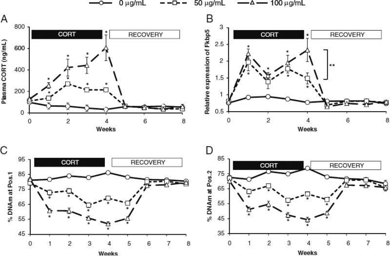
Plasma corticosterone levels varied in a dose-dependent manner, as shown in Fig. 1a. In the first experiment (experiment 1), mean plasma corticosterone levels in the group receiving 100 μg/ml of corticosterone in their drinking water (“CORT100”) varied from 254.2 to 606.7 ng/ml, and peaked at 606.7±119.2 ng/ml (mean±SEM) during the fourth week of CORT treatment. Plasma CORT levels in the group receiving 50 μg/ml of corticosterone (“CORT50”) varied from 137.2 to 270.0 ng/ml and peaked at 270.0±30.2 ng/ml during the second week. For both treatment groups, plasma CORT returned to baseline levels once treatment ended. The group receiving only vehicle solution without corticosterone (“CONTROL”) had mean plasma corticosterone levels ranging from 31.4 to 64.7 ng/ml for the first 4 weeks with no deviation from that range during the recovery period.
Fig. 1.
Plasma corticosterone, expression, and DNAm vs. time by treatment group. Mice were given different doses of corticosterone [100 μg/ml (triangle), 50 μg/ml (square), or vehicle solution (circle)] in their drinking water for 4 weeks (depicted by black horizontal bar with “CORT”), after which they were allowed to recover for an additional 4 weeks (depicted by white horizontal bar with “RECOVERY”). a Weekly plasma CORT levels in the two corticosterone-treated groups (100 μg/ml dose in triangles and 50 μg/ml dose in squares, with dashed lines) compared to vehicle-treated groups (circles, with solid lines). b Expression levels of Fkbp5 in blood across the 8-week experiment. Dose-dependent expression differences were observed only at the fourth week. c and d A time-course change (weeks) in blood DNAm at Fkbp5 intron 1, CpG position 1 c and CpG position 2 d, in which dose-dependent methylation differences were observed between corticosterone-treated and vehicle-treated groups during the 4-week-treatment period and early in the recovery period. Single asterisks (*) indicate differences that are statistically significant (P<0.05). Double asterisks (**) indicate the only time point at week 4 where a statistically significant difference (P<0.05) in expression levels of the Fkbp5 gene was observed between the two CORT-treated groups
We assessed expression changes in the Fkbp5 gene during CORT treatment and recovery periods. The greatest increase in expression was observed in the CORT100 group (range 66.0% to 202.1%, P≤0.0016). A significant increase in expression was also observed in the CORT50 group (46.7% to 113.7%, P≤0.05). However, expression levels did not show a clear dose-dependent change, except at week 4, where the CORT100 group showed a 56.7% increase in expression over that of the CORT50 group (P=0.045). A time-course change in Fkbp5 expression is shown in Fig. 1b.
In contrast, we observed a more pronounced dosedependent decrease in DNAm at the intron 1 region of Fkbp5 (CpG-1) with the greatest overall decrease seen in the CORT100 group (range 20.9% to 34.2%, P<0.0003) and a lesser decrease seen in the CORT50 group (range 8.8% to 18.4%, P≤0.0001), with only minor fluctuations seen in the CONTROL group (Fig. 1c). At the CpG-2 position, the greatest decrease in DNAm was observed again in the CORT100 group (range 20.1% to 34.4%, P< 0.0001), with a smaller decrease in the CORT50 group (range 8.1% to 17.2%, P<0.0003; Fig. 1d). Intriguingly, DNAm changes persisted for both treatment groups in the first week of recovery (at week 5), with a return to baseline in the following weeks.
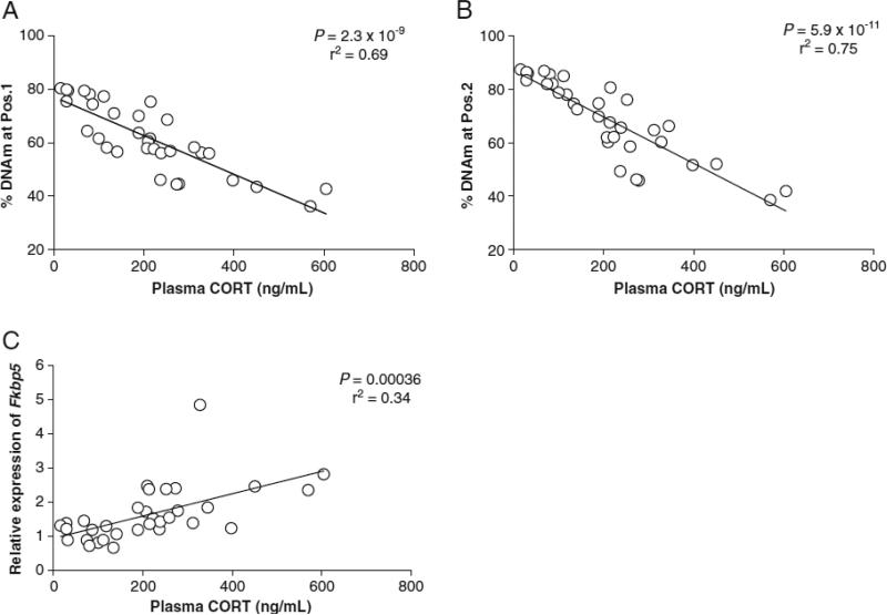
Plasma corticosterone levels determined from blood drawn on a weekly basis show a dynamic range of corticosterone levels among individuals (mean plasma CORT levels from 15.4 to 604.8 ng/ml), as well as intra-individual differences from week-to-week blood draws (e.g., weekly ranges from 204.2 to 1,009.3 ng/ml for the highest exposed CORT-treated mouse and weekly ranges from 7.9 to 25.3 ng/ml for the least varying control mouse). This wide range of CORT levels within a given mouse shows how unreliable a single serum corticosterone level can be as a marker of chronic glucocorticoid exposure, even when glucocorticoids are being administered in the drinking water. We asked whether either of two measures, percent DNAm changes in the intron 1 region or Fkbp5 expression level, could be used to accurately reflect the mean 4-week corticosterone burden imposed on the animals. For both CpG-1 and CpG-2 positions, DNAm measured at week 4 in each animal correlated significantly with the corticosterone burden in the animal as determined by taking an average of plasma CORT levels from weeks 1 to 4. For CpG-1 and CpG-2, the squares of the correlation coefficient were determined to be r2=0.69 (P=2.3×10⁻⁹) and r2=0.75 (P=5.9×10⁻¹¹), respectively (Fig. 2a and b). On the other hand, correlation between the expression levels of Fkbp5 and the mean CORT levels were not as strong as those observed for the two CpG positions (r2=0.34, P=0.00036; Fig. 2c).
Fig. 2.
Regression analysis of mean corticosterone levels vs. methylation and expression. A strong correlation was observed between week-4-blood DNAm of Fkbp5 intron 1, CpG position 1 (a) and CpG position 2 (b) vs. mean corticosterone levels from weeks 1–4. c A more modest correlation was observed between expression of Fkbp5 and mean corticosterone levels
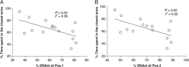
Chronic exposure to glucocorticoids is accompanied by physiological and metabolic changes that include significant reductions in the mass of the thymus, spleen, and adrenal glands, as well as an increase in adipose tissue deposition. In the first experiment (experiment 1), we observed a complete atrophy of the thymus tissues and a significant reduction in adrenal gland mass in animals that were treated with either 50 or 100 μg/ml of CORT (Fig. 3a). Since the spleen and adipose tissues varied with a wider range of masses, we asked whether these changes correlated with the DNAm values at week 4. We found a significant correlation for both spleen (r2=0.56, P=2.8×10⁻⁵) and percent fat (r2=0.51, P=9.0×10⁻⁵) when compared to DNAm at CpG-1 (Fig. 3b and c). Similar results were obtained for CpG-2 (spleen: r2=0.60, P= 1.05×10⁻⁵;% fat: r2=0.65, P=2.1×10⁻⁶; Fig. 3d and e). We observed that glucose, one of the metabolites commonly elevated in the Cushingoid state, was also moderately correlated with DNAm at CpG-2 (r2=0.48, P=0.0010). In addition, we found a modest but statistically significant relationship between DNAm and the percent of time the mice spent in the closed arms of the elevated plus maze (EPM, r2=0.35, P=0.02 for both CpG-1 and CpG-2), reflecting anxiety-like behavior. The correlation between the two measurements is shown in Fig. 4a and b.
Fig. 3.
Organ weights and regression analysis of DNAm vs. spleen mass and percent visceral fat. a Thymus and adrenal glands were weighed for corticosterone-treated (gray and black bars for 50 and 100 μg/ml, respectively) and vehicle-treated (white bars) mice. The absence of data for thymic mass of corticosterone-treated mice reflects a complete atrophy of the organ after 4 week of treatment with corticosterone. Asterisks indicate differences that are statistically significant (P<0.05). A strong correlation was observed between week-4 DNAm of Fkbp5 intron 1, CpG position 1 vs. spleen mass (b), and DNAm vs. percentage of visceral tat (c). Similar results were observed for CpG position 2 vs. spleen mass (d) and percentage of visceral fat (e). All of the organ weights are expressed as weight per gram total body weight of the mice
Fig. 4.
Regression analysis of DNAm vs. percentage of time spent in the closed arms on the elevated plus maze (EPM). After 4 weeks of CORT treatment, mice were challenged with the EPM to test for anxiety-like symptoms. Mice that exhibit higher anxiety-like symptoms spent more time in the closed arms of the apparatus. The percentage of time (10 min total) that the animals spent in the closed arms vs. the open arms and the center was calculated and correlated to the week 4 DNAm of the two CpG positions in Fkbp5 intron 1
We next sought to extend our results in a second experiment (experiment 2), in which we reasoned that we might be able to: (a) more accurately assess mean CORT burden by performing daily rather than weekly blood draws; and (b) more accurately calculate correlation of DNAm with plasma CORT levels by using a broader range of CORT dose exposures. To this end, we divided 20 mice into five groups of four animals each and gave them different doses of corticosterone in their drinking water (0, 25, 50, 75, 100 μg/ml). Daily plasma CORT levels determined for 25 days were used to establish mean CORT values for each mouse. The range of mean plasma CORT values we achieved was less than that observed for the first experiment (first experiment inter-individual range of 15.4 to 604.8 ng/ml vs. second experiment inter-individual range of 34.1 to 186.3 ng/ml). In the second experiment, the animals drank less water, suggesting that the reduced range of CORT may have been due to the reduced CORT intake via the drinking water. For instance, while the mean daily intake of water for the mice administered 100 μg/ml of CORT was 7.8 ml per mouse in the first experiment, the mean daily intake of water for the same group in the second cohort was only 3.5 ml. The reduction in water consumption in the second cohort may reflect the stress of taking blood samples 5 days per week.
Nonetheless, a strong correlation was again observed between DNAm and the mean CORT levels during the 25-day period for both CpG-1 (r2=0.60, P=6.6×10⁻⁵, Suppl. Fig. 1a) and CpG-2 (r2=0.63, P=3.3×10⁻⁵, Suppl. Fig. 1b). We also determined the various organ weights of the second cohort of mice and correlated spleen mass and percent fat to DNAm at week four. Once again, we observed a complete atrophy of the thymus at CORT doses greater than 25 μg/ml and a dose-dependent decrease in adrenal gland mass (Suppl. Fig. 2a). There was a significant correlation between spleen mass vs. DNAm at CpG-1 (r2=0.48, P=0.0011) and percent fat vs. DNAm (r2=0.35, P=0.0076) (Suppl. Fig. 2b and c, respectively). DNAm at CpG-2 showed similar correlations with spleen mass (r2=0.48, P=0.0010, Suppl. Fig. 2d) and percent fat (r2=0.33, P=0.011, Suppl. Fig. 2e).
Discussion
The present study was designed to determine whether the degree of DNAm of the Fkbp5 gene in a model of glucocorticoid excess could serve as a peripheral biomarker of glucocorticoid burden (cortisol concentration × duration of exposure). We selected the Fkbp5 gene as our candidate based on a previous report showing that of a number of genes tested, only Fkbp5 showed changes in DNAm levels as a function of the degree of glucocorticoid exposure (Lee et al. 2010). The FKBP5 protein regulates glucocorticoid receptor (GR) sensitivity, as binding of the protein to the receptor complex reduces the latter's affinity for cortisol and translocation to the nucleus (Binder 2009). One of the consequences of nuclear translocation of activated GR is transactivation of the Fkbp5 gene. Prolonged exposure to glucocorticoids has the effect of increasing the levels of the FKBP5 protein that in turn limits the translocation activity of GR, thus completing a short negative feedback loop that ultimately downregulates Fkbp5 activation. Another consequence of chronic glucocorticoid exposure is epigenetic changes that occur at intronic sequences of Fkbp5, which might be interpreted as the cells trying to reestablish a new steady-state level to counteract elevated levels of glucocorticoids (Lee et al. 2010).
We observed a strong dose-dependent relationship of DNAm response to corticosterone exposure. This relationship was also time-dependent, i.e., there was limited durability of DNAm changes, which allowed Fkbp5 methylation to be a useful marker reflecting dynamic changes in blood corticosterone levels. For our study, this period was approximately a week's time following 4 weeks of glucocorticoid exposure. This makes assessment of DNAm particularly useful for determining glucocorticoid burden in the immediate past, especially if detrimental effects and symptoms of chronic exposure can be reversible. It would also be interesting to see whether that period could be extended to a larger time frame if the animals were treated for a longer duration. Our study was a minimal assessment; the predictive power of DNAm may go back even further in time with longer glucocorticoid exposure. As with the measurement of glycosylated hemoglobin (HbA1c) as an integrated measure of 90-day glucose exposure, only a single blood draw followed by a simple assay was necessary in our study to obtain an assessment of previous glucocorticoid burden, suggesting that DNAm measured in blood samples may be a biomarker with potential clinical value.
The strong correlation of Fkbp5 DNAm with metabolic consequences of corticosterone exposure supports the validity of the hypothesis that DNAm can reflect glucocorticoid burden. In both experiments, we observed the expected atrophy of the thymus and adrenal glands, and a dose-dependent decrease in spleen mass and increase in percentage of visceral fat. These physiological/metabolic changes are classic symptoms of chronic glucocorticoid exposure (Karatsoreos et al. 2010), and DNAm was found to be highly correlated with these measurements. In addition, we observed a modest correlation between DNAm and anxiety-like behavior on the elevated plus maze, suggesting the potential application of our finding in predicting the behavioral consequences of glucocorticoid burden. More detailed experiments, such as those exploring a longer treatment as well as the recovery period, are necessary to further elucidate this relationship.
We were surprised to observe the wide range of CORT levels within a given mouse during the treatment period. Our findings show how unreliable a single serum corticosterone level can be as a measure of chronic glucocorticoid exposure even when glucocorticoids are being administered, and are not the product of a stress paradigm. Although glucocorticoid administration is not without its own limitations, such as the varying frequency with which the animal might consume the glucocorticoid, this finding nonetheless underscores the problems of using one or more serum corticosterone levels to assess glucocorticoid burden and stresses the need to find an accurate biomarker. We and other groups have considered using expression levels of Fkbp5 as a measurement for glucocorticoid burden. One prior study showed increased Fkbp5 expression in lymphoblast cell lines as concentration of glucocorticoids rose (Vermeer et al. 2003), consistent with our results. However, our animal model suggests that expression levels are not as predictive as DNAm changes for the following reasons: (1) expression levels drop to baseline immediately following cessation of glucocorticoid exposure; (2) the increase in expression is not dose-dependent; and (3) the correlations with glucocorticoid burden are not as robust for expression levels as they are for DNAm. We posit that whereas the expression pattern of Fkbp5 reflects glucocorticoid exposure in the previous several hours, DNAm reflects exposure over a longer period. Thus decreased DNAm at a given point may not correlate with expression at that same point, but might correlate with the history of expression pattern and hormone exposure in the preceding weeks.
Our findings raise questions regarding the mechanism behind the methylation/demethylation process. We speculate that, since the intronic regions are experimentally and bioinformatically determined GREs and enhancers, it is possible that constant binding of activated GR homodimer may recruit demethylating complexes of proteins, such as the ones associated with the putative demethylase Gadd45b (Ma et al. 2009). Once the region has lost some of its DNA methylation, by a demethylase, replication-dependent loss, or other base-excision-mediated processes, the genomic loci then can be occupied by proteins that promote transcriptional activation. Loss of methylation and subsequent occupancy by transcription factors may allow the GRE to more quickly and/or repeatedly activate Fkbp5, allowing the cell to achieve a more robust response to glucocorticoids.
Taken together, epigenetic changes observed in the two cohorts of mice in response to glucocorticoid administration reveal a strong inverse relationship between cumulative plasma stress hormone levels and DNAm of Fkbp5. This correlation was stronger in the first cohort that had greater exposure to glucocorticoids, as evidenced by the increased CORT-water intake and the subsequently higher levels of CORT observed in the serum.
This study should be viewed in light of several limitations. We mentioned the difference in the range of corticosterone levels between cohorts. We attributed this difference to the reduced amount of fluid that the mice in the second cohort had consumed over the treatment period. The variations in the amount of fluid consumed by the mice may also play a significant role in the week-to-week and day-to-day fluctuations observed in the plasma CORT levels. While circadian rhythm most certainly contributes to the variations, the drinking pattern, or deviations from it, must also be considered as a factor. Another limitation of this study is the supraphysiological levels of plasma corticosterone levels observed in a subset of the treated animals. Nevertheless, we decided to test our hypothesis with a Cushing's model before a stress-based model because it allowed for examination of dose-dependent effects of glucocorticoids on the biomarker. Thus, an important next step would be to observe similar correlations among DNAm, plasma CORT levels, and various physiological measurements that accompany a physiologically relevant model of stress such as chronic social stress. Third, it is possible that metabolites and electrolytes that accompany the Cushingoid state may themselves be responsible for the loss of methylation observed in Fkbp5. To test these, animal models (e.g., hyperglycemia and hypokalemia) could be used to assess their impact on Fkbp5. In summary, a whole-blood assessment of Fkbp5 gene methylation is a biomarker that integrates 4 weeks of investigator-administered glucocorticoid exposure and may be a useful measure in states of excess exposure.
Supplementary Material
Acknowledgments
This study was funded by NIH grants AA10158 (GSW), HD055030 (KLKT), and T32MH015330 (RSL); the Kenneth Lattman Foundation (GSW), a NARSAD Young Investigator Award (RSL); and the Margaret Ann Price Investigatorship (JBP and RSL).
Footnotes
Disclosure statement The authors have nothing to disclose.
Contributor Information
R. S. Lee, Department of Psychiatry and Behavioral Sciences Johns Hopkins University School of Medicine Baltimore, MD, USA
K. L. K. Tamashiro, Department of Psychiatry and Behavioral Sciences Johns Hopkins University School of Medicine Baltimore, MD, USA
R. H. Purcell, Department of Psychiatry and Behavioral Sciences Johns Hopkins University School of Medicine Baltimore, MD, USA
Y. Huo, Department of Psychiatry and Behavioral Sciences Johns Hopkins University School of Medicine Baltimore, MD, USA
M. Rongione, Department of Psychiatry and Behavioral Sciences Johns Hopkins University School of Medicine Baltimore, MD, USA
J. B. Potash, Department of Psychiatry and Behavioral Sciences, Center for Epigenetics Johns Hopkins University School of Medicine Baltimore, MD, USA
X. Yang, Department of Medicine Johns Hopkins University School of Medicine Baltimore, MD, USA
G. S. Wand, Department of Medicine Johns Hopkins University School of Medicine Baltimore, MD, USA gwand@jhmi.edu
References
- Binder EB. The role of FKBP5, a co-chaperone of the glucocorticoid receptor in the pathogenesis and therapy of affective and anxiety disorders. Psychoneuroendocrinology. 2009;34(Suppl 1):S186–195. doi: 10.1016/j.psyneuen.2009.05.021. [DOI] [PubMed] [Google Scholar]
- Clegg DJ, Brown LM, Woods SC, Benoit SC. Gonadal hormones determine sensitivity to central leptin and insulin. Diabetes. 2006;55:978–987. doi: 10.2337/diabetes.55.04.06.db05-1339. [DOI] [PubMed] [Google Scholar]
- Colella S, Shen L, Baggerly KA, Issa JP, Krahe R. Sensitive and quantitative universal Pyrosequencing methylation analysis of CpG sites. Biotechniques. 2003;35:146–150. doi: 10.2144/03351md01. [DOI] [PubMed] [Google Scholar]
- Faggiano A, Pivonello R, Spiezia S, De Martino MC, Filippella M, Di Somma C, Lombardi G, Colao A. Cardiovascular risk factors and common carotid artery caliber and stiffness in patients with Cushing's disease during active disease and 1 year after disease remission. J Clin Endocrinol Metab. 2003;88:2527–2533. doi: 10.1210/jc.2002-021558. [DOI] [PubMed] [Google Scholar]
- Hansen AM, Garde AH, Skovgaard LT, Christensen JM. Seasonal and biological variation of urinary epinephrine, norepinephrine, and cortisol in healthy women. Clin Chim Acta. 2001;309:25–35. doi: 10.1016/s0009-8981(01)00493-4. [DOI] [PubMed] [Google Scholar]
- Haskett RF. Diagnostic categorization of psychiatric disturbances in Cushing's syndrome. Am J Psychiatry. 1985;142:911–916. doi: 10.1176/ajp.142.8.911. [DOI] [PubMed] [Google Scholar]
- Karatsoreos IN, Bhagat SM, Bowles NP, Weil ZM, Pfaff DW, McEwen BS. Endocrine and physiological changes in response to chronic corticosterone: a potential model of the metabolic syndrome in mouse. Endocrinology. 2010;151:2117–2127. doi: 10.1210/en.2009-1436. [DOI] [PMC free article] [PubMed] [Google Scholar]
- Kelly JJ, Mangos G, Williamson PM, Whitworth JA. Cortisol and hypertension. Clin Exp Pharmacol Physiol Suppl. 1998;25:S51–56. doi: 10.1111/j.1440-1681.1998.tb02301.x. [DOI] [PubMed] [Google Scholar]
- Kelly WF, Kelly MJ, Faragher B. A prospective study of psychiatric and psychological aspects of Cushing's syndrome. Clin Endocrinol. 1996;45:715–720. doi: 10.1046/j.1365-2265.1996.8690878.x. [DOI] [PubMed] [Google Scholar]
- Kunnecke B, Verry P, Benardeau A, von Kienlin M. Quantitative body composition analysis in awake mice and rats by magnetic resonance relaxometry. Obes Res. 2004;12:1604–1615. doi: 10.1038/oby.2004.200. [DOI] [PubMed] [Google Scholar]
- Lee RS, Tamashiro KL, Yang X, Purcell RH, Harvey A, Willour VL, Huo Y, Rongione M, Wand GS, Potash JB. Chronic corticosterone exposure increases expression and decreases deoxyribonucleic acid methylation of Fkbp5 in mice. Endocrinology. 2010;151:4332–4343. doi: 10.1210/en.2010-0225. [DOI] [PMC free article] [PubMed] [Google Scholar]
- Ma DK, Jang MH, Guo JU, Kitabatake Y, Chang ML, Pow-Anpongkul N, Flavell RA, Lu B, Ming GL, Song H. Neuronal activity-induced Gadd45b promotes epigenetic DNA demethylation and adult neurogenesis. Science. 2009;323:1074–1077. doi: 10.1126/science.1166859. [DOI] [PMC free article] [PubMed] [Google Scholar]
- Magee JA, Chang LW, Stormo GD, Milbrandt J. Direct, androgen receptor-mediated regulation of the FKBP5 gene via a distal enhancer element. Endocrinology. 2006;147:590–598. doi: 10.1210/en.2005-1001. [DOI] [PubMed] [Google Scholar]
- McEwen BS. Interacting mediators of allostasis and allostatic load: towards an understanding of resilience in aging. Metabolism. 2003;52:10–16. doi: 10.1016/s0026-0495(03)00295-6. [DOI] [PubMed] [Google Scholar]
- McEwen BS. Protection and damage from acute and chronic stress: allostasis and allostatic overload and relevance to the pathophysiology of psychiatric disorders. Ann NYAcad Sci. 2004;1032:1–7. doi: 10.1196/annals.1314.001. [DOI] [PubMed] [Google Scholar]
- Nuber UA, Kriaucionis S, Roloff TC, Guy J, Selfridge J, Steinhoff C, Schulz R, Lipkowitz B, Ropers HH, Holmes MC, Bird A. Up-regulation of glucocorticoid-regulated genes in a mouse model of Rett syndrome. Hum Mol Genet. 2005;14:2247–2256. doi: 10.1093/hmg/ddi229. [DOI] [PubMed] [Google Scholar]
- Paakinaho V, Makkonen H, Jaaskelainen T, Palvimo JJ. Glucocorticoid receptor activates poised FKBP51 locus through long-distance interactions. Mol Endocrinol. 2010;24:511–525. doi: 10.1210/me.2009-0443. [DOI] [PMC free article] [PubMed] [Google Scholar]
- Pearce G, Tabensky DA, Delmas PD, Baker HW, Seeman E. Corticosteroid-induced bone loss in men. J Clin Endocrinol Metab. 1998;83:801–806. doi: 10.1210/jcem.83.3.4621. [DOI] [PubMed] [Google Scholar]
- Resmini E, Farkas C, Murillo B, Barahona MJ, Santos A, Martinez-Momblan MA, Roig O, Ybarra J, Geli C, Webb SM. Body composition after endogenous (Cushing's syndrome) and exogenous (rheumatoid arthritis) exposure to glucocorticoids. Horm Metab Res. 2010;42:613–618. doi: 10.1055/s-0030-1255032. [DOI] [PubMed] [Google Scholar]
- Sapolsky RM, Uno H, Rebert CS, Finch CE. Hippocampal damage associated with prolonged glucocorticoid exposure in primates. J Neurosci. 1990;10:2897–2902. doi: 10.1523/JNEUROSCI.10-09-02897.1990. [DOI] [PMC free article] [PubMed] [Google Scholar]
- Seeman TE, Singer BH, Rowe JW, Horwitz RI, McEwen BS. Price of adaptation– allostatic load and its health consequences. MacArthur studies of successful aging. Arch Intern Med. 1997;157:2259–2268. [PubMed] [Google Scholar]
- Taicher GZ, Tinsley FC, Reiderman A, Heiman ML. Quantitative magnetic resonance (QMR) method for bone and wholebody-composition analysis. Anal Bioanal Chem. 2003;377:990–1002. doi: 10.1007/s00216-003-2224-3. [DOI] [PubMed] [Google Scholar]
- Tauchmanova L, Rossi R, Biondi B, Pulcrano M, Nuzzo V, Palmieri EA, Fazio S, Lombardi G. Patients with subclinical Cushing's syndrome due to adrenal adenoma have increased cardiovascular risk. J Clin Endocrinol Metab. 2002;87:4872–4878. doi: 10.1210/jc.2001-011766. [DOI] [PubMed] [Google Scholar]
- Vermeer H, Hendriks-Stegeman BI, van der Burg B, van Buul-Offers SC, Jansen M. Glucocorticoid-induced increase in lymphocytic FKBP51 messenger ribonucleic acid expression: a potential marker for glucocorticoid sensitivity, potency, and bioavailability. J Clin Endocrinol Metab. 2003;88:277–284. doi: 10.1210/jc.2002-020354. [DOI] [PubMed] [Google Scholar]
- Wochnik GM, Ruegg J, Abel GA, Schmidt U, Holsboer F, Rein T. FK506-binding proteins 51 and 52 differentially regulate dynein interaction and nuclear translocation of the glucocorticoid receptor in mammalian cells. J Biol Chem. 2005;280:4609–4616. doi: 10.1074/jbc.M407498200. [DOI] [PubMed] [Google Scholar]
Associated Data
This section collects any data citations, data availability statements, or supplementary materials included in this article.


