Abstract
A 53-year-old man developed a widespread erythematous eruption which rapidly evolved into fluid-filled bulla mostly involving the distal areas of all four limbs and erosions on the oral as well as anogenital mucosa. Based on clinical presentation, chronology of drug exposure, past events and histopathology as diagnosis of widespread bullous fixed drug eruption was made over Steven Johnson-toxic epidermal necrolysis syndrome. Steroids were deferred and the lesions healed with minimal pigmentation within a week. Differentiating between the two entities has been historically difficult, and yet can have significant therapeutic and prognostic implications.
Background
Although fixed drug eruptions (FDEs) are not very uncommon adverse drug reactions, the widespread bullous variant are comparatively rare and may be difficult to identify early.1 Wide spread bullous FDEs are a rare variant of FDEs that can be clinically confused with Steven-Johnson syndrome-toxic epidermal necrolysis (SJS-TEN).2–4 These two entities differ significantly with respect to management and prognosis. A biopsy can be a useful tool in making an accurate diagnosis. Identifying and withdrawing the culprit medications is paramount. The case highlights the importance of differentiating these two entities that resemble each other closely and yet differ in terms of management and prognosis.
Case presentation
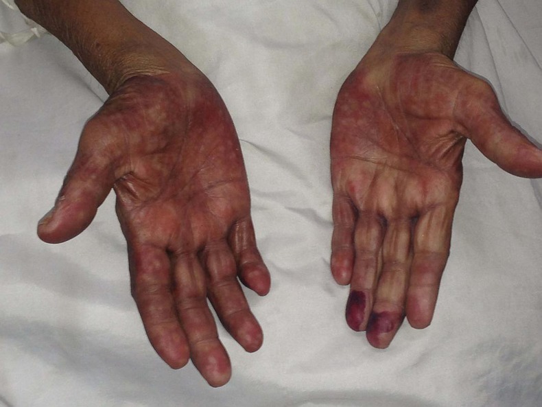
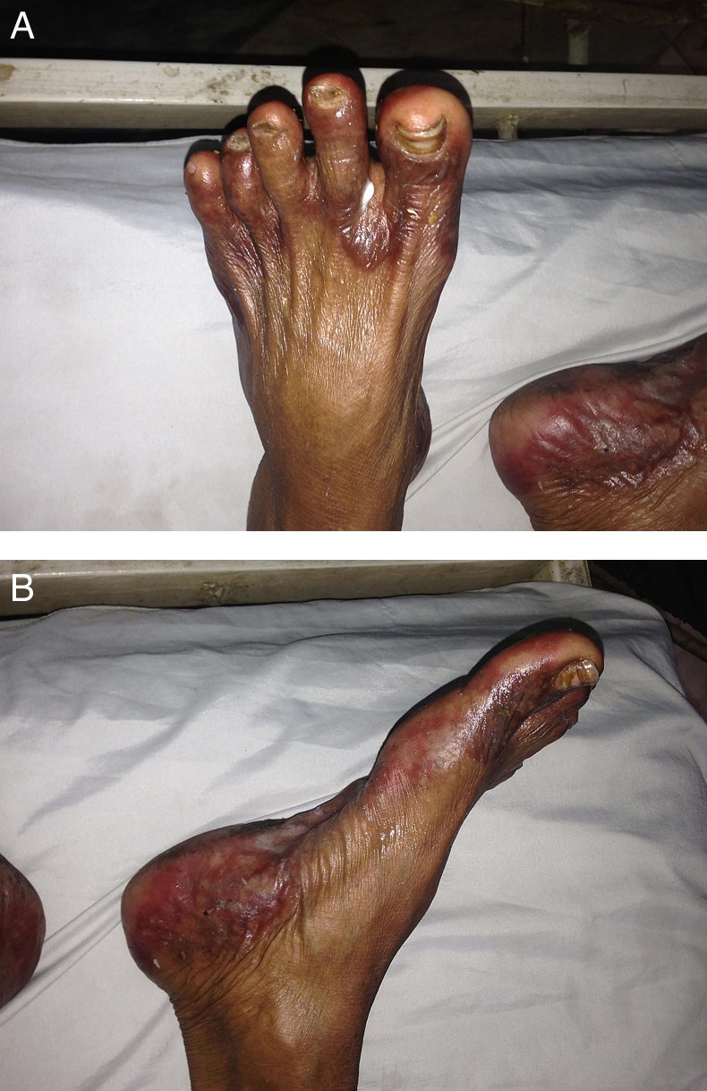
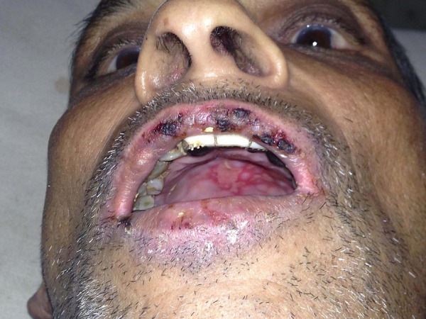
A 53-year-old man with a known history of chronic pancreatitis, diabetes mellitus and prior alcohol misuse presented to the clinic with severe abdominal pain, vomiting and fever. He was treated with non-steroidal anti-inflammatory drugs (NSAIDs; diclofenac), antiemetics (ondansetron), antipyretics (acetaminophen) opioids (tramadol) and intravenous fluid with normal saline. On the second day following admission (around 20 h after the administration of diclofenac and ondansetron and 12 h of the tramadol), he reported of an itchy tender rash on his palms and soles and painful oral ulcers. An examination revealed multiple erythematous and bullous plaques ranging in size from 2×2 cm to the largest of almost 5×5 cms, on both palms and soles (figures 1 and 2A,B). The bullae contained clear serous fluid. Skin erosions were also noted over the genital and perianal region with multiple ulcers and crusts in the oral cavity (figure 3). A working diagnosis of SJS versus widespread FDE was made and skin biopsies were sent for histopathological examination. A review of his medical history revealed a similar occurrence 3 years ago when he received diclofenac during a hospital admission within a day of administration, at the time he was diagnosed with SJS-TEN; blistering erythematous lesions on both palms and soles as well as oral sores were mentioned, but further details including exact treatment received were unavailable; biopsy was, however, not performed during the prior event. The lesions as per the patient healed in less than a week without consequence.
Figure 1.

Symmetrical bullous erythematous lesions noted on palms.
Figure 2.

(A and B) bullous erythematous lesions seen on the feet, including soles and intertriginous areas.
Figure 3.

Oral mucosa and lips have erosions and crusting.
Investigations
The histopathology assessment showed intraepidermal and some subepidermal vesicles with necrosis of epidermal keratinocytes. Infiltration of polymorphonuclear, eosinophilic and mononuclear cells was noted with interphase dermatitis and perivascular distribution. The picture was consistent with FDE.
Differential diagnosis
Generalised FDE and SJS-TEN were the two major differentials.
Treatment
Dermatology was consulted and a decision to defer systemic steroids was made. Local measures to prevent infection and emollients over the raw areas were instituted.
Outcome and follow-up
The patient was discharged a week later with hyper-pigmented lesions and appropriate treatment for pancreatitis. Diclofenac was deemed as the most likely culprit agent based on his history and its profile. He was explained the need to avoid diclofenac and educated about the importance of highlighting his allergic drug history in all future contacts with the medical care system. A patch testing to confirm the culprit drug was considered but it was not possible due to logistic and financial constraints.
Discussion
FDE are a part of a spectrum of hypersensitivity skin reactions following the ingestion of certain drugs (most commonly antibacterials, antimalarials, NSAIDs, paracetamol and barbiturates). Characteristics include dusky red or violaceous plaques occurring over a fixed area of the body within hours of exposure to medications and lesions typically reoccurring over the same region on re-exposure to the culprit medications.1 Rare variants of FDE include non-pigmenting and bullous lesions, the latter resembling SJS and TEN. The incidence of drug-induced skin reactions varies from 2% to 5% for inpatients to 1% for outpatients. FDEs contribute to a significant portion of these accounting for up to 9–21% in some series.1 5
FDEs usually appear as solitary, erythematous, bright red or dusky macules that may evolve into an oedematous plaque or bullous-type lesions. They may develop within 24 h after ingestion of the drug and are often painful. Residual hyperpigmentation is common. Rechallenge or patch testing at a previous location site (positive in 43% patients) can be used to identify the precise culprit medication in case of uncertainity.6 Historically differentiating between widespread bullous FDE and SJS-TEN has been difficult. Paucity of constitutional symptoms, well-demarcated blisters and erythematous patches, absence of target lesions, relative sparing of the mucosa, history of similar eruption and rapid onset since exposure support a diagnosis of FDE over SJS-TEN. Histopathology, especially detailed histopathological examination from early stage lesions or perilesional skin,7 may be helpful. While SJS-TEN lesions affect the dermoepidermal junction with lymphocytic infiltrate, FDEs are identified by vacuolar interface dermatitis with epidermal necrosis and a superficial and deep perivascular infiltrate of lymphocytes, eosinophils and neutrophils.2 Despite these differentiating features, precise delineation remains a challenge and often relies on the acumen and experience of the clinician and dermatopathologist.3 4
The management of widespread FDEs, besides immediate withdrawal of the offending agent, involves antiseptic solutions and emollients to promote the healing of local ulcers and supportive measures for pain and infection prevention. Oral antiseptic rinses for mucosal erosions and local ointments for perianal lesions are usually used. The role of steroids is unproven but are resorted to in severe cases.6 Most dermatologists consider widespread bullous FDEs have a more favourable outcome as compared to SJS/TEN often with complete resolution of symptoms and lesions on withdrawing the offending agent2 although a recent paper questions its benign reputation.8
Learning points.
Generalised bullous fixed drug eruption is a rare variant of fixed drug eruptions and may be confused with Steven Johnson syndrome/toxic epidermal necrolysis.
Various clinical and histopathological differences can guide the clinician to the right diagnosis.
Differentiating between the two entities is important as it may have important prognostic and management implications.
Acknowledgments
The authors would like to acknowledge Dr YS Marfatiya and his team from the Department of Dermatology, Medical College Baroda and SSG Hospital, Vadodara for their assistance and help.
Footnotes
Contributors: All the authors were involved in patient care, reviewing the literature and preparing the manuscript.
Competing interests: None.
Patient consent: Obtained.
Provenance and peer review: Not commissioned; externally peer reviewed.
References
- 1.Lee AY. Fixed drug eruptions. Incidence, recognition, and avoidance. Am J Clin Dermatol 2000;1:277–85 [DOI] [PubMed] [Google Scholar]
- 2.Lin TK, Hsu MM, Lee JY. Clinical resemblance of widespread bullous fixed drug eruption to Stevens Johnson syndrome or toxic epidermal necrolysis: report of two cases. J Formos Med Assoc 2002;101:572–6 [PubMed] [Google Scholar]
- 3.Baird BJ, De Villez RL. Widespread bullous fixed drug eruption mimicking toxic epidermal necrolysis. Int J Dermatol 1988;27:170. [DOI] [PubMed] [Google Scholar]
- 4.Rai R, Jain R, Kaur I, et al. Multifocal bullous fixed drug eruption mimicking Stevens-Johnson syndrome. Indian J Dermatol Venereol Leprol 2002;68:175–6 [PubMed] [Google Scholar]
- 5.Krahenbuhl-Melcher A, Schlienger R, Lampert M, et al. Drug-related problems in hospitals: a review of the recent literature. Drug Saf 2007;30:379–407 [DOI] [PubMed] [Google Scholar]
- 6.Shear NH, Knowles SR. Chapter 41. Cutaneous reactions to drugs. In: Goldsmith LA, Katz SI, Gilchrest BA, Paller AS, Leffell DJ, Dallas NA. eds Fitzpatrick's dermatology in general medicine. 8th edn New York: McGraw-Hill, 2012. http://www.accessmedicine.com.ezpprod1.hul.harvard.edu/content.aspx?aID=56033404 (accessed 22 July 2013). [Google Scholar]
- 7.Lee C-H, Chen Y-C, Cho Y-T, et al. Fixed-drug eruption: a retrospective study in a Single Referral Center in Northern Taiwan. Dermatol Sin 2012;30:11–5 [Google Scholar]
- 8.Lipowicz S, Sekula P, Ingen-Housz-Oro S, et al. Prognosis of generalized bullous fixed drug eruption: comparison with Stevens–Johnson syndrome and toxic epidermal necrolysis. Br J Dermatol 2013;168:726–32 [DOI] [PubMed] [Google Scholar]


