
Figure 3.
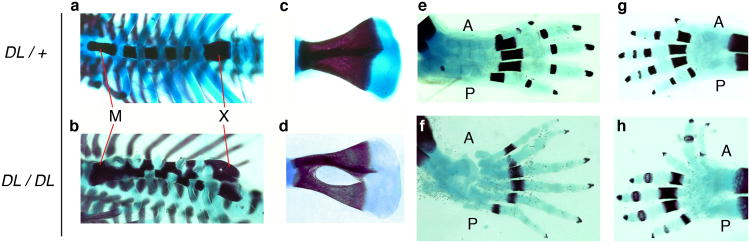
Additional skeletal abnormalities in Ptch1DL homozygous mutants.
E18.5 wild-type (a, c, e, g) and Ptch1DL mutants (b, d, f, h) showing ribcage in ventral view (a, b), scapula (c, d), hindlimb (e, f) and forelimb (g, h). Note the ribs have been cut by the investigators on one side of the sternum in the mutant sample. A, anterior; M, manubrium; P, posterior; X, xiphoid process.