Abstract
Cytoplasmic dynein is a microtubule-based motor required for intracellular transport and cell division. Its movement involves coupling cycles of track binding and release with cycles of force-generating nucleotide hydrolysis. How this is accomplished given the ~25 nm separating dynein’s track- and nucleotide-binding sites is not understood. Here, we present a sub-nanometer-resolution structure of dynein’s microtubule-binding domain bound to microtubules by cryo-electron microscopy that was used to generate a pseudo-atomic model of the complex with molecular dynamics. We identified large rearrangements triggered by track binding and specific interactions, confirmed by mutagenesis and single molecule motility assays, which tune dynein’s affinity for microtubules. Our results provide a molecular model for how dynein’s binding to microtubules is communicated to the rest of the motor.
Dyneins are ATP-driven molecular motors that move towards the minus ends of microtubules (MTs) (1). The superfamily includes axonemal dyneins, which power the movements of cilia, and those that transport cargo, which include cytoplasmic dyneins 1 (“cytoplasmic”) and 2 (“intraflagellar”) (2). The transport of organelles, ribonucleoprotein complexes and proteins by cytoplasmic dynein is required for cellular homeostasis, cell-cell communication, cell division, and cell migration (3) and defects in these processes result in neurological disease in humans (4). Despite cytoplasmic dynein’s role in such diverse activities and recent advances in characterizing its structure and motility, many aspects of its molecular mechanism remain poorly understood.
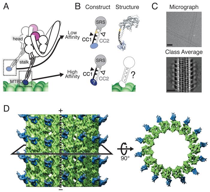
The core of the cytoplasmic dynein holoenzyme is a homodimer of ~500 kDa motor-containing subunits (Fig. 1A). The major functional elements include: (a) a “tail” domain required for dimerization and cargo binding (5); (b) the force-generating “head” or “motor domain” (6, 7), a ring containing six AAA+ ATPase domains (8-10); (c) a “linker” connecting the head and tail, required for motility (6, 7, 11, 12); (d) the “stalk”, a long antiparallel coiled-coil that emerges from AAA4 (13, 14), and (e) the MT-binding domain (MTBD), a small alpha-helical domain at the end of the stalk responsible for binding the MT track (15-17). Unlike the other cytoskeletal molecular motors, kinesin and myosin, dynein does not have its ATPase and polymer track binding sites located within a single domain. With 25 nm separating AAA1, the main site of ATP hydrolysis, and the MTBD, an unresolved question is how dynein coordinates the cycles of nucleotide hydrolysis and MT binding and release required for its motion.
Fig. 1.
Cryo-EM reconstruction of the cytoplasmic dynein high-affinity MTBD bound to a MT. (A) Schematic of dimeric cytoplasmic dynein. Major features relevant to this study are indicated. The MTBD is depicted in its low- (light blue) and high- (dark blue) affinity states during a step along the MT. (B) Schematic of the fusion constructs between the MTBD and seryl-tRNA synthetase (SRS) that fix the heptad registry of the stalk. The low-affinity construct has an additional 4 amino acids (yellow) inserted in CC1 (black) relative to the high-affinity construct. (C) Cryo-EM image of MTs highly decorated with the high-affinity SRS-MTBD construct (top) and a class average generated from segments of decorated 14-protofilament MTs (bottom). Scale bars: 25 nm. (D) Three-dimensional reconstruction of the MTBD-MT complex, filtered to the calculated resolution of 9.7 Å (Fig. S1). The black solid line represents a slice through the volume, which is shown on the right viewed from the minus end of the MT. The MT polarity is indicated and the dashed line shows the location of the MT seam. The SRS has been omitted due to its lower resolution (Fig. S1).
The mechanism coupling nucleotide hydrolysis to MT affinity has been suggested to be sliding in which the two helices in the stalk’s coiled-coil adopt different registries by using alternative sets of hydrophobic heptad repeats (17-19). Dynein’s stalk can be fixed in a specific registry by fusing it to a coiled-coil of known structure, such as that of seryl-tRNA synthetase (SRS) (18). Changing the length of the first stalk helix (CC1) relative to the second (CC2) shifts their alignment between high-affinity (“α”) and low-affinity (“β+”) registries, and alters the affinity of the MTBD for MTs by up to twenty-fold (Fig. 1B) (18). Engineered disulfide cross-linking in the monomeric dynein motor domain showed that its stalk explores multiple registries in solution, and that a given registry is coupled to a specific MT affinity (19). Fixing the stalk registry also uncoupled the nucleotide state of the head from MT affinity (19).
Understanding the molecular mechanism by which stalk sliding is coupled to nucleotide hydrolysis and MT affinity would be aided by a structure of dynein’s MTBD bound to MTs. Crystal structures are available for a low-affinity dynein MTBD fused to SRS through a short segment of the stalk (Fig. 1B) (17), and an ADP-bound, presumably high-affinity dynein monomer (20). Only low-resolution structures of the MT-bound form have been reported (17, 21). Here we describe a cryo-electron microscopy (cryo-EM) reconstruction of the MTBD bound to MTs at sub-nanometer resolution, using a high-affinity version of the SRS-MTBD construct (18).
We collected images of MTs highly decorated with the SRS-MTBD under cryogenic conditions (Fig. 1C, Fig. S1A), and adapted an image processing method (22, 23) to solve its structure bound to 14-protofilament MTs. The SRS-MTBD binds to α-tubulin and β-tubulin at the intradimer interface and is positioned to the side of the protofilament, as previously reported (17, 21) (Fig. 1D, Fig. S1B, Movie S1). In the reconstruction, the portion including the MT, the MTBD and the beginning of the stalk has a resolution of 9.7 Å (Fig. S1D), where α-helices become visible.
We used molecular dynamics (MD) and our cryo-EM reconstruction to obtain pseudo-atomic models of the low- and high-affinity states of the MTBD bound to MTs. First, we rigid-body docked the atomic-resolution structures of the tubulin dimer (24, 25) and the low-affinity MTBD into our map (Figs. 2A, C, and S2A, Movies S1 and S2) and used MD to resolve steric clashes between a helix (H1) in the MTBD and the MT (Figs. 2A, C and S2A, B, Movies S1 and S2). We then performed explicit-solvent molecular dynamics flexible fitting (MDFF) to shift the low-affinity MTBD structure to the high-affinity conformation in our reconstruction (Fig. S2C). In addition to the MD force field, MDFF uses a potential energy term derived from the experimental map and restraints on secondary structure to drive conformational changes that better fit the map (26, 27). The initial MDFF model agreed well within the experimental density of the MTBD (Fig. S2C); however, the stalk remained in the low-affinity β+ registry present in the starting model. MDFF likely did not shift the registry of the coiled-coil due to the lower resolution at the tip of the stalk segment (Fig. S1); a movement of the CC1 helix to the α registry would make it protrude from the map, incurring a penalty in the simulations. We achieved the shift by applying targeted molecular dynamics (TMD) (28) to the tip of the coiled-coil (Fig. S2D), using the Cα coordinates of the half-heptad shift in our construct to guide the final position of the stalk during the simulations. This final model (Fig. 2B, D and Movie S3), which we refer to as the high-affinity MTBD, has the highest cross-correlation with the experimental map (Figs. S2 and S3, Movies S4 and S5).
Fig. 2.
The high-affinity, MT-bound state of the dynein MTBD is characterized by repositioning of helices H1 and CC1. (A) Rigid-body docking of the low-affinity MTBD structure into our cryo-EM density. (B) Pseudo-atomic model of the high-affinity MTBD bound to MTs generated by Molecular Dynamics Flexible Fitting (MDFF) and Targeted Molecular Dynamics (TMD) (see text for details). (C) Close-up of the structure shown in (A), with its orientation indicated in panel (A). (D) Close-up of the structure shown in (B), with its orientation indicated in panel (B). The cryo-EM map is shown as a transparent grey mesh. The MTBD is colored following the scheme shown at the bottom of the figure and structural elements are indicated in the different views. H1 (orange/red) is the element with the largest movement in the transition to the high-affinity conformation; H3 and H6 (dark blue) are major contact points with the MT (green). α- and β-tubulin are indicated (green). MT polarity is indicated in panels A and B. H1 protrudes from the cryo-EM map and clashes with the MT in the rigid-body docked low-affinity state (A and C).
We repeated the MDFF calculations using the MTBD from the recent crystal structure of an ADP-bound dynein monomer (20) (Fig. S4). The only difference in the resulting pseudo-atomic model is in the stalk, where the dynein monomer’s structure is missing density for one of the helices next to the MTBD (Fig. S4A, B). The similarity in the crystal structures of the MTBD in the low-affinity and ADP-bound states is likely due to the absence of MTs; our results suggest that the conformation we observe in our MT-bound high-affinity structure is stabilized by its interactions with β-tubulin (Fig. S5A, Tables S1 and S2). MDFF of the ADP-bound dynein monomer’s MTBD into our cryo-EM map results in a large change in the angle between the MTBD and the stalk in the dynein monomer structure (Fig. S4A, B). This change makes the docking of the dynein monomer into our map compatible with previously reported measurements of the MT-stalk angle (17, 21) and our two-dimensional analysis of images of monomer-decorated MTs (Fig. S4D, E).
The cryo-EM map shows three points of continuous density between the MT and dynein’s MTBD: the H1-H2 loop and helices H3 and H6 (Fig. 2B). Several parts of the structure are unchanged by its interaction with the MT, especially helices H6, H5, and CC2, with root mean square deviations (RMSD) between the low- and high-affinity models of 1.4 Å, 1.4 Å, and 1.8 Å, respectively. The largest changes are the repositioning of H1 (RMSD = 10.1 Å) and an opening of CC1 in the coiled-coil next to the MTBD (RMSD = 8.1 Å) (Fig. 2A-D and Movies S1, S2, S3 and S6), a movement anchored at the proline kink present in CC1. The final position of H1 is stabilized by multiple interactions with an acidic patch in H12 of β-tubulin not fully occupied in the low-affinity state (Fig. S5A). This patch also stabilizes the high-affinity state of kinesin (29).
We monitored hydrogen bonds and salt bridges formed between the MTBD and the MT during MD simulations (Tables S1 and S2); the high-affinity MTBD formed more hydrogen bonds with the MT (Fig. S6) and electrostatic interactions at H1, H3, and H6 (Fig. S5, Table S1). Nearly all of these residues are highly conserved, and mutating them results in defects in MT binding (18, 30) (Fig. S5). The importance of salt bridges to the MTBD-MT interaction is consistent with the sensitivity of dynein’s motility to ionic strength (Fig. S7).
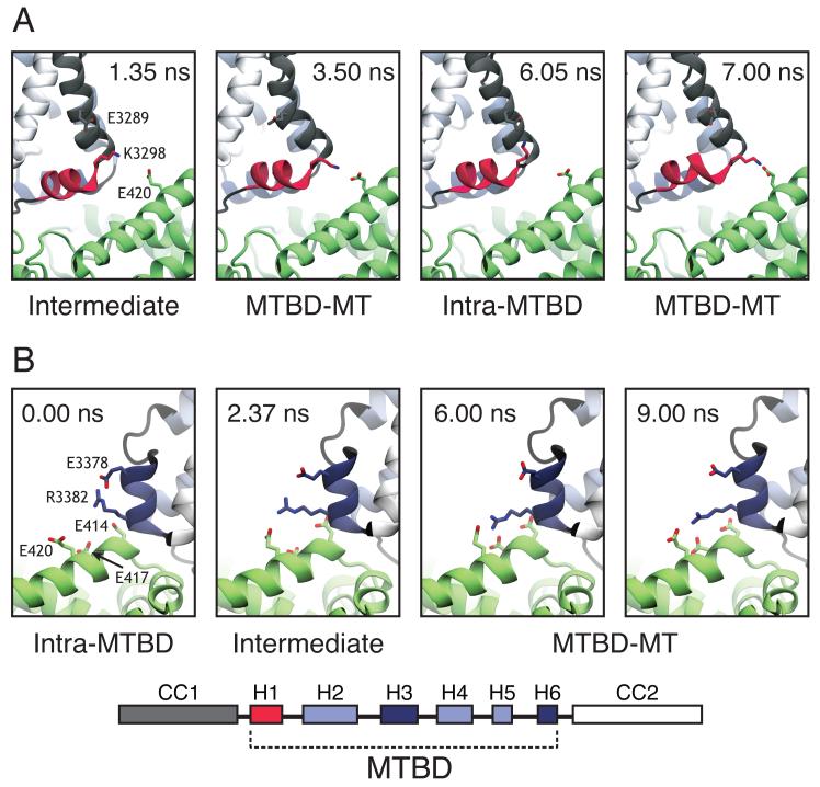
Our structural analysis suggested that the MTBD contains residues that lower its own affinity for the MT. In the MD simulations, basic residues in H1 and H6 formed salt bridges that alternated between intramolecular and intermolecular partners. In the MT-bound high-affinity conformation, H1-K3298 switched between a glutamate on β-tubulin and a conserved glutamate in CC1 (E3289) (Figs. 3A, S8A); neither contact can be formed by H1-K3298 in the low-affinity conformation (Fig. S5A). H6-R3382 switched from an intramolecular interaction with a conserved glutamate in the same helix (H6-E3378) in the low-affinity unbound state to an intermolecular interaction with a cluster of glutamates on α-tubulin upon binding (Fig. 3B, S8B); the intramolecular interaction might weaken the MTBD-MT interaction in both the low- and high-affinity conformations. The importance of the two MTBD glutamates involved in the intramolecular salt bridges had previously been recognized; substitution of CC1-E3289 and H6-E3378 with alanine increased dynein’s MT-binding affinity (18, 30) and reduced its ATP-stimulated release from MTs, respectively (30). We hypothesized that these phenotypes resulted from the competition between MT and MTBD residues in CC1 and H6 for salt bridge formation with the basic residues K3298 and R3382 in the MTBD.
Fig. 3.
Behavior of dynamic salt bridges in the MTBD as determined by MD. (A) K3298 in H1 of the MTBD alternates between an intermolecular salt bridge with E420 on β-tubulin (MTBD-MT) and an intramolecular salt bridge with E3289 on CC1 of the MTBD (intra-MTBD). (B) R3382 in H6 of the MTBD alternates between an intermolecular salt bridge with E414 and E420 on α-tubulin (MTBD-MT) and an intramolecular salt bridge with E3378 in H6 (intra-MTBD). Single letter amino acid code and number are indicated for Bos taurus tubulin and Mus musculus cytoplasmic dynein. Time stamps for frames from MD simulations are indicated. Intermediate refers to a position midway between MTBD-MT and intra-MTBD salt bridges.
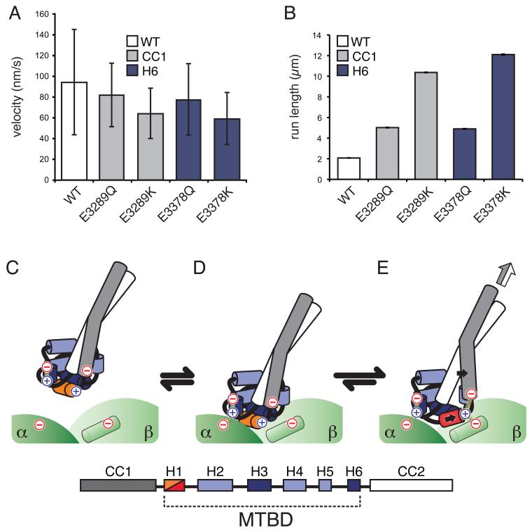
To test this prediction we mutated the residues equivalent to E3289 and E3378 in CC1 and H6 of Saccharomyces cerevisiae dynein to either an isosteric but neutral (Q) or a basic amino acid (K) to disrupt the salt bridge (Q) or introduce an intramolecular charge repulsion (K) that may favor intermolecular interactions between H1-K3298 or H6-R3382 and acidic residues on the MT surface. Single molecule motility assays that monitored the movement of purified mutant dyneins showed significant increases in dynein’s run length and small decreases in velocity that paralleled the severity of the mutation (E -> Q -> K) (Fig. 4A, B and Fig. S9). Most dramatically, the basic substitutions CC1-E3289K and H6-E3378K increased dynein’s run length by five-fold and six-fold, respectively (Fig 4B), and the double mutant even further (Fig. S10). These results suggest that cytoplasmic dynein has been selected for sub-maximal processivity. The effects observed with the mutants are not due to a strengthened interaction with the unstructured carboxyl-terminal tails of tubulin (E-hooks). Although their removal decreased the run length of all constructs tested, in agreement with previous studies (31), the trend of increasing processivity (WT -> E3289K -> E3378K) remained (Fig. S11).
Fig. 4.
Dynamic salt briges reduce dynein motility. Bar graphs of (A) mean velocities and (B) characteristic run lengths of fluorescently labeled Saccharomyces cerevisiae dynein bearing the equivalent of the indicated Mus musculus mutations moving on MTs. Error bars: standard deviation, SD (A) and standard error of the mean, SE (B). Velocity and run length differences between WT and Q mutants, as well as between Q and K mutations at the same position, are statistically significant (t-test, P < 0.01 for velocity, and two-tailed KS-test, P < 0.01 for run length). The data for the double mutant (E->K at both CC1 and H6) was omitted because run lengths could only be determined under more stringent motility conditions (Fig. S10). (C-E) Molecular model for the coordination of nucleotide state and MT binding by dynein (see text for details). (C) Unbound dynein in the low affinity conformation, H1 is colored orange. (D) Initial interaction with a new binding site. (E) Repositioning of H1 (now in red) leads to the formation of new ionic interactions with β-tubulin (green cylinder) that stabilize the high-affinity state of the MTBD. The repositioning of H1 is accompanied by a movement in CC1; both movements are indicated by solid black arrows. The conformational change in the MTBD biases the registry of the coiled-coil towards the high-affinity α state, a change that can propagate to the motor domain (white/grey arrow). Ionic interactions are indicated with dashed lines, The identities of the helices in the MTBD are indicated by the key.
These findings provide a molecular model for how dynein couples its affinity for MTs with the nucleotide state of the motor domain (Fig. 4C-E and Movie S6). We describe the transition from low to high affinity, but suggest that the proposed changes are reversible. During a diffusive search for its next binding site (Fig. 4C) an unbound MTBD is in the low-affinity conformation with its stalk in the β+ registry, H1 oriented perpendicular to the MT axis and intramolecular salt bridges at key MT-binding residues. Consistent with this, an NMR study found that an unconstrained, minimal MTBD in solution exists in the β+ registry and displays low affinity for MTs (32). Upon binding (Fig. 4D), transition to a high-affinity conformation involves a large displacement of H1, stabilized by new salt bridges with β-tubulin, and an opening of CC1 at the base of the stalk (Fig. 4E). The movements of H1 and CC1 likely constraint the registries that can be explored by the stalk, biasing the distribution towards the high-affinity α registry (Fig. 4E). Propagation of this signal to the head would elicit conformational changes that produce a movement of the linker domain, and a displacement of dynein towards the MT minus end.
Our analysis of dynamic salt bridges reveals that cytoplasmic dynein has been selected for sub-maximal processivity. While kinesin has diversified its functional repertoire through gene duplication and divergence (33), cytoplasmic dynein is expressed from a single locus and may have evolved sub-optimal processivity to increase the dynamic range of its regulation. High processivity could also be detrimental when multiple dyneins and kinesins must balance their actions on a single cargo (34). Consistent with this idea, intraflagellar dyneins, responsible for long, unidirectional transport within cilia (35, 36), contain neutral or basic residues at the equivalent of H6-E3378 (Fig. S12), which would likely increase their processivity.
Supplementary Material
Acknowledgements
We thank A. Carter (LMB-MRC) for reagents and advice, C. Sindelar (Yale), V. Ramey (UC Berkeley), E. Egelman (U of Virginia), and R. Sinkovits (UCSD) for sharing processing scripts and helpful advice, M. Sotomayor (Harvard) and R. Gaudet (Harvard) for advice concerning MD, J. Hogle (Harvard), M. Strauss (Harvard) and M. Wolf (OIST) for help with film and the use of a film scanner, E. Nogales (UC-Berkeley), N. Francis (Harvard), and D. Pellman (Harvard) for critically reading the manuscript, as well as all the members of the Leschziner and Reck-Peterson Labs for advice and helpful discussions. EM data was collected at the Center for Nanoscale Systems (CNS), a member of the National Nanotechnology Infrastructure Network (NNIN), which is supported by the National Science Foundation under NSF award no. ECS-0335765. CNS is part of Harvard University. MD simulations were run on the Odyssey cluster supported by the FAS Science Division Research Computing Group, Harvard University. SRP is funded by the Rita Allen Foundation, the Harvard Armenise Foundation, and a NIH New Innovator award (1 DP2 OD004268-1). AEL was funded in part by a Research Fellowship from the Alfred P. Sloan Foundation. RHL was supported in part by CONACYT and Fundacion Mexico en Harvard. The cryo-EM map was deposited at the EM Data Bank (EMDB-5439) and pseudo-atomic models at the Protein Data Bank (PDB-3J1T and -3J1U).
References and Notes
- 1.Gibbons I. Dynein family of motor proteins: present status and future questions. Cell Motil Cytoskeleton. 1995;32:136–144. doi: 10.1002/cm.970320214. [DOI] [PubMed] [Google Scholar]
- 2.Höök P, Vallee RB. The dynein family at a glance. J Cell Sci. 2006;119:4369–4371. doi: 10.1242/jcs.03176. [DOI] [PubMed] [Google Scholar]
- 3.Vale RD. The molecular motor toolbox for intracellular transport. Cell. 2003;112:467–480. doi: 10.1016/s0092-8674(03)00111-9. [DOI] [PubMed] [Google Scholar]
- 4.Vallee RB, Seale GE, Tsai J-W. Emerging roles for myosin II and cytoplasmic dynein in migrating neurons and growth cones. Trends Cell Biol. 2009;19:347–355. doi: 10.1016/j.tcb.2009.03.009. [DOI] [PMC free article] [PubMed] [Google Scholar]
- 5.Vallee RB, Williams JC, Varma D, Barnhart LE. Dynein: An ancient motor protein involved in multiple modes of transport. J Neurobiol. 2004;58:189–200. doi: 10.1002/neu.10314. [DOI] [PubMed] [Google Scholar]
- 6.Burgess SA, Walker ML, Sakakibara H, Knight PJ, Oiwa K. Dynein structure and power stroke. Nature. 2003;421:715–718. doi: 10.1038/nature01377. [DOI] [PubMed] [Google Scholar]
- 7.Roberts AJ, et al. AAA+ Ring and linker swing mechanism in the dynein motor. Cell. 2009;136:485–495. doi: 10.1016/j.cell.2008.11.049. [DOI] [PMC free article] [PubMed] [Google Scholar]
- 8.Gibbons IR, Gibbons BH, Mocz G, Asai DJ. Multiple nucleotide-binding sites in the sequence of dynein beta heavy chain. Nature. 1991;352:640–643. doi: 10.1038/352640a0. [DOI] [PubMed] [Google Scholar]
- 9.Kon T, Nishiura M, Ohkura R, Toyoshima YY, Sutoh K. Distinct functions of nucleotide-binding/hydrolysis sites in the four AAA modules of cytoplasmic dynein. Biochemistry. 2004;43:11266–11274. doi: 10.1021/bi048985a. [DOI] [PubMed] [Google Scholar]
- 10.Reck-Peterson SL, Vale RD. Molecular dissection of the roles of nucleotide binding and hydrolysis in dynein’s AAA domains in Saccharomyces cerevisiae. Proc Natl Acad Sci USA. 2004;101:1491–1495. doi: 10.1073/pnas.2637011100. [DOI] [PMC free article] [PubMed] [Google Scholar] [Retracted]
- 11.Reck-Peterson SL, et al. Single-molecule analysis of dynein processivity and stepping behavior. Cell. 2006;126:335–348. doi: 10.1016/j.cell.2006.05.046. [DOI] [PMC free article] [PubMed] [Google Scholar]
- 12.Shima T, Kon T, Imamula K, Ohkura R, Sutoh K. Two modes of microtubule sliding driven by cytoplasmic dynein. Proc Natl Acad Sci USA. 2006;103:17736–17740. doi: 10.1073/pnas.0606794103. [DOI] [PMC free article] [PubMed] [Google Scholar]
- 13.Carter AP, Cho C, Jin L, Vale RD. Crystal structure of the dynein motor domain. Science. 2011;331:1159–1165. doi: 10.1126/science.1202393. [DOI] [PMC free article] [PubMed] [Google Scholar]
- 14.Kon T, Sutoh K, Kurisu G. X-ray structure of a functional full-length dynein motor domain. Nat Struct Mol Biol. 2011;18:638. doi: 10.1038/nsmb.2074. [DOI] [PubMed] [Google Scholar]
- 15.Koonce MP. Identification of a Microtubule-binding Domain in a Cytoplasmic Dynein Heavy Chain. Journal of Biological Chemistry. 1997;272:19714–19718. doi: 10.1074/jbc.272.32.19714. [DOI] [PubMed] [Google Scholar]
- 16.Gee MA, Heuser JE, Vallee RB. An extended microtubule-binding structure within the dynein motor domain. Nature. 1997;390:636. doi: 10.1038/37663. [DOI] [PubMed] [Google Scholar]
- 17.Carter AP, et al. Structure and functional role of dynein’s microtubule-binding domain. Science. 2008;322:1691–1695. doi: 10.1126/science.1164424. [DOI] [PMC free article] [PubMed] [Google Scholar]
- 18.Gibbons IR, et al. The affinity of the dynein microtubule-binding domain is modulated by the conformation of its coiled-coil stalk. J Biol Chem. 2005;280:23960–23965. doi: 10.1074/jbc.M501636200. [DOI] [PMC free article] [PubMed] [Google Scholar]
- 19.Kon T, et al. Helix sliding in the stalk coiled coil of dynein couples ATPase and microtubule binding. Nat Struct Mol Biol. 2009;16:325–333. doi: 10.1038/nsmb.1555. [DOI] [PMC free article] [PubMed] [Google Scholar]
- 20.Kon T, et al. The 2.8 Å crystal structure of the dynein motor domain. Nature. 2012 doi: 10.1038/nature10955. [DOI] [PubMed] [Google Scholar]
- 21.Mizuno N, et al. Dynein and kinesin share an overlapping microtubule-binding site. EMBO J. 2004;23:2459–2467. doi: 10.1038/sj.emboj.7600240. [DOI] [PMC free article] [PubMed] [Google Scholar]
- 22.Sindelar CV, Downing KH. The beginning of kinesin’s force-generating cycle visualized at 9-A resolution. J Cell Biol. 2007;177:377–385. doi: 10.1083/jcb.200612090. [DOI] [PMC free article] [PubMed] [Google Scholar]
- 23.Sindelar CV, Downing KH. An atomic-level mechanism for activation of the kinesin molecular motors. Proc Natl Acad Sci USA. 2010;107:4111. doi: 10.1073/pnas.0911208107. [DOI] [PMC free article] [PubMed] [Google Scholar]
- 24.Wells DB, Aksimentiev A. Mechanical properties of a complete microtubule revealed through molecular dynamics simulation. Biophysical Journal. 2010;99:629–637. doi: 10.1016/j.bpj.2010.04.038. [DOI] [PMC free article] [PubMed] [Google Scholar]
- 25.Löwe J, Li H, Downing KH, Nogales E. Refined structure of alpha beta-tubulin at 3.5 A resolution. J Mol Biol. 2001;313:1045–1057. doi: 10.1006/jmbi.2001.5077. [DOI] [PubMed] [Google Scholar]
- 26.Trabuco LG, Villa E, Mitra K, Frank J, Schulten K. Flexible fitting of atomic structures into electron microscopy maps using molecular dynamics. Structure. 2008;16:673–683. doi: 10.1016/j.str.2008.03.005. [DOI] [PMC free article] [PubMed] [Google Scholar]
- 27.Trabuco LG, Villa E, Schreiner E, Harrison CB, Schulten K. Molecular dynamics flexible fitting: a practical guide to combine cryo-electron microscopy and X-ray crystallography. Methods. 2009;49:174–180. doi: 10.1016/j.ymeth.2009.04.005. [DOI] [PMC free article] [PubMed] [Google Scholar]
- 28.Schlitter J, Engels M, Krüger P. Targeted molecular dynamics: a new approach for searching pathways of conformational transitions. J Mol Graph. 1994;12:84–89. doi: 10.1016/0263-7855(94)80072-3. [DOI] [PubMed] [Google Scholar]
- 29.Uchimura S, Oguchi Y, Hachikubo Y, Ishiwata S, Muto E. Key residues on microtubule responsible for activation of kinesin ATPase. EMBO J. 2010;29:1167. doi: 10.1038/emboj.2010.25. [DOI] [PMC free article] [PubMed] [Google Scholar]
- 30.Koonce MP, Tikhonenko I. Functional elements within the dynein microtubule-binding domain. Mol Biol Cell. 2000;11:523–529. doi: 10.1091/mbc.11.2.523. [DOI] [PMC free article] [PubMed] [Google Scholar]
- 31.Wang Z, Sheetz MP. The C-terminus of tubulin increases cytoplasmic dynein and kinesin processivity. Biophysical Journal. 2000;78:1955–1964. doi: 10.1016/S0006-3495(00)76743-9. [DOI] [PMC free article] [PubMed] [Google Scholar]
- 32.Mcnaughton L, Tikhonenko I, Banavali NK, Lemaster DM, Koonce MP. A Low Affinity Ground State Conformation for the Dynein Microtubule Binding Domain. Journal of Biological Chemistry. 2010;285:15994–16002. doi: 10.1074/jbc.M109.083535. [DOI] [PMC free article] [PubMed] [Google Scholar]
- 33.Dagenbach EM, Endow SA. A new kinesin tree. J Cell Sci. 2004;117:3–7. doi: 10.1242/jcs.00875. [DOI] [PubMed] [Google Scholar]
- 34.Welte MA. Bidirectional transport along microtubules. Curr Biol. 2004;14:R525–37. doi: 10.1016/j.cub.2004.06.045. [DOI] [PubMed] [Google Scholar]
- 35.Iomini C, Babaev-Khaimov V, Sassaroli M, Piperno G. Protein particles in Chlamydomonas flagella undergo a transport cycle consisting of four phases. J Cell Biol. 2001;153:13–24. doi: 10.1083/jcb.153.1.13. [DOI] [PMC free article] [PubMed] [Google Scholar]
- 36.Laib JA, Marin JA, Bloodgood RA, Guilford WH. Proceedings of the National Academy of Sciences; 2009; pp. 3190–3195. [DOI] [PMC free article] [PubMed] [Google Scholar]
- 37.Ludtke SJ, Baldwin PR, Chiu W. EMAN: semiautomated software for high-resolution single-particle reconstructions. J Struct Biol. 1999;128:82–97. doi: 10.1006/jsbi.1999.4174. [DOI] [PubMed] [Google Scholar]
- 38.Ramey VH, Wang H-W, Nogales E. Ab initio reconstruction of helical samples with heterogeneity, disorder and coexisting symmetries. J Struct Biol. 2009;167:97–105. doi: 10.1016/j.jsb.2009.05.002. [DOI] [PMC free article] [PubMed] [Google Scholar]
- 39.Alushin GM, et al. The Ndc80 kinetochore complex forms oligomeric arrays along microtubules. Nature. 2010;467:805–810. doi: 10.1038/nature09423. [DOI] [PMC free article] [PubMed] [Google Scholar]
- 40.Grigorieff N. FREALIGN: high-resolution refinement of single particle structures. J Struct Biol. 2007;157:117–125. doi: 10.1016/j.jsb.2006.05.004. [DOI] [PubMed] [Google Scholar]
- 41.Mindell JA, Grigorieff N. Accurate determination of local defocus and specimen tilt in electron microscopy. J Struct Biol. 2003;142:334–347. doi: 10.1016/s1047-8477(03)00069-8. [DOI] [PubMed] [Google Scholar]
- 42.Grigorieff N, Harrison SC. Near-atomic resolution reconstructions of icosahedral viruses from electron cryo-microscopy. Curr Opin Struct Biol. 2011;21:265–273. doi: 10.1016/j.sbi.2011.01.008. [DOI] [PMC free article] [PubMed] [Google Scholar]
- 43.Zhang X, et al. Near-atomic resolution using electron cryomicroscopy and single-particle reconstruction. Proc Natl Acad Sci USA. 2008;105:1867–1872. doi: 10.1073/pnas.0711623105. [DOI] [PMC free article] [PubMed] [Google Scholar]
- 44.Egelman EH. A robust algorithm for the reconstruction of helical filaments using single-particle methods. Ultramicroscopy. 2000;85:225–234. doi: 10.1016/s0304-3991(00)00062-0. [DOI] [PubMed] [Google Scholar]
- 45.Pettersen EF, et al. UCSF Chimera--a visualization system for exploratory research and analysis. J Comput Chem. 2004;25:1605–1612. doi: 10.1002/jcc.20084. [DOI] [PubMed] [Google Scholar]
- 46.Phillips JC, et al. Scalable molecular dynamics with NAMD. J Comput Chem. 2005;26:1781–1802. doi: 10.1002/jcc.20289. [DOI] [PMC free article] [PubMed] [Google Scholar]
- 47.Mackerell AD. Empirical force fields for biological macromolecules: overview and issues. J Comput Chem. 2004;25:1584–1604. doi: 10.1002/jcc.20082. [DOI] [PubMed] [Google Scholar]
- 48.Jorgensen WL, Chandrasekhar J, Madura JD, Impey RW, Klein ML. Comparison of simple potential functions for simulating liquid water. J. Chem. Phys. 1983;79:926. [Google Scholar]
- 49.Humphrey W, Dalke A, Schulten K. VMD: visual molecular dynamics. J Mol Graph. 1996;14:33–8. 27–8. doi: 10.1016/0263-7855(96)00018-5. [DOI] [PubMed] [Google Scholar]
- 50.Qiu W, et al. Dynein achieves processive motion using both stochastic and coordinated stepping. Nat Struct Mol Biol. 2012 doi: 10.1038/nsmb.2205. doi:10.1038/nsmb.2205. [DOI] [PMC free article] [PubMed] [Google Scholar]
- 51.Case RB, Pierce DW, Hom-Booher N, Hart CL, Vale RD. The directional preference of kinesin motors is specified by an element outside of the motor catalytic domain. Cell. 1997;90:959–966. doi: 10.1016/s0092-8674(00)80360-8. [DOI] [PubMed] [Google Scholar]
- 52.Katoh K, Misawa K, Kuma K-I, Miyata T. MAFFT: a novel method for rapid multiple sequence alignment based on fast Fourier transform. Nucleic Acids Res. 2002;30:3059–3066. doi: 10.1093/nar/gkf436. [DOI] [PMC free article] [PubMed] [Google Scholar]
- 53.Waterhouse AM, Procter JB, Martin DMA, Clamp M, Barton GJ. Jalview Version 2--a multiple sequence alignment editor and analysis workbench. Bioinformatics. 2009;25:1189–1191. doi: 10.1093/bioinformatics/btp033. [DOI] [PMC free article] [PubMed] [Google Scholar]
Associated Data
This section collects any data citations, data availability statements, or supplementary materials included in this article.


