

Abstract
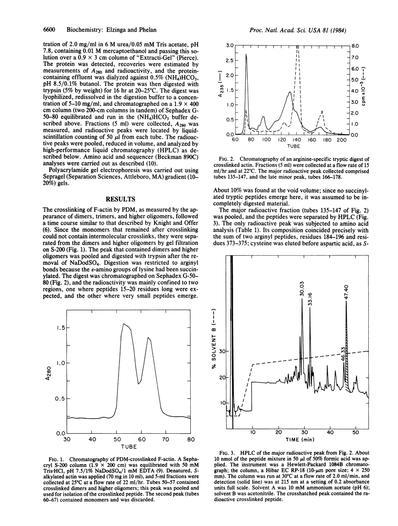
The bifunctional reagent N,N'-p-phenylenedimaleimide (PDM) is being used in an attempt to measure distances between specific side chains in adjacent monomers within F-actin. [14C]PDM was synthesized and was used to crosslink F-actin. Uncrosslinked actin was removed by gel filtration, and, from an arginine-specific tryptic digest of the covalently crosslinked dimers and higher oligomers, one radioactive crosslinked peptide was obtained in high yield. Amino acid composition and sequence analysis indicated that it comprises residues 184-196 of one monomer and 373-375 of an adjacent actin molecule, bridged by PDM through Cys-374 and Lys-191. Thus, these groups are shown to be 1.2-1.4 nm apart in adjacent actin monomers in F-actin. This information may be crucial in establishing the orientation of actin monomers within F-actin.
Full text
PDF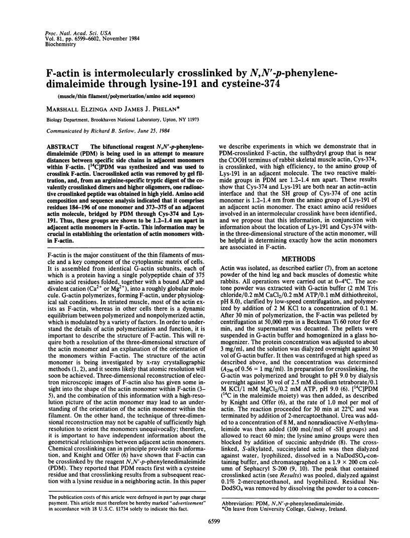

Selected References
These references are in PubMed. This may not be the complete list of references from this article.
- Aebi U., Smith P. R., Isenberg G., Pollard T. D. Structure of crystalline actin sheets. Nature. 1980 Nov 20;288(5788):296–298. doi: 10.1038/288296a0. [DOI] [PubMed] [Google Scholar]
- Collins J. H., Elzinga M. The primary structure of actin from rabbit skeletal muscle. Completion and analysis of the amino acid sequence. J Biol Chem. 1975 Aug 10;250(15):5915–5920. [PubMed] [Google Scholar]
- Elzinga M. Amino acid sequence studies on rabbit skeletal muscle actin. Cyanogen bromide cleavage of the protein and determination of the sequences of seven of the resulting peptides. Biochemistry. 1970 Mar 17;9(6):1365–1374. doi: 10.1021/bi00808a010. [DOI] [PubMed] [Google Scholar]
- Elzinga M., Collins J. H. The primary structure of actin from rabbit skeletal muscle. Five cyanogen bromide peptides, including the NH2 and COOH termini. J Biol Chem. 1975 Aug 10;250(15):5897–5905. [PubMed] [Google Scholar]
- Hartman F. C., Wold F. Cross-linking of bovine pancreatic ribonuclease A with dimethyl adipimidate. Biochemistry. 1967 Aug;6(8):2439–2448. doi: 10.1021/bi00860a021. [DOI] [PubMed] [Google Scholar]
- Hitchcock-De Gregori S. E., Mandala S., Sachs G. A. Changes in actin lysine reactivities during polymerization detected using a competitive labeling method. J Biol Chem. 1982 Nov 10;257(21):12573–12580. [PubMed] [Google Scholar]
- Kartha G., Bello J., Harker D. Tertiary structure of ribonuclease. Nature. 1967 Mar 4;213(5079):862–865. doi: 10.1038/213862a0. [DOI] [PubMed] [Google Scholar]
- Knight P., Offer G. p-NN'-phenylenebismaleimide, a specific cross-linking agent for F-actin. Biochem J. 1978 Dec 1;175(3):1023–1032. doi: 10.1042/bj1751023. [DOI] [PMC free article] [PubMed] [Google Scholar]
- Lu R. C., Szilagyi L. Change of reactivity of lysine residues upon actin polymerization. Biochemistry. 1981 Sep 29;20(20):5914–5919. doi: 10.1021/bi00523a040. [DOI] [PubMed] [Google Scholar]
- Mockrin S. C., Korn E. D. Isolation and characterization of covalently cross-linked actin dimer. J Biol Chem. 1981 Aug 10;256(15):8228–8233. [PubMed] [Google Scholar]
- Moore P. B., Huxley H. E., DeRosier D. J. Three-dimensional reconstruction of F-actin, thin filaments and decorated thin filaments. J Mol Biol. 1970 Jun 14;50(2):279–295. doi: 10.1016/0022-2836(70)90192-0. [DOI] [PubMed] [Google Scholar]
- Mornet D., Bertrand R., Pantel P., Audemard E., Kassab R. Structure of the actin-myosin interface. Nature. 1981 Jul 23;292(5821):301–306. doi: 10.1038/292301a0. [DOI] [PubMed] [Google Scholar]
- Mornet D., Pantel P., Bertrand R., Audemard E., Kassab R. Isolation and characterization of the trypsin-modified myosin -S1 derivatives. FEBS Lett. 1981 Jan 12;123(1):54–58. doi: 10.1016/0014-5793(81)80018-x. [DOI] [PubMed] [Google Scholar]
- Sakabe N., Sakabe K., Sasaki K., Kondo H., Ema T., Kamiya N., Matsushima M. Crystallographic studies of the chicken gizzard G-actin X DNase I complex at 5A resolution. J Biochem. 1983 Jan;93(1):299–302. doi: 10.1093/oxfordjournals.jbchem.a134168. [DOI] [PubMed] [Google Scholar]
- Suck D., Kabsch W., Mannherz H. G. Three-dimensional structure of the complex of skeletal muscle actin and bovine pancreatic DNAse I at 6-A resolution. Proc Natl Acad Sci U S A. 1981 Jul;78(7):4319–4323. doi: 10.1073/pnas.78.7.4319. [DOI] [PMC free article] [PubMed] [Google Scholar]
- Sutoh K. Actin-actin and actin-deoxyribonuclease I contact sites in the actin sequence. Biochemistry. 1984 Apr 24;23(9):1942–1946. doi: 10.1021/bi00304a009. [DOI] [PubMed] [Google Scholar]
- Sutoh K. Identification of myosin-binding sites on the actin sequence. Biochemistry. 1982 Jul 20;21(15):3654–3661. doi: 10.1021/bi00258a020. [DOI] [PubMed] [Google Scholar]
- Tong S. W., Elzinga M. The sequence of the NH2-terminal 204-residue fragment of the heavy chain of rabbit skeletal muscle myosin. J Biol Chem. 1983 Nov 10;258(21):13100–13110. [PubMed] [Google Scholar]
- Wakabayashi T., Huxley H. E., Amos L. A., Klug A. Three-dimensional image reconstruction of actin-tropomyosin complex and actin-tropomyosin-troponin T-troponin I complex. J Mol Biol. 1975 Apr 25;93(4):477–497. doi: 10.1016/0022-2836(75)90241-7. [DOI] [PubMed] [Google Scholar]