Abstract
A 29-year-old primigravida was admitted to the urology ward with acute urinary retention. The patient underwent cystoscopy, MRI (magnetic resonance imaging), and tissue biopsy, which consequently led to the diagnosis of bladder neck leiomyoma that obstructed urine outflow. Subsequent to a cesarean section, a successful transurethral resection was performed. Here the diagnostic complexity in the pregnant patient, clinical course, and outcome are described. One year after successful treatment both mother and daughter are in good condition.
Keywords: bladder tumor, bladder leiomyoma, pregnancy
INTRODUCTION
Benign bladder tumors are rare entities, especially in pregnant patients. Only several cases were described so far. Diagnostic means are limited in such situations as are treatment options.
CASE REPORT
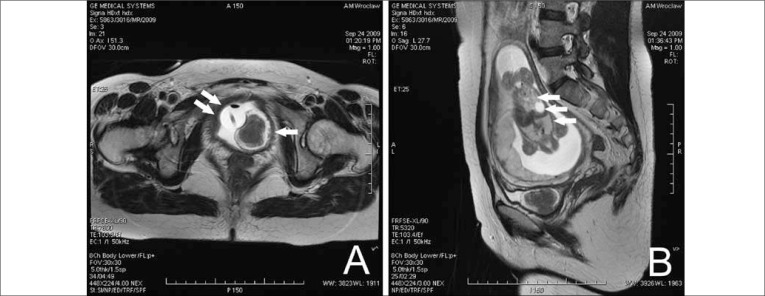
A 29-year-old primigravida, 18-weeks pregnant, was experiencing urinary retention. The first voiding difficulties appeared in the 9th gestational week. There were no urological complaints before pregnancy. Ultrasonography (USG) of the bladder neck revealed a well circumscribed, 4.5-centimeter exophytic lesion with rich flow in Doppler mode. The patient was catheterized then a cystoscopy (Fig. 1a) and MRI (Fig. 2a,b) followed. Biochemical investigations did not reveal any significant abnormalities. Neoplastic markers, CA125, CA19-9, CA15.3, and AFP, were within normal ranges. The specimen from the transvaginal biopsy revealed smooth muscle tissue indicating a bladder neck leiomyoma. In the 36th week, a healthy girl was delivered via c-section. Some weeks later, during the transurethral resection (TUR), after resecting the main body of the leiomyoma a superficial cut (less than the resectoscope's loop depth) into the neck was performed to excise as much of the leiomyoma's pedicle as possible without damaging the sphincter (Fig. 1b). Postoperative histopathology confirmed the preoperational diagnosis. Subsequently, the patient voided without difficulties and had no urine leakage. Cystoscopy (Fig. 1c) and MRI showed no regrowth after six months. One year after cesarean delivery and simultaneous excision of two small uterine leiomyomas both mother and daughter are well.
Fig. 1.
Cystoscopy A – a knob-shaped bladder neck tumor covered with healthy mucosa. The “head” covers the whole bladder neck surface stopping urine outflow in a flap-like manner and the “leg” (arrow) protrudes from the bladder neck from 2 to 5-o'clock. Cystoscopy B – during TUR, note the yellow-whitish color of the tissue. Cystoscopy C – control cystoscopy after six months.
Fig. 2.
T2-weighted MRI image of bladder tumor (arrow), A – axial and B – sagittal planes; the pedicle invades the bladder neck without crossing its whole thickness. The Foley catheter (double arrow) and fetus (triple arrow) can also be seen.
DISCUSSION
Benign bladder and urethral tumors are rare in pregnant women [1, 2, 3]. The etiology of bladder leiomyomas in pregnant women is not clear. Several hypotheses such as inflammatory metaplasia and embryonic residual tumors have been proposed. Hormonally stimulated growth has also been suspected since women in childbearing age are predominant in this group of patients [1]. Conservative treatment is the method of choice after demonstrating benign histology. Although some have been described, the safety of TUR, even bipolar TUR, in pregnancy has not been confirmed [4]. Prolonged catheterization carries the risk of bacterial infection. Repeated urine cultures allow for early detection of dangerous flora. The tumor can be safely monitored with USG and MRI [5]. The patient should be informed about the elevated risk of early termination of pregnancy.
REFERENCES
- 1.Mizuno K, Sasaki S, Tozawa K, et al. Leiomyoma of the urinary bladder during pregnancy. Int J Urol. 2003;10:407–409. doi: 10.1046/j.1442-2042.2003.00641.x. [DOI] [PubMed] [Google Scholar]
- 2.Alvarado-Cabrero I, Candanedo-Gonzalez F, Sosa-Romero A. Leiomyoma of the urethra in a Mexican woman: a rare neoplasm associated with the expression of estrogen receptors by immunohistochemistry. Arch Med Res. 2001;32:88–90. doi: 10.1016/s0188-4409(00)00239-3. [DOI] [PubMed] [Google Scholar]
- 3.Gaynor-Krupnick DM, Kreder KJ. Bladder neck leiomyoma presenting as voiding dysfunction. J Urol. 2004;172:249–250. doi: 10.1097/01.ju.0000129009.94436.ca. [DOI] [PubMed] [Google Scholar]
- 4.Castrillo AH, Pena AV, de Diego Rodriguez E, et al. Hematuria durante de la gestación debida a tumor vesical. Presentación de 2 casos. Actas Urol Esp. 2005;29:981–984. doi: 10.1016/s0210-4806(05)73381-7. [DOI] [PubMed] [Google Scholar]
- 5.Loughlin KR. Urologic radiology during pregnancy. Urol Clin North Am. 2007;34:23–26. doi: 10.1016/j.ucl.2006.10.009. [DOI] [PubMed] [Google Scholar]


