Abstract
We shall discuss the case of a female patient, aged 64 years, who was suffering from long–term purulent inflammation of the vaginal fornix that later involved the vaginal stump. This inflammatory process spread to the bladder trigone and resulted in vesicovaginal fistula (VVF) formation together with a bilateral hydronephrosis that required the placement of a temporary percutaneous nephrostomy. A non–cicatrized inflammatory reaction occurred at the right–sided insertion of the nephrostomy, which has yet to be successfully treated despite intensive dermatological and surgical approaches that included skin grafting. In the course of five–year treatment we observed a gradual regression of the inflammatory infiltration of both the trigone of the bladder and the vagina as well as a gradual closing of the VVF. The extremely long–lasting and uncommon local inflammatory reactions in the vagina, bladder, and dermal layers mandated the application of conservative treatment. The possibility of difficulties and defective healing of tissues that could result from surgical correction of the VVF are discouraging for both the patient and medical staff.
Keywords: urinary bladder, vesicovaginal fistula (VVF), vagina purulent inflammation of vaginal fornix
CASE REPORT
The patient, aged 64, underwent surgery in 1989 in the regional hospital. The surgery entailed removing the uterine body and the left appendages due to myoma. In September 2003, the patient was admitted to the Department of Gynecology at the Medical University of Lublin due to genital bleeding combined with cervicitis and vaginitis (for a 1 year). After several days of antibiotic treatment, the uterine cervix and the right appendages were removed. The post–surgical analysis revealed no complications. The histopathological examination revealed erosion and a bloated atypical partly purulent inflammatory process with an extended necrosis of the endocervical mucosa.
In December 2003, the patient was admitted to the Department of Gynecology, due to purulent inflammation of the vaginal stump, which was treated conservatively. Histopathological analysis of vaginal samples revealed ulceration and chronic inflammation. The urine culture for TB gave negative results, but a CT of the pelvis revealed inflammatory infiltration in the vaginal stump with possible metastasis to the rectum. Examination of the vaginal excretion showed the presence of microorganisms from the Enterococcus group as well as Gram–negative bacteria.
In September 2004, a diagnostic laparotomy was performed in the Department of Gynecology, because of the continually defective healing of the vaginal stump (surgery showed that the organs of the lesser pelvis were normal). However, the laparotomy caused atony of the urinary bladder, which it necessary to insert a Foley catheter. After two weeks of treatment with catheter and Ubretid (5 mg x day), the action of the urinary bladder was restored. The cystoscopy showed no changes.
In March 2005, the patient was admitted to the Department of Urology at the Medical University of Lublin due to LUTS and an early stage of a bilateral hydronephrosis with continued purulent inflammation of the vaginal stump that transposed into the corpus of the urinary bladder. The examination revealed a nodular inflammatory infiltration in the trigone and the vesical cervix. Due to a suspected bladder tumor, tissue samples were collected and reported a bloated and chronic inflammatory processes involving nonspecific granulation.
In May 2005, the patient was again admitted to the Department of Urology due to a bilateral hydronephrosis. The CT performed showed an inflammatory infiltration in the bladder wall and in the posterior bladder area and consequently bilateral percutaneous nephrostomies were performed. In July 2005, bilateral DJ catheters were inserted. In the trigone of the bladder a VVF with a diameter of approx. 1.5 cm was diagnosed, which resulted from the chronic inflammation process in the vaginal stump. The patient was discharged home with the Foley catheter.
In September 2005, the DJ catheters were removed and anti–inflammatory treatment was applied (Doxycycline and Urosept). In November 2005, the cystoscopy performed revealed closure of the vesicovaginal fistula. Conservative treatment continued.
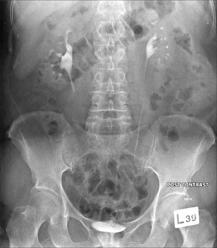
In May 2006, in cystoscopy, an inflammatory change was observed in the area of the left ureter orifice (the macroscopic image triggered suspicions of a tumor that was resected). A histopathological examination revealed a bloated chronic/acute inflammatory process involving granulation, as well as an unspecific inflammatory infiltration comprising lymphocytes, plasmacytes, and both acidophilic and neutrophilic granulocytes. It was also observed that the inflammatory reaction in the vaginal stump had intensified, and so had the inflammatory reaction with dermal necrosis in the area of the former right–sided nephrostomy. A urography showed no abnormality (Fig. 1).
Figure 1.
Intravenous urography.
In November 2006, a slight VVF was identified in the trigone of the bladder. The urethra was calibrated and the Foley catheter was placed once again.
The continual inflammation in the right lumbar area was treated conservatively for one year. This treatment proved unsuccessful despite the surgical wound preparation and skin grafting.
In May 2007, the patient was treated in the Department of Dermatology at the Medical University in Lublin, which involved collecting skin samples that revealed nonspecific inflammatory infiltration with ulceration (unknown etiology).
The examinations performed in October 2007 in the Department of Urology revealed a narrowed inflexible urethra and stress urinary incontinence (SUI). The last medical follow up in November 2010 revealed a limited capacity of the urinary bladder, SUI, and a narrowed inflexible urethra together with an average inflammatory vaginal reaction and fistula with a diameter of 1–2 mm in the trigone.
DISCUSSION
We have focused on this case due to its atypical course, as well as to the difficulties we encountered while diagnosing and treating the patient. We performed a full set of image examinations, such as ultrasonography, CT scan, and urography. The urinary tract condition was assessed several times with cystoscopy. Such analyses as urinary culture and vaginal swab (including for TB) were performed. Each histopathological examination revealed a nonspecific chronic inflammatory reaction, whereas the urinary culture and vaginal swabs indicated changing bacterial floras (Enterococcus sp, Gram positive cocci, Staphylococcus aureus, and Streptococcus pyogenes) with 50% of the cultures performed being negative.
The one–year gynecological treatment, comprising eight hospital stays and two surgeries, combined with the general and local anti–inflammatory therapy based on the urinary culture results proved ineffective. The vaginal inflammatory process additionally expanded to the bladder and urethra.
The four–year therapy, generally recommended in treating vesicovaginal fistulas and bladder inflammation, comprising nine diagnostic and treatment stays in the Department of Urology, as well as regular checks in the Urological Out–Patient Department, prevented the further development of the disease [1]. However, its outcomes, such as fibrotic changes in the urinary bladder and urethra, SUI, a persistent micro–fistula, and dermal inflammation in the post–nephrostomy area, have notably lowered the patient's living comfort.
The case analyzed poses a significant dilemma for the medical staff as to whether they should apply surgical treatment or whether such treatment would lead to intensified inflammatory reactions in the operated area, as was the case with previous gynecological surgeries. Infectious complications still remain the challenge for gynecological surgery [2].
Inflammatory infiltration of the urinary bladder, resulting from a long–term purulent inflammation of the vaginal stump that is accompanied by a VVF and a bilateral hydronephrosis arising from infiltration on the ureter orifice, is an uncommon disease of the lower urinary tract. The authors did not find a similar case in the literature. It is known that approx. 75% of all VVF cases result from previous gynecological surgery [3]. Other reasons for such fistulas are radiotherapy, advanced cancer processes in the lesser pelvis, tuberculosis, and genital injuries connected with childbirth or sexual activity [4].
All these reasons were excluded in the analyzed case. The patient underwent gynecological surgeries without any iatrogenic injuries. The VVF originated from the local chronic inflammatory condition of the vaginal stump, which occurred 10 months after the previous gynecological surgery.
The spontaneous closure of VVF without surgical intervention has already been encountered in clinical practice [5]. In this case, it took six months until the fistula was reduced from 1.5 cm to 1–2 mm. The history of the surgical treatment of vesicovaginal fistulas has always recognized the problem of timing the reconstruction surgery–based treatment [6]. It has been assumed that under proper healing conditions, the fistula surgery should not be performed until the fistula edges have healed, which usually takes place within three to six months. In the case analyzed, since diagnosis, i.e. for the last three years, no conditions have occurred that would allow the performance of surgical treatment for the micro–fistula and SUI. Taking into account the distorted healing processes, the authors are afraid that the reconstructive surgery may not bring satisfactory effects.
References
- 1.Rovner ES. Urinary tract fistula. In: Walsh PC, editor. Cambell's Urology. ed 9. Philadelphia: Saunders; 2007. pp. 2232–2360. [Google Scholar]
- 2.Faro S, Phillips LE, Martens MG. Perspectives on the bacteriology of postoperative obstetric–gynecologic infections. Am J Obstet Gynecol. 1988;158:694–700. doi: 10.1016/s0002-9378(16)44529-1. [DOI] [PubMed] [Google Scholar]
- 3.Lee RA, Symmonds RE, Williams TJ. Current status of genitourinary fistula. Obstet Gynecol. 1988;72:313–319. [PubMed] [Google Scholar]
- 4.Gerber GS, Schoenberg HW. Female urinary tract fistulas. J Urol. 1993;149:229–236. doi: 10.1016/s0022-5347(17)36045-7. [DOI] [PubMed] [Google Scholar]
- 5.Bazi T. Spontaneous closure of vesicovaginal fistulas after bladder drainage alone: review of the evidence. Int Urogynecol J Pelvic Floor Dysfunct. 2007;18:329–333. doi: 10.1007/s00192-006-0194-7. [DOI] [PubMed] [Google Scholar]
- 6.Blaivas JG, Heritz DM, Romanzi LJ. Early versus late repair of vesicovaginal fistulas: Vaginal and abdominal approaches. J Urol. 1995;153:1110–1113. [PubMed] [Google Scholar]


