Abstract
IMPORTANCE
We previously reported a 90% decreased risk in major depression, assessed prospectively, in adult offspring of depressed probands who reported that religion or spirituality was highly important to them. Frequency of church attendance was not significantly related to depression risk. Our previous brain imaging findings in adult offspring in these high-risk families also revealed large expanses of cortical thinning across the lateral surface of the right cerebral hemisphere.
OBJECTIVE
To determine whether high-risk adults who reported high importance of religion or spirituality had thicker cortices than those who reported moderate or low importance of religion or spirituality and whether this effect varied by family risk status.
DESIGN, SETTING, AND PARTICIPANTS
Longitudinal, retrospective cohort, familial study of 103 adults (aged 18–54 years) who were the second- or third-generation offspring of depressed (high familial risk) or nondepressed (low familiar risk) probands (first generation). Religious or spiritual importance and church attendance were assessed at 2 time points during 5 years, and cortical thickness was measured on anatomical images of the brain acquired with magnetic resonance imaging at the second time point.
MAIN OUTCOMES AND MEASURES
Cortical thickness in the parietal regions by risk status.
RESULTS
Importance of religion or spirituality, but not frequency of attendance, was associated with thicker cortices in the left and right parietal and occipital regions, the mesial frontal lobe of the right hemisphere, and the cuneus and precuneus in the left hemisphere, independent of familial risk. In addition, the effects of importance on cortical thickness were significantly stronger in the high-risk than in the low-risk group, particularly along the mesial wall of the left hemisphere, in the same region where we previously reported a significant thinner cortex associated with a familial risk of developing depressive illness. We note that these findings are correlational and therefore do not prove a causal association between importance and cortical thickness.
CONCLUSIONS AND RELEVANCE
A thicker cortex associated with a high importance of religion or spirituality may confer resilience to the development of depressive illness in individuals at high familial risk for major depression, possibly by expanding a cortical reserve that counters to some extent the vulnerability that cortical thinning poses for developing familial depressive illness.
We previously reported a 90% decreased risk, assessed prospectively for 10 years, of developing major depressive disorder (MDD) in adult offspring of depressed probands (high familial risk [HR]) who said that religion or spirituality was highly important to them.1 Attendance at religious services and religious denomination did not decrease the risk of MDD. Among the same participants in our 25-year, longitudinal, multigenerational study of MDD who underwent magnetic resonance imaging (MRI),we identified large expanses of cortical thinning across the lateral surface of the right cerebral hemisphere and mesial wall of the left hemisphere in adult offspring of the HR group.2 These findings led us to explore whether the regions where cortical thinning was located in the HR adults would be thicker in those who report a high personal importance of religion or spirituality and whether these findings would be significantly more prominent in persons at HR compared with low familial risk (LR) for MDD. A relatively thicker cortex in these regions could potentially account for the protection against depression that religion or spirituality seem to afford. (For ease of reading, we will refer to the personal importance of religion or spirituality simply as importance.)
Numerous studies have found an inverse association between religiosity and depression, and additional studies have attempted to identify a neurobiological basis for religious and spiritual experiences.3–9 In healthy individuals, for example, transcranial magnetic stimulation of the temporoparietal regions evoked feelings of sensed presence.10 A study11 on older adults using structural MRI prospectively associated born-again status, life-changing religious experiences, and Catholicism with subsequent greater atrophy in the hippocampus. Several functional neuroimaging studies2,12–15 of healthy adults using functional MRI and single-photon emission computed tomography revealed that the intensity of self-evoked religious experiences during MRI was associated with increased blood flow in various subregions of the prefrontal and parietal cortices. These neurobiological correlates of religious and spiritual experiences, however, have yet to be investigated in terms of the risk and protective benefits that they confer for MDD.
In the present study, we followed adults for more than 30 years adults were at either HR or LR for MDD, during which time the participants self-reported importance and frequency of attendance at services and were assessed for symptoms of depression. We assessed the associations of importance with measures of cortical thickness measured on MRIs of the brain acquired at the 25-year follow-up. In addition to reporting thinner cortices in HR adults that averaged nearly 30%across the lateral surface of the right hemisphere and mesial wall of the left, we also previously reported that thinner cortices in the HR group were state independent (ie, a thinner cortex was independent of whether participants were ever depressed and therefore was likely an endophenotype for MDD) and that the cortical thickness mediated the associations of familial risk for MDD with inattention and difficulty recalling social stimuli, cognitive disturbances that in turn were associated with increased symptoms of anxiety and depression.2,15
We therefore hypothesized that adults with self-reported high importance compared with those with low or moderate importance would have thicker cortices in brain regions, which was previously identified as an endophenotype for familial MDD. Because we have previously found that the effects of religious importance in protecting against MDD are greater in HR compared with LR adults, we further hypothesized that the HR compared with the LR participants would have larger expanses of the brain in which cortical thickness correlated positively with religious importance.
Methods
Participants
The 103 adults (mean [SD] age at MRI,37.4 [9.5]years; age range, 18–54 years; 40 men) in this study are part of an ongoing 3-generation study and have been followed up for more than 30 years.16,17 Sample characteristics, assessed when the MRIs were performed, were as follows: 61.2% were women, approximately half (47.6%) were married, 36.9% were single, 13.6% were separated or divorced, and 1.9% did not report marital status. Most (57.3%) had greater than a high school education, 37.9%had a high school diploma or general educational development certificate, 3.9%had not graduated high school, and 1.0% did not report educational attainment. Annual income was below $20 000 for 25.2% of the sample, between $20 000 and $39 999 for 25.2%, and above $40 000 for 40.8%, with 8.7% not reporting annual income. The distribution of religious denomination was as follows: 48.5% were Catholic, 8.7% were Protestant, 5.8%were Jewish, 23.3% espoused personal religious or spiritual beliefs not affiliated with any institutionalized religion, and 13.6% gave another response or no response. There were no differences in demographics by risk groups at 20 years (eTable 1 in the Supplement).
The first generation (G1, or the probands) consisted of adults with no current or past history of depression (LR) or who were clinically identified as having current or past (HR)MDD. Their biological offspring composed the second generation (G2), and their grandchildren composed the third generation (G3).We defined the familial risk for depression of G2 and G3 members based on the clinical diagnosis of probands. The 67 adult offspring of G1 with MDD were defined as being at HR for depression, and the 36 adult offspring of healthy G1 members were defined as being at LR for depression. The participants were assessed at baseline and then 2, 10, 20, and 25 years later (T2, T10, T20, and T25, respectively). A 30-year follow-up is underway. Assessments of religiosity at T20 and T25 were used to be closer to the time of the MRI evaluation, which was at T25. In the clinical study described previously,1 we reported on religiosity at T10 and T20. A detailed description of the study, especially the participant ascertainment and diagnostic assessment, has been previously published.16,17 The diagnostic interviews across all waves were conducted using the Schedule for Affective Disorders and Schizophrenia–Lifetime Version for adults.18,19 To determine current clinical symptoms at the time of the scan, the Hamilton depression and anxiety rating scales were used.20 The diagnoses and religious variables were assessed by investigators masked to familial risk status and MRI results.
Anatomical MRIs of the brain were acquired at T25.Acomplete description of participant ascertainment into the MRI portion of the overall longitudinal study has been published elsewhere.2 Of relevance to the present study is that of the 131 participants in the previously published study identifying an endophenotype for depression, 103 met criteria for this study by being older than 18 years, the age at which the questions on religion were administered as self-report rather than as parental report. Of these 103 participants, 85 participants were G2 and 18 participants were G3.
Religion and Spirituality Measures
The questions on religiosity were determined at T20 and T25 by asking the following 3 questions, which traditionally have been the most frequently used in research on religion and health6,7: (1) “How important to you is religion or spirituality?” with a response ranging from1 (not important at all) to 4 (highly important); (2) “How often, if at all, do you attend church, synagogue, or other religious or spiritual services?” with a response ranging 4 categories from never to once a week or more; and (3) “How would you describe your current religious beliefs? Is there a particular denomination or religious organization that you are part of?” with 10 denominations specified, including an opportunity to specify others. Level of personal importance and frequency of attendance across studies tend to correlate moderately,6,7 with a Spearman rank correlation coefficient of0.59 (N = 87) at T20 in the current sample. The direction of response on the frequently used single-item question for importance correlates well with direction of response on the Brief Multidimensional Measure of Religiousness/Spirituality (originally developed by the Fetzer Institute) on the 12 items that represent relational spirituality, the dimensions of daily spiritual experience, forgiveness, and positive religious coping (Pearson r = 0.70).21,22 The responses to the importance and the frequency of attendance were independently dichotomized as either high (≥3) or low importance and either high or low attendance, respectively. At T20, a total of 22 participants reported high importance and 66 reported low importance. At T25, a total of 25 participants reported high importance, and 78 reported low importance. Level of personal importance of religion or spirituality at both T20 and T25 did not differ by risk group. Frequent attendance of religious service did not differ by risk group at T20, but at T25 a higher percentage of participants attended at least once a month in the LR group (eTable 2 in the Supplement).
Participants designated as stable high were those who reported high importance at both T20 and T25 (12 participants); stable low, those who reported low importance at both times (53 adults); or unstable, those who reported high importance at one time and low importance at the other time point (17 participants). These 3 groups defined by stability of high importance did not differ significantly in demographic characteristics (eTable 3 in the Supplement).With respect to attendance of religious services, at T20, frequent attendance was reported by 10 of 12 adults (83.3%) in the high stable group, 16 of 56 adults (28.6%) in the low stable group, and 12 of 19 adults (63.2%) in the unstable group (, P < .001).Means of raw data scores on personal importance and frequency of attendance by the 3 groups are displayed longitudinally at T10 (initial time that all religion questions were included in the study), T20, and T25 (eFigure 1 and eFigure 2 in the Supplement).
MRI Pulse Sequence
The anatomical MRI data were acquired using a Sonata 1.5-T scanner (Siemens AG). A 3-dimensional magnetization-prepared rapid acquisition with gradient-echo sequence (repetition time, 24 milliseconds; echo time, 2.96 milliseconds;45° flip; 256 × 192 matrix; field of vision, 30 cm; phase field of view, 100%; 2 excitations; slice thickness, 1.2 mm; and 124 contiguous slices) provided nearly isotropic voxels (1.17 × 1.17 × 1.2 mm) for all participants.
Image Preprocessing
Nonuniformities in image intensities were corrected using an automated program,23 and the brain was isolated from non-brain tissue using semiautomated procedures. Brain tissue was segmented as either gray matter or white matter using protocols detailed elsewhere.2 We used a 2-step procedure to select as a template a single brain that was morphologically representative of the brains of the LR adults (eMethods in the Supplement).
Cortical Thickness Analyses and Surface Morphometry
Each brain was first coregistered to the template brain using a similarity transformation,24 and then a high-dimensional, nonlinear warping algorithm25 was applied to establish point-by-point correspondence across the surface of the template and each brain. The nonlinear warping then was reversed to bring each structure into the previous similarity registration with the template structure, thereby establishing point-by-point correspondence across the surface of the 2 brains. For each participant, cortical thickness was measured in template space using a 3-dimensional morphologic operator and mapped onto the surface of the template brain (eMethods in the Supplement), thereby permitting statistical analyses of cortical thickness measures at each point across the participants in our study. Use of a single brain as the template rather than a unique template brain for each group permitted us to compare cortical thicknesses between groups and to correlate thickness with measures of religious importance and ratings of anxiety or depression. We correlated cortical thickness with the importance and the frequency of church attendance while controlling for age, sex, and familial risk for MDD (eMethods in the Supplement).
Statistical Analysis
First, we studied the effects of importance at T25 on brain measures and assessed whether these effects differed across the HR and LR groups. Second, we studied the effects on our brain measures of stable high vs stable low importance for 5 years (ie, from T20 to T25).We assessed independently the effects of importance and the frequency of attendance at religious services on brain measures using product-moment correlations because their ordinal measures were significantly intercorrelated at both time points (at T20: r = 0.58; at T25: r = 0.50; both P < .001). Finally, we correlated brain measures with normalized measures (z scores) for anxiety and/or depression. The z score for current depressive symptoms for each participant was summed and was subsequently correlated with brain measures. We controlled for familial risk for MDD in all our statistical analyses to identify the neuroanatomical correlates of importance and the interactions of importance with familial risk that are independent of familial risk. In addition, all statistics controlled for age and sex of the participants. We, however, did not explicitly include whole-brain volume or intracranial volume as covariates in our statistical models because cortical thickness was scaled by brain size when the participant brains were transformed into the coordinate space of the template brain.
In testing our a priori hypotheses in which we associated cortical thickness with the importance at T25, stable importance during a 5-year period, and frequency of church attendance, we corrected for multiple testing of statistical hypotheses at each point across the surface of the brain using gaussian random fields (GRFs).26 The GRF-based correction is conservative and minimizes type I errors when testing multiple hypotheses, but it also increases the likelihood of type II error (ie, increases the number of false-negative findings). Therefore, in our exploratory analyses in which we assessed the interaction of risk by importance, whereby stratifying individuals by familial risk decreased our statistical power, we applied the less conservative False Discovery Rate corrected for multiple comparisons to minimize type II errors while permitting a prespecified rate (rate of 0.05) of false-positive findings.
Results
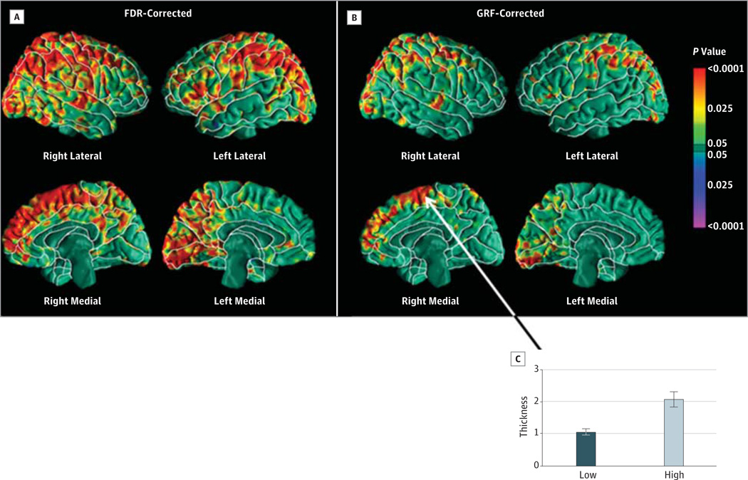
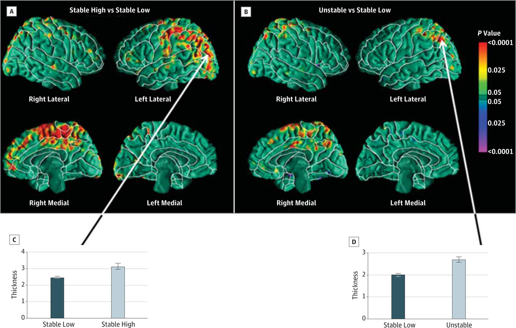
Adults who reported high importance at T25 had thicker cortices in the left and right parietal and occipital regions, the mesial frontal lobe of the right hemisphere, and the cuneus and precuneus in the left hemisphere (Figure 1). Adults who reported stable high importance had thicker cortices in the same regions, independent of familial risk (Figure 2), showing the association of sustained high importance over 5 years on cortical thickness. In addition, adults who had high importance only at 1 of the 2 time points also had thicker cortices across large expanses of both hemispheres than those who had moderate to low importance at both time points (eFigure 4 in the Supplement), although participants with high importance at T25 (time of scan) had significant associations across much larger expanses of the cortical surface. Controlling for education increased the expanse of regions with significant associations. The current level of depressive symptoms, measured using Hamilton Depression Rating Scale scores at the time of MRI, did not alter the association between cortical thickness and importance. Although persons who reported frequent attendance at religious services had thicker cortices in the same regions as the adults with stable high importance, the associations were not statistically significant after controlling for multiple comparisons (eFigure 5 in the Supplement), which is consistent with our previous finding that, when controlling for importance, attendance at religious services was not significantly associated with increased risk for depression.1
Figure 1. Associations of Religious and Spiritual Importance With Cortical Thickness.
Methods for false discovery rate (FDR) (A) and gaussian random field (GRF) (B) were applied to correct for the multiple comparisons. The GRF method is more conservative but has lower statistical power than the FDR method; therefore, the GRF-corrected maps have fewer significant findings. The corrected P values were color encoded and displayed on the surface. The color bar shows the color encoding for the P values. Red and yellow indicate positive correlations, and violet and blue indicate negative correlations between the measures of cortical thickness and importance of religion. C, The plot of the mean thicknesses and SEs (error bars) for a region in the mesial right hemisphere showed that the mean cortex was 1-mm thicker in the individuals with high compared with low importance for religion. These maps show that the higher importance for religion was associated with thicker cortical mantle bilaterally across the occipital cortex and superior parietal lobe, in the mesial right hemisphere, and in the precuneus and cuneus of the left hemisphere.
Figure 2. The Effects of Stable Importance During a 5-Year Period.
A and B, Although the participants with either stable high or unstable importance for religion had thicker cortical mantle in the left posterior parietal and occipital cortex and mesial right hemisphere, the expanse of regions with a thicker cortex was smaller in the participants with unstable than those with stable high importance. C and D, The plots of the mean thickness showed that the cortex was approximately 0.69-mm thicker in the occipital lobe for the stable high participants and in the posterior parietal lobe for the unstable participants compared with participants with stable low importance for religion.
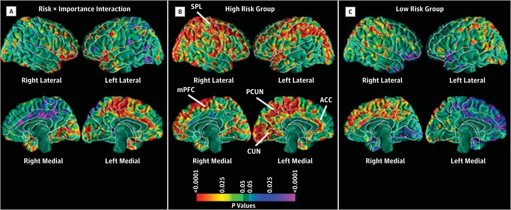
Exploratory analyses indicated that the effects of importance were significantly stronger in the HR than in the LR group, particularly in the mesial wall of the left hemisphere (Figure 3), in the same region where we previously reported a thinner cortex to be significantly associated with a familial risk for developing MDD.2 Symptom severity at the time of MRI correlated inversely with cortical thickness in the parietal lobes bilaterally in the HR group more prominently than in the LR group, suggesting that thicker cortices in these regions proportionately protected against depressive symptoms (eFigure 6 in the Supplement).
Figure 3. Differing Effects of Religious and Spiritual Importance in the High-Compared With Low-Risk Group.
The interaction map of familial risk by religious importance at 25 years (T25) shows differing effects of religious importance on cortical thickness in the high-compared with low-risk group across the orbital frontal cortex and the mesial left and right hemispheres. In the 67 high-risk adults, bilateral increases in cortical thickness across the entire brain were associated with higher importance for religion at T25. In the 36 low-risk adults, thinning in the orbital frontal cortex and mesial left hemisphere and thickening in the right mesial hemisphere were associated with high importance for religion. We applied the false discovery rate to correct for multiple comparisons. P values were color encoded and plotted on the surface of the brain. ACC indicates anterior cingulate cortex; CUN, cuneus; mPFC, mesial prefrontal cortex; PCUN, precuneus; SPL, superior parietal lobe.
Discussion
Our findings indicate that high importance of religion or spirituality was associated with thicker cortices throughout the superior parietal and occipital lobes of the brain, especially in the brain regions where a thinner cortex was found to be a morphologic endophenotype of familial risk for MDD.2,15 Although a high frequency of attendance of religious services was associated with a high personal importance of religion or spirituality, the association between attendance and cortical thickness was not statistically significant, a contrasting set of findings that warrants comment. Although some individuals may attend services to cultivate personal spirituality, others may attend for other important reasons, whether or not they place a high importance on personal spirituality. In our cohort, 49 participants reported high attendance, yet only 21 of those also reported high importance. The remaining 28 participants may be attending services for a host of reasons, which may include seeking community and social support. In a previous study27 using this sample, we found that the participants who frequently attended religious services were in fact at increased risk for depression after controlling for personal importance, suggesting that a subset of participants may attend religious services for comfort or management of depressive symptoms. Thus, although frequent attendance may express, sustain, and cultivate personal importance, our findings suggest that religious beliefs and experiences, and not overt behavior, are associated with cortical thickness.
The associations between importance and cortical thickness were generally similar in direction and spatial location across the 2 risk groups (Figure 3), suggesting that the morphologic effects of high importance may hold in the general population. However, the association of cortical thickness with importance was strongest and spatially most extensive in HR adults than in LR adults (Figure 3), in whom our previous study1 demonstrated that high importance conferred a 90% reduction in relative risk for depression during a 10-year period. The large magnitude and spatially extensive degree of thinning previously documented in the HR group may have afforded a larger potential effect of religiosity on cortical thickness than in the LR group, where ceiling effects presumably constrained to some extent the morphologic effects of religiosity. Stable reports of high importance during 5 years rather than reporting a high level of spirituality at only one point in time were associated with thicker cortices across a greater spatial extent of the cerebral surface (Figure 2), suggesting that structural differences are associated with a more sustained use of the cortical region over time. A report of high importance contemporaneous with the MRI correlated with cortical thickening more robustly than did a report of high importance 5 years earlier, suggesting the relevance of current importance to cortical thickening and, presumably, the resilience that it confers against depressive illness. Consistent with this interpretation, the severity of depressive symptoms at the time of MRI correlated inversely with cortical thickness in the parietal lobes bilaterally in the HR group more prominently than in the LR group. Because individuals with thicker cortices had less severe depressive symptoms, increasing cortical thickness in these regions likely conferred a proportionate degree of protection against depressive symptoms.
Our findings are consistent with and add to the findings of imaging studies of spiritual and religious experience, as well as findings on the effects of the sustained practice of meditation. Functional neuroimaging studies using functional MRI and single-photon emission computed tomography associated increased blood flow in various subregions of prefrontal and parietal cortices with an increased intensity of religious experience.12–14 Adults who regularly meditated had a thicker prefrontal cortex and right anterior insula28,29 as well as increased functional activity in the prefrontal cortex.30,31 Davidson and McEwen32 identified a neural pathway from attention to social empathy that was associated with sustained meditation and mediated by the activity of the prefrontal cortex. Although these prior studies cannot establish a causal relationship between meditation and morphologic changes in the brain, an additional imaging study33 combined with an experimental intervention using mindfulness meditation suggested that meditation may cause these morphologic changes in the brain. That study found that gray matter density increased in the left hippocampus, posterior cingulate cortex, and temporoparietal junction in 16 healthy, meditation-naive participants who underwent meditation training for 8 weeks. Consistent with the major findings of these prior studies, the locations of the strongest effects of cortical thickening that we observed predominantly support sensory and spatial processing, with the parietal lobe mediating the sense of self, navigation in space, and spatial orientation34,35 and the precuneus supporting reflective self-awareness.36
Taken together with these previous reports, our finding that the cortex was thicker in those with high importance in both risk groups suggests that high importance may be related to structures in the brain that confer a relative resilience against developing depression. The thicker cortices were located in the same general regions as the morphologic endophenotype, conferring familial risk for depression but much more strongly in the mirror-image locations of the risk endophenotype (Figures 1–3) in regions that we previously interpreted as providing cortical reserve that compensated for the presence of cortical thinning within the risk endophenotype. The importance of religion or spirituality therefore likely reinforces persons who are at greater familial and neuroanatomical risk for developing depression against actually becoming ill by providing reserve in the regions within the endophenotype and in the mirror-image locations. The importance of religion or spirituality appears to confer a neuroanatomical resilience in those who are otherwise predisposed to developing depressive illness. The findings should not be interpreted as identifying a single location in the brain that is the site of religious experience or spirituality.
The present study is limited by the collection of MRI data at only one time point, thereby preventing us from assessing a temporal association between importance and cortical thickening. Therefore, we cannot determine whether the high importance altered cortical thickness or whether individuals who had a thicker cortex were disproportionately attracted toward spiritual or religious practice. Nevertheless, we note that the correlation of cortical thickness with importance was stronger for ratings of importance that were contemporaneous with MRI compared with ratings from 5 years earlier, suggesting the presence of a temporal association between importance and cortical morphologic findings. The temporal association between importance and cortical thickness would be more firmly established in a prospective, longitudinal study design, and causality could be more firmly established through imaging before and after a randomized assignment to an intervention that deepens spiritual experience. Nevertheless, the present study is, to our knowledge, the first published report to identify the neural correlates of the highly robust protective effects that high importance confers against developing MDD in adults who are at HR for developing MDD.1 Our findings therefore may identify a neural pathway through which the personal importance of spirituality or religion protects against MDD in people who are otherwise predisposed to developing it. Future studies should assess whether the cortical thickening and greater cortical reserve associated with the importance of religion or spirituality is found in offspring at HR for other disorders and whether it is located in the same regions of the brain as in offspring at HR for MDD.
Supplementary Material
Acknowledgments
During the past 3 years, Dr Miller has received funding from the Templeton Foundation, the Klingenstein Fund, the Pritchard Foundation, and Goldman Sachs Gives and has received payment from Oxford University Press and St. Martin’s Press. Dr Weissman has received funding from the National Institute of Mental Health, the National Institute on Drug Abuse, the Brain and Behavior Research Foundation, the Sackler Foundation, and the Templeton Foundation and receives royalties from Oxford University Press, Perseus Books, American Psychiatric Association Press, and MultiHealth Systems. Dr Peterson has received funding from the National Institute of Mental Health, the National Institute on Drug Abuse, the National Institute of Environmental and Health Science, the Brain and Behavior Research Foundation, the Sackler Foundation, the Simons Foundation, and the Templeton Foundation and holds a patent (No. 8,143,890) entitled “Spectral Resolution Enhancement of Magnetic Resonance Spectroscopic Imaging.”
Funding/Support: These analyses and manuscript preparation were supported by the John Templeton Foundation (Dr Weissman, principal investigator). The data collection was funded by grant 2 R01 MH36197 from the National Institute of Mental Health (Dr Weissman, principal investigator). The study was also supported by grants MH36295 and MH94356 from the National Institutes of Health (Dr Tenke).
Role of the Sponsor: The funding sources had no role in the design and conduct of the study; collection, management, analysis, and interpretation of the data; and preparation, review, or approval of the manuscript; and decision to submit the manuscript for publication.
Footnotes
Author Contributions: Drs Miller and Bansal had full access to all the data in the study and take responsibility for the integrity of the data and the accuracy of the data analysis.
Study concept and design: Miller, Bansal, Wickramaratne, Weissman, Peterson.
Acquisition of data: Weissman, Peterson.
Analysis and interpretation of data: All authors.
Drafting of the manuscript: Miller, Bansal, Tenke, Weissman, Peterson.
Critical revision of the manuscript for important intellectual content: All authors.
Statistical analysis: Bansal, Tenke, Peterson.
Obtained funding: Miller, Weissman, Peterson.
Administrative, technical, and material support: Hao, Tenke, Peterson.
Study supervision: Weissman, Peterson.
Conflict of Interest Disclosures: No other disclosures were reported.
REFERENCES
- 1.Miller L, Wickramaratne P, Gameroff MJ, Sage M, Tenke CE, Weissman MM. Religiosity and major depression in adults at high risk: a ten-year prospective study. Am J Psychiatry. 2012;169(1):89–94. doi: 10.1176/appi.ajp.2011.10121823. [DOI] [PMC free article] [PubMed] [Google Scholar]
- 2.Peterson BS, Warner V, Bansal R, et al. Cortical thinning in persons at increased familial risk for major depression. Proc Natl Acad Sci U S A. 2009;106(15):6273–6278. doi: 10.1073/pnas.0805311106. [DOI] [PMC free article] [PubMed] [Google Scholar]
- 3.Braam AW, Beekman AT, Deeg DJ, Smit JH, van Tilburg W. Religiosity as a protective or prognostic factor of depression in later life; results from a community survey in the Netherlands. Acta Psychiatr Scand. 1997;96(3):199–205. doi: 10.1111/j.1600-0447.1997.tb10152.x. [DOI] [PubMed] [Google Scholar]
- 4.Kendler KS, Liu XQ, Gardner CO, McCullough ME, Larson D, Prescott CA. Dimensions of religiosity and their relationship to lifetime psychiatric and substance use disorders. Am J Psychiatry. 2003;160(3):496–503. doi: 10.1176/appi.ajp.160.3.496. [DOI] [PubMed] [Google Scholar]
- 5.Koenig HG, George LK, Peterson BL. Religiosity and remission of depression in medically ill older patients. Am J Psychiatry. 1998;155(4):536–542. doi: 10.1176/ajp.155.4.536. [DOI] [PubMed] [Google Scholar]
- 6.Koenig HG, King DE, Carson VB. Handbook of Religion and Health. 2nd ed. New York, NY: Oxford University Press; 2012. [Google Scholar]
- 7.Larson DB, Larson SS. Spirituality's potential relevance to physical and emotional health: a brief review of quantitative research. J Psychol Theol. 2003;31:37–51. [Google Scholar]
- 8.McCullough ME, Larson DB. Religion and depression: a review of the literature. Twin Res. 1999;2(2):126–136. doi: 10.1375/136905299320565997. [DOI] [PubMed] [Google Scholar]
- 9.Smith TB, McCullough ME, Poll J. Religiousness and depression: evidence for a main effect and the moderating influence of stressful life events. Psychol Bull. 2003;129(4):614–636. doi: 10.1037/0033-2909.129.4.614. [DOI] [PubMed] [Google Scholar]
- 10.St-Pierre LS, Persinger MA. Experimental facilitation of the sensed presence is predicted by the specific patterns of the applied magnetic fields, not by suggestibility: re-analyses of 19 experiments. Int J Neurosci. 2006;116(9):1079–1096. doi: 10.1080/00207450600808800. [DOI] [PubMed] [Google Scholar]
- 11.Owen AD, Hayward RD, Koenig HG, Steffens DC, Payne ME. Religious factors and hippocampal atrophy in late life. PLoS One. 2011;6(3):e17006. doi: 10.1371/journal.pone.0017006. [DOI] [PMC free article] [PubMed] [Google Scholar]
- 12.Azari NP, Nickel J, Wunderlich G, et al. Neural correlates of religious experience. Eur J Neurosci. 2001;13(8):1649–1652. doi: 10.1046/j.0953-816x.2001.01527.x. [DOI] [PubMed] [Google Scholar]
- 13.Beauregard M, Paquette V. Neural correlates of a mystical experience in Carmelite nuns. Neurosci Lett. 2006;405(3):186–190. doi: 10.1016/j.neulet.2006.06.060. [DOI] [PubMed] [Google Scholar]
- 14.Newberg A, Pourdehnad M, Alavi A, d’Aquili EG. Cerebral blood flow during meditative prayer: preliminary findings and methodological issues. Percept Mot Skills. 2003;97(2):625–630. doi: 10.2466/pms.2003.97.2.625. [DOI] [PubMed] [Google Scholar]
- 15.Peterson BS, Weissman MM. A brain-based endophenotype for major depressive disorder. Annu Rev Med. 2011;62:461–474. doi: 10.1146/annurev-med-010510-095632. [DOI] [PMC free article] [PubMed] [Google Scholar]
- 16.Weissman MM, Wickramaratne P, Nomura Y, Warner V, Pilowsky D, Verdeli H. Offspring of depressed parents: 20 years later. Am J Psychiatry. 2006;163(6):1001–1008. doi: 10.1176/ajp.2006.163.6.1001. [DOI] [PubMed] [Google Scholar]
- 17.Weissman MM, Wickramaratne P, Nomura Y, et al. Families at high and low risk for depression: a 3-generation study. Arch Gen Psychiatry. 2005;62(1):29–36. doi: 10.1001/archpsyc.62.1.29. [DOI] [PubMed] [Google Scholar]
- 18.Kaufman J, Birmaher B, Brent D, et al. Schedule for Affective Disorders and Schizophrenia for School-Age Children-Present and Lifetime Version (K-SADS-PL): initial reliability and validity data. J Am Acad Child Adolesc Psychiatry. 1997;36(7):980–988. doi: 10.1097/00004583-199707000-00021. [DOI] [PubMed] [Google Scholar]
- 19.Mannuzza S, Fyer AJ, Klein DF, Endicott J. Schedule for Affective Disorders and Schizophrenia–Lifetime Version modified for the study of anxiety disorders (SADS-LA): rationale and conceptual development. J Psychiatr Res. 1986;20(4):317–325. doi: 10.1016/0022-3956(86)90034-8. [DOI] [PubMed] [Google Scholar]
- 20.Hamilton M. Development of a rating scale for primary depressive illness. Br J Soc Clin Psychol. 1967;6(4):278–296. doi: 10.1111/j.2044-8260.1967.tb00530.x. [DOI] [PubMed] [Google Scholar]
- 21.Desrosiers A, Miller L. Relational spirituality and depression in adolescent girls. J Clin Psychol. 2007;63(10):1021–1037. doi: 10.1002/jclp.20409. [DOI] [PubMed] [Google Scholar]
- 22.Idler EL, Musick MA, Ellison CG, et al. Measuring multiple dimensions of religion and spirituality for health research: conceptual background and findings from the 1998 General Social Survey. Res Aging. 2003;25:327–365. [Google Scholar]
- 23.Sled JG, Zijdenbos AP, Evans AC. A nonparametric method for automatic correction of intensity nonuniformity in MRI data. IEEE Trans Med Imaging. 1998;17(1):87–97. doi: 10.1109/42.668698. [DOI] [PubMed] [Google Scholar]
- 24.Wells WM, III, Viola P, Atsumi H, Nakajima S, Kikinis R. Multi-modal volume registration by maximization of mutual information. Med Image Anal. 1996;1(1):35–51. doi: 10.1016/s1361-8415(01)80004-9. [DOI] [PubMed] [Google Scholar]
- 25.Christensen GE, Joshi SC, Miller MI. Volumetric transformation of brain anatomy. IEEE Trans Med Imaging. 1997;16(6):864–877. doi: 10.1109/42.650882. [DOI] [PubMed] [Google Scholar]
- 26.Benjamini Y, Hochberg Y. Controlling the false discovery rate: a practical and powerful approach to multiple testing. J R Stat Soc B. 1995;57(1):289–300. [Google Scholar]
- 27.Kasen S, Wickmaratne P, Gameroff MJ, Weissman MM. Religiosity and resilience in persons at high risk for major depression. Psychol Med. 2012;42(3):509–519. doi: 10.1017/S0033291711001516. [DOI] [PMC free article] [PubMed] [Google Scholar]
- 28.Lazar SW, Kerr CE, Wasserman RH, et al. Meditation experience is associated with increased cortical thickness. Neuroreport. 2005;16(17):1893–1897. doi: 10.1097/01.wnr.0000186598.66243.19. [DOI] [PMC free article] [PubMed] [Google Scholar]
- 29.Xiong GL, Doraiswamy PM. Does meditation enhance cognition and brain plasticity? Ann N Y Acad Sci. 2009;1172:63–69. doi: 10.1196/annals.1393.002. [DOI] [PubMed] [Google Scholar]
- 30.Lutz A, Slagter HA, Dunne JD, Davidson RJ. Attention regulation and monitoring in meditation. Trends Cogn Sci. 2008;12(4):163–169. doi: 10.1016/j.tics.2008.01.005. [DOI] [PMC free article] [PubMed] [Google Scholar]
- 31.Short EB, Kose S, Mu Q, et al. Regional brain activation during meditation shows time and practice effects: an exploratory FMRI study. Evid Based Complement Alternat Med. 2010;7(1):121–127. doi: 10.1093/ecam/nem163. [DOI] [PMC free article] [PubMed] [Google Scholar]
- 32.Davidson RJ, McEwen BS. Social influences on neuroplasticity: stress and interventions to promote well-being. Nat Neurosci. 2012;15(5):689–695. doi: 10.1038/nn.3093. [DOI] [PMC free article] [PubMed] [Google Scholar]
- 33.Hölzel BK, Carmody J, Vangel M, et al. Mindfulness practice leads to increases in regional brain gray matter density. Psychiatry Res. 2011;191(1):36–43. doi: 10.1016/j.pscychresns.2010.08.006. [DOI] [PMC free article] [PubMed] [Google Scholar]
- 34.Kjaer TW, Nowak M, Lou HC. Reflective self-awareness and conscious states: PET evidence for a common midline parietofrontal core. Neuroimage. 2002;17(2):1080–1086. [PubMed] [Google Scholar]
- 35.Lou HC, Luber B, Crupain M, et al. Parietal cortex and representation of the mental Self. Proc Natl Acad Sci U S A. 2004;101(17):6827–6832. doi: 10.1073/pnas.0400049101. [DOI] [PMC free article] [PubMed] [Google Scholar]
- 36.Vogt BA, Laureys S. Posterior cingulate, precuneal and retrosplenial cortices: cytology and components of the neural network correlates of consciousness. Prog Brain Res. 2005;150:205–217. doi: 10.1016/S0079-6123(05)50015-3. [DOI] [PMC free article] [PubMed] [Google Scholar]
Associated Data
This section collects any data citations, data availability statements, or supplementary materials included in this article.


