

Abstract
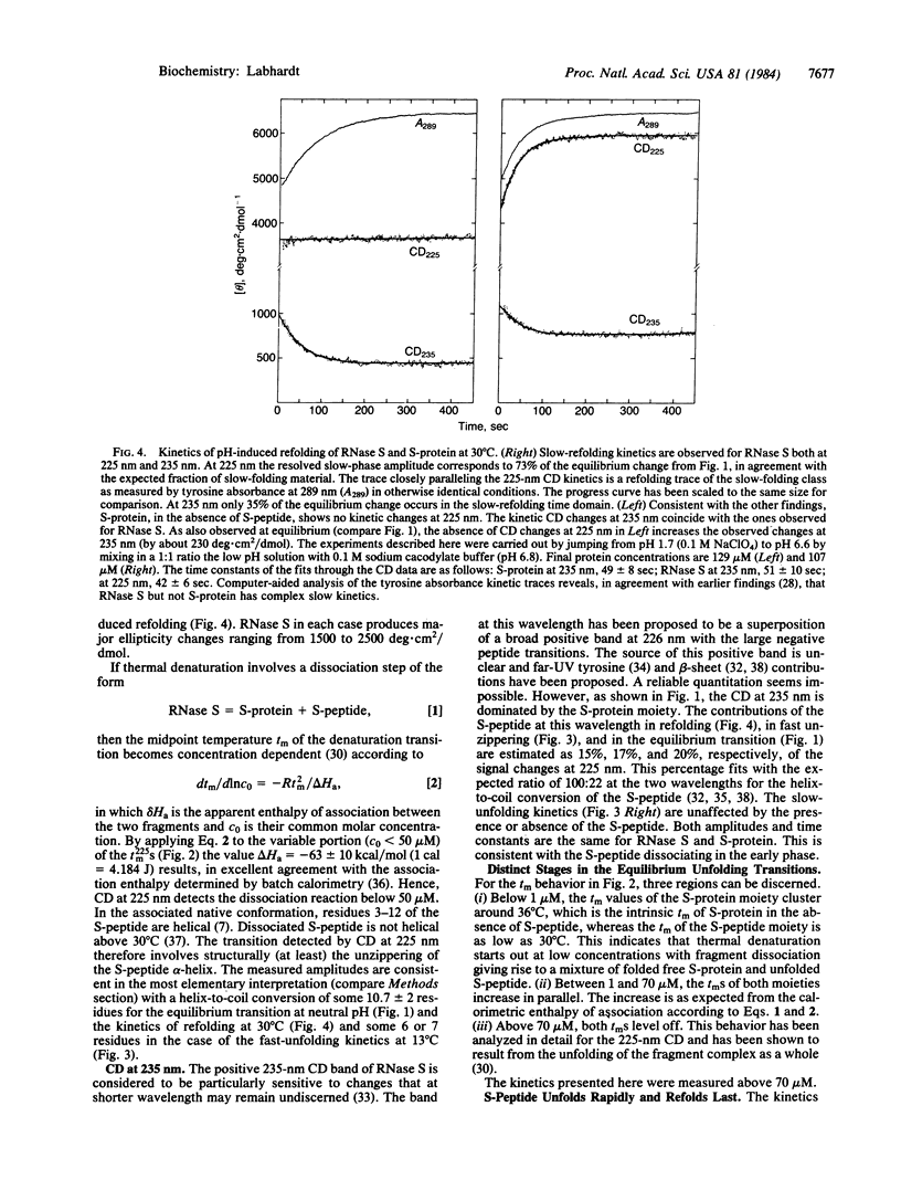
It is shown that circular dichroism (CD) can distinguish between the S-peptide and the S-protein fragments of RNase S at 225 nm and 235 nm. The conformational source for the strong CD at 225 nm is the S-peptide alpha-helix. The structural assignment of the CD at 235 nm is not clear but it is shown to be largely due to the S-protein moiety. This situation is utilized to monitor the kinetics of pH-induced unfolding and refolding of the two moieties. It is observed that major changes occur both in the fast and slow phases of unfolding as well as refolding. Specifically, the S-peptide alpha-helix unzippering is a fast reaction, followed by slow kinetics only at 235 nm. These latter kinetics parallel the appearance of the slow-folding species commonly attributed to the accumulation of non-native proline isomers. In refolding, a large fraction of the CD of S-protein at 235 nm recovers rapidly. The S-peptide alpha-helix zippers up last. These results are unexpected and their implications for the folding mechanism of ribonuclease are discussed.
Full text
PDF


Selected References
These references are in PubMed. This may not be the complete list of references from this article.
- Bierzynski A., Kim P. S., Baldwin R. L. A salt bridge stabilizes the helix formed by isolated C-peptide of RNase A. Proc Natl Acad Sci U S A. 1982 Apr;79(8):2470–2474. doi: 10.1073/pnas.79.8.2470. [DOI] [PMC free article] [PubMed] [Google Scholar]
- Borkakoti N. The active site of ribonuclease A from the crystallographic studies of ribonuclease-A-inhibitor complexes. Eur J Biochem. 1983 Apr 15;132(1):89–94. doi: 10.1111/j.1432-1033.1983.tb07329.x. [DOI] [PubMed] [Google Scholar]
- Brandts J. F., Halvorson H. R., Brennan M. Consideration of the Possibility that the slow step in protein denaturation reactions is due to cis-trans isomerism of proline residues. Biochemistry. 1975 Nov 4;14(22):4953–4963. doi: 10.1021/bi00693a026. [DOI] [PubMed] [Google Scholar]
- Chang C. T., Wu C. S., Yang J. T. Circular dichroic analysis of protein conformation: inclusion of the beta-turns. Anal Biochem. 1978 Nov;91(1):13–31. doi: 10.1016/0003-2697(78)90812-6. [DOI] [PubMed] [Google Scholar]
- Chen Y. H., Yang J. T., Martinez H. M. Determination of the secondary structures of proteins by circular dichroism and optical rotatory dispersion. Biochemistry. 1972 Oct 24;11(22):4120–4131. doi: 10.1021/bi00772a015. [DOI] [PubMed] [Google Scholar]
- Cook K. H., Schmid F. X., Baldwin R. L. Role of proline isomerization in folding of ribonuclease A at low temperatures. Proc Natl Acad Sci U S A. 1979 Dec;76(12):6157–6161. doi: 10.1073/pnas.76.12.6157. [DOI] [PMC free article] [PubMed] [Google Scholar]
- Garel J. R., Baldwin R. L. A physical difference between the fast- and slow-refolding forms of nitrotyrosyl ribonuclease A: the pK values of the nitrotyrosyl groups. J Mol Biol. 1975 Jun 5;94(4):621–632. doi: 10.1016/0022-2836(75)90326-5. [DOI] [PubMed] [Google Scholar]
- Garel J. R., Nall B. T., Baldwin R. L. Guanidine-unfolded state of ribonuclease A contains both fast- and slow-refolding species. Proc Natl Acad Sci U S A. 1976 Jun;73(6):1853–1857. doi: 10.1073/pnas.73.6.1853. [DOI] [PMC free article] [PubMed] [Google Scholar]
- Hagerman P. J., Baldwin R. L. A quantitative treatment of the kinetics of the folding transition of ribonuclease A. Biochemistry. 1976 Apr 6;15(7):1462–1473. doi: 10.1021/bi00652a017. [DOI] [PubMed] [Google Scholar]
- Hagerman P. J., Schmid F. X., Baldwin R. L. Refolding behavior of a kinetic intermediate observed in the low pH unfolding of ribonuclease A. Biochemistry. 1979 Jan 23;18(2):293–297. doi: 10.1021/bi00569a009. [DOI] [PubMed] [Google Scholar]
- Hearn R. P., Richards F. M., Sturtevant J. M., Watt G. D. Thermodynamics of the binding of S-peptide to S-protein to form ribonuclease S.. Biochemistry. 1971 Mar 2;10(5):806–817. doi: 10.1021/bi00781a013. [DOI] [PubMed] [Google Scholar]
- Kartha G., Bello J., Harker D. Tertiary structure of ribonuclease. Nature. 1967 Mar 4;213(5079):862–865. doi: 10.1038/213862a0. [DOI] [PubMed] [Google Scholar]
- Kim P. S., Baldwin R. L. Specific intermediates in the folding reactions of small proteins and the mechanism of protein folding. Annu Rev Biochem. 1982;51:459–489. doi: 10.1146/annurev.bi.51.070182.002331. [DOI] [PubMed] [Google Scholar]
- Kim P. S., Baldwin R. L. Structural intermediates trapped during the folding of ribonuclease A by amide proton exchange. Biochemistry. 1980 Dec 23;19(26):6124–6129. doi: 10.1021/bi00567a027. [DOI] [PubMed] [Google Scholar]
- Labhardt A. M., Baldwin R. L. Recombination of S-peptide with S-protein during folding of ribonuclease S. I. Folding pathways of the slow-folding and fast-folding classes of unfolded S-protein. J Mol Biol. 1979 Nov 25;135(1):231–244. doi: 10.1016/0022-2836(79)90349-8. [DOI] [PubMed] [Google Scholar]
- Labhardt A. M., Baldwin R. L. Recombination of S-peptide with S-protein during folding of ribonuclease S. II. Kinetic characterization of a stable folding intermediate shown by S-protein at pH 1.7. J Mol Biol. 1979 Nov 25;135(1):245–254. doi: 10.1016/0022-2836(79)90350-4. [DOI] [PubMed] [Google Scholar]
- Labhardt A. M., Ridge J. A., Lindquist R. N., Baldwin R. L. Measurement of the refolding combination reaction between S-peptide and S-protein. Biochemistry. 1983 Jan 18;22(2):321–327. doi: 10.1021/bi00271a014. [DOI] [PubMed] [Google Scholar]
- Labhardt A. M. Secondary structure in ribonuclease. I. Equilibrium folding transitions seen by amide circular dichroism. J Mol Biol. 1982 May 15;157(2):331–355. doi: 10.1016/0022-2836(82)90238-8. [DOI] [PubMed] [Google Scholar]
- Labhardt A. M. Secondary structure in ribonuclease. II. Relations between folding kinetics and secondary structure elements. J Mol Biol. 1982 May 15;157(2):357–371. doi: 10.1016/0022-2836(82)90239-x. [DOI] [PubMed] [Google Scholar]
- Levitt M., Greer J. Automatic identification of secondary structure in globular proteins. J Mol Biol. 1977 Aug 5;114(2):181–239. doi: 10.1016/0022-2836(77)90207-8. [DOI] [PubMed] [Google Scholar]
- Lin L. N., Brandts J. F. Determination of cis-trans proline isomerization by trypsin proteolysis. Application to a model pentapeptide and to oxidized ribonuclease A. Biochemistry. 1983 Feb 1;22(3):553–559. doi: 10.1021/bi00272a005. [DOI] [PubMed] [Google Scholar]
- Lin L. N., Brandts J. F. Isomerization of proline-93 during the unfolding and refolding of ribonuclease A. Biochemistry. 1983 Feb 1;22(3):559–563. doi: 10.1021/bi00272a006. [DOI] [PubMed] [Google Scholar]
- Lin L. N., Brandts J. F. Mechanism for the unfolding and refolding of ribonuclease A. Kinetic studies utilizing spectroscopic methods. Biochemistry. 1983 Feb 1;22(3):564–573. doi: 10.1021/bi00272a007. [DOI] [PubMed] [Google Scholar]
- Lin L. N., Brandts J. F. Mechanism for the unfolding and refolding of ribonuclease A. Simulations using a simple model with no structural intermediates. Biochemistry. 1983 Feb 1;22(3):573–580. doi: 10.1021/bi00272a008. [DOI] [PubMed] [Google Scholar]
- Nall B. T., Garel J. R., Baldwin R. L. Test of the extended two-state model for the kinetic intermediates observed in the folding transition of ribonuclease A. J Mol Biol. 1978 Jan 25;118(3):317–330. doi: 10.1016/0022-2836(78)90231-0. [DOI] [PubMed] [Google Scholar]
- Pflumm M. N., Beychok S. Optical activity of cystine-containing proteins. II. Circular dichroism spectra of pancreatic ribonuclease A, ribonuclease S, and ribonuclease S-protein. J Biol Chem. 1969 Jul 25;244(14):3973–3981. [PubMed] [Google Scholar]
- Rehage A., Schmid F. X. Fast- and slow-refolding forms of unfolded ribonuclease A differ in tyrosine fluorescence. Biochemistry. 1982 Mar 30;21(7):1499–1505. doi: 10.1021/bi00536a006. [DOI] [PubMed] [Google Scholar]
- Rosa J. H., Richards F. M. Hydrogen exchange from identified regions of the S-protein component of ribonuclease as a function of temperature, pH, and the binding of S-peptide. J Mol Biol. 1981 Feb 5;145(4):835–851. doi: 10.1016/0022-2836(81)90318-1. [DOI] [PubMed] [Google Scholar]
- Schmid F. X. A native-like intermediate on the ribonuclease A folding pathway. 1. Detection by tyrosine fluorescence changes. Eur J Biochem. 1981;114(1):105–109. doi: 10.1111/j.1432-1033.1981.tb06179.x. [DOI] [PubMed] [Google Scholar]
- Schmid F. X., Baldwin R. L. Acid catalysis of the formation of the slow-folding species of RNase A: evidence that the reaction is proline isomerization. Proc Natl Acad Sci U S A. 1978 Oct;75(10):4764–4768. doi: 10.1073/pnas.75.10.4764. [DOI] [PMC free article] [PubMed] [Google Scholar]
- Schmid F. X., Baldwin R. L. The rate of interconversion between the two unfolded forms of ribonuclease A does not depend on guanidinium chloride concentration. J Mol Biol. 1979 Sep 15;133(2):285–287. doi: 10.1016/0022-2836(79)90536-9. [DOI] [PubMed] [Google Scholar]
- Schmid F. X., Blaschek H. A native-like intermediate on the ribonuclease A folding pathway. 2. Comparison of its properties to native ribonuclease A. Eur J Biochem. 1981;114(1):111–117. doi: 10.1111/j.1432-1033.1981.tb06180.x. [DOI] [PubMed] [Google Scholar]
- Wlodawer A., Bott R., Sjölin L. The refined crystal structure of ribonuclease A at 2.0 A resolution. J Biol Chem. 1982 Feb 10;257(3):1325–1332. [PubMed] [Google Scholar]
- Wlodawer A., Sjölin L. Structure of ribonuclease A: results of joint neutron and X-ray refinement at 2.0-A resolution. Biochemistry. 1983 May 24;22(11):2720–2728. doi: 10.1021/bi00280a021. [DOI] [PubMed] [Google Scholar]