Abstract
Background
Thyroid nodules with cystic content or mixed sponge-like aspect on ultrasonography and a concordant cytology are strongly predictive of benignity.
Objectives
We present 8 patients with honeycomb-like papillary thyroid carcinoma with multiple small cysts on ultrasonography.
Methods
The patients were 6 women and 2 men aged between 30 and 57 years. The tumors of these patients showed honeycomb-like multiple small cysts that were aggregated in some area of the thyroid gland on ultrasonography. Histopathological examination indicated a well-differentiated type of papillary thyroid carcinoma with multiple small cysts and a small solid lesion. The cysts were lined with papillary carcinoma cells, and normal thyroid tissue lay between the cysts.
Results
There is a peculiar type of papillary thyroid carcinoma that histopathologically shows honeycomb-like multiple small cysts in the thyroid gland. Ultrasonography can be used to identify characteristic features of honeycomb-like multiple small cysts in the thyroid gland in such patients.
Conclusions
One should be aware of this peculiar type of papillary thyroid carcinoma with honeycomb-like multiple small cysts on ultrasonography, although thyroid nodules with cystic lesions are generally regarded as benign in clinical management.
Key Words : Thyroid, Papillary carcinoma, Cyst, Honeycomb, Ultrasonography
Introduction
Ultrasonography and cytology are very useful for the diagnosis and management of patients with thyroid nodules [1,2,3,4]. Some researchers [5,6,7,8] reported that a solid hypoechoic appearance, irregular or microlobulated margins, taller-than-wide shape, and intranodular vascularity on ultrasonography are important characteristics in differentiating malignancies of the thyroid from benign nodules. Cystic lesions in thyroid tumors are benign in most cases [9], and malignant tumors have few cystic lesions [10]. Therefore, general sonologists, radiologists, and clinicians are likely to diagnose lesions as benign when thyroid nodules show cystic lesions partially or totally on ultrasonography.
We herein report eight patients with honeycomb-like papillary thyroid carcinoma showing multiple small cysts, and describe the characteristic features on ultrasonography.
Methods and Patients
Ultrasonographic examination was performed by well-trained registered ultrasonographers, using a TOSHIBA Aplio SSA-770A ultrasound system with PLT-1204AX (7-14 MHz) and PLT-805AT (5-12 MHz) linear probes. Surgical samples of the thyroid gland and lymph nodes were cut before fixation. Specimens were fixed in buffered formalin and embedded in paraffin, and HE and immunohistochemical stainings were performed. All related pathological specimens were reviewed (by M.H.), and the histopathological diagnosis was included in this study.
We discovered 8 patients with papillary thyroid carcinoma whose nodules present honeycomb-like multiple small cysts on ultrasonography and histopathology from 2006 to 2011 at the Kuma hospital. Table 1 shows the clinical characteristics of these patients, including ultrasonographic, cytological, and histopathological findings. All the patients presented negative anti-thyroglobulin antibody in serum, well-differentiated type of papillary thyroid carcinoma and lymph node metastasis on histopathology. Cervical lymph nodes swelling indicating metastasis were observed in 7 patients on preoperative ultrasonography.
Table 1.
Patients of papillary thyroid carcinoma with honeycomb-like multiple small cysts on ultrasonography
| Patient No. | Serum |
Ultrasonography |
FNAB | Pre-op. Dx | Surgery | Histopathology |
||||
| Tg | TgAb | right/left | size, mm | LNS | diagnosis | LNM | ||||
| 1 | 46 F | 80.4, (−) | right | 27 | (–) | malignancy | PC | TT, CND | WD PC | (+) |
| 2 | 31 F | 49.1, (–) | left | 57 | (+) | malignancy | PC | TT, MND | WD PC | (+) |
| 3 | 31 F | 145.3, (–) | right | 43 | (+) | malignancy | PC | TT, CND | WD PC | (+) |
| 4 | 30 F | 237.6, (–) | right | 44 | (+) | malignancy | PC | TT, MND | WD PC | (+) |
| 5 | 38 M | 19.6, (–) | right | 23 | (+) | malignancy | PC | TT, CND | WD PC | (+) |
| 6 | 34 F | 55.5, (–) | left | 47 | (+) | malignancy | PC | TT, MND | WD PC | (+) |
| 7 | 57 M | 48.2, (–) | right | 35 | (+) | malignancy | PC | TT, CND | WD PC | (+) |
| 8 | 42 F | 123.7, (–) | right | 30 | (+) | malignancy | PC | TT, MND | WD PC | (+) |
Tg = Thyroglobulin (reference range: <35 ng/ml); TgAb = antithyroglobulin antibody; (–) = negative; size = maximum length of the tumor on ultrasonography; LNS = lymph node swelling; cervical lymph nodes swelling indicating metastasis on preoperative ultrasonography; (+) = positive; FNAB = fine-needle aspiration biopsy; Pre-op. Dx = preoperative diagnosis; PC = papillary carcinoma; TT = total thyroidectomy; CND = central neck dissection; MND = modified neck dissection; WD PC = well-differentiated type of papillary carcinoma; LNM = lymph nodes metastasis.
Histopathologically, multiple small cysts were aggregated and normal thyroid tissue lay between the cysts. The cysts were lined with papillary carcinoma cells, and the small solid lesion showed conventional papillary carcinoma with a papillary structure and desmoplastic change. Metastatic papillary carcinomas that were seen in lymph nodes presented conventional papillary architecture without cysts.
Patient 1
A 46-year-old woman had noticed an elastic mass in the right anterior neck nine months previously. The neck mass was diagnosed as a benign thyroid cyst by ultrasonography without performing cytological examination at another hospital.
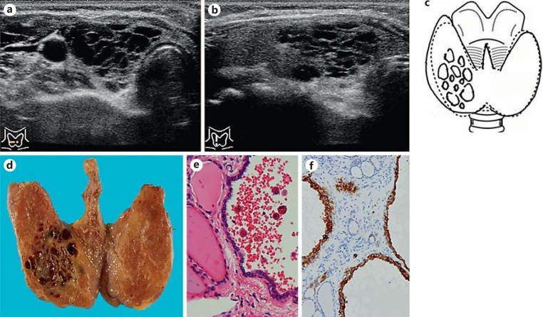
The serum concentration of thyroid-stimulating hormone was 2.4 μIU/ml (reference range 0.3-5.0), free thyroxine 0.98 ng/dl (0.70-1.60), and thyroglobulin 80.4 ng/ml (<35). Antithyroglobulin autoantibody was negative. Ultrasonography showed multiple small cysts aggregated in the lower region of the right lobe of the thyroid gland. These multiple small cysts presented a honeycomb-like appearance, were 27 × 16 × 21 mm in size, and there were no solid lesions on ultrasonography (Fig. 1a-c). No swollen lymph nodes in the neck were observed. Power-Doppler ultrasonography revealed moderate blood flow signals at the sites of the multiple cysts. An ultrasonography-guided fine-needle aspiration biopsy from the cysts revealed atypical cells with intranuclear cytoplasmic pseudoinclusions and foam cells. The cytological diagnosis was papillary thyroid carcinoma associated with multiple cystic lesions. Total thyroidectomy and central nodes dissection were performed. A coronal section of the whole thyroid gland is shown in Figure 1d, in which honeycomb-like multiple small cysts aggregated in the right lobe can be observed.
Fig. 1.
a Ultrasonography of a transverse section of the right lobe of the thyroid. Honeycomb-like multiple small cysts are present. b Ultrasonography of a longitudinal section of the right lobe of the thyroid. Honeycomb-like multiple small cysts are present. No solid lesions are observed. c Schema of ultrasonography. d Coronal section of the whole thyroid gland. The neoplasm shows honeycomb-like multiple small cysts in the right lobe of the thyroid. e Multiple small cysts are aggregated in the right lobe. The cystic lesion is lined with cuboidal to columnar atypical cells, that is, papillary carcinoma cells, and normal thyroid tissue lies between the cysts. HE. f Immunohistochemical staining for cytokeratin 19. Neoplastic cells, that is, papillary carcinoma cells, lining the cyst wall are strongly positive for cytokeratin 19.
Histopathologically, multiple small cysts were aggregated in the right lobe. Normal thyroid tissue lay between the cysts. The cysts were lined with cuboidal to columnar atypical cells (Fig. 1e). The nuclei showed clearing, ground-glass appearance, irregular nuclear contours, grooves, and pseudo-inclusions. Psammoma bodies were not observed. The small solid lesion showed features of conventional papillary thyroid carcinoma. The neoplastic cells were immunopositive for thyroglobulin, thyroid transcription factor-1, cytokeratin 19 (Fig. 1f) [11], and galectin-3, and partially positive for HBME-1. Metastatic papillary carcinomas that were seen in lymph nodes presented a conventional papillary architecture without cysts. The histopathological diagnosis was a well-differentiated type of papillary thyroid carcinoma characterized by multiple cystic lesions. The patient is currently doing well 5 years and 6 months after surgery.
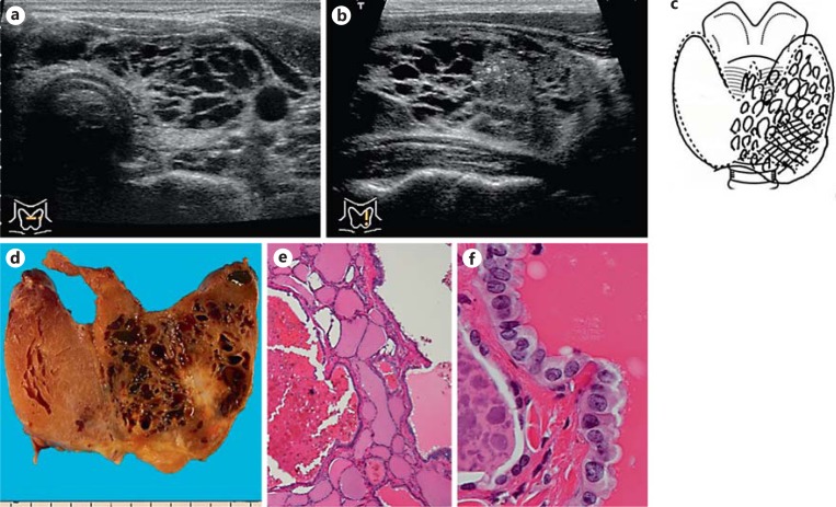
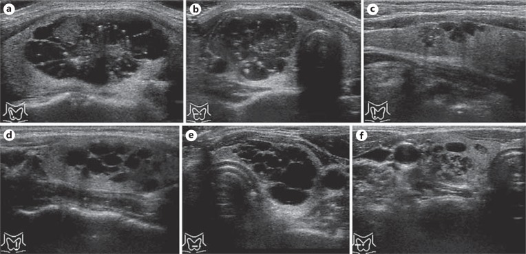
Figure 2 shows the ultrasonography, the specimen and the histopathological features of patient 2. Figure 3 shows the ultrasonography of patient 3-8.
Fig. 2.
a Ultrasonography of a transverse section of the left lobe and isthmus of the thyroid. Honeycomb-like multiple small cysts are present. b Ultrasonography of a longitudinal section of the left lobe of the thyroid. Honeycomb-like multiple small cysts in the periphery of the neoplasm and a solid lesion with micro-calcifications in the center are observed. c Schema of ultrasonography. d Coronal section of the whole thyroid gland. The neoplasm shows honeycomb-like multiple small cysts in the periphery and a solid lesion in the center in the left lobe and the isthmus of the thyroid. e Multiple small cysts are aggregated in the left lobe. The cystic lesion is lined with cuboidal to columnar atypical cells, that is, papillary carcinoma cells, and normal thyroid tissue lies between the cysts. HE. f The cystic lesion is lined with papillary carcinoma cells. HE.
Fig. 3.
a Ultrasonography of patient 3. b Ultrasonography of patient 4. c Ultrasonography of patient 5. d Ultrasonography of patient 6. e Ultrasonography of patient 7. f Ultrasonography of patient 8. These ultrasonographic features present honeycomb-like multiple small cysts in the thyroid gland.
Discussion
The strategic value of ultrasound as well as fine-needle aspiration biopsy cytology in preoperative surgical planning of patients with thyroid nodules has become increasingly appreciated [1,2,3,4]. Ultrasonographic findings in thyroid nodules such as a solid hypoechoic appearance, irregular or microlobulated margins, taller-than-wide shape, and intranodular vascularity can be important in differentiating between benign and malignant nodules [5,6,7,8]. Conversely, nodules with cystic content or mixed sponge-like aspect and a concordant cytology are strongly predictive of benignity [9]. For this reason, thyroid nodules that present cystic lesions either partially or totally on ultrasonography are likely to be regarded as benign.
Besides the above-mentioned facts, there is a peculiar type of papillary thyroid carcinoma that shows honeycomb-like multiple small cysts in the thyroid gland. Histopathologically, these neoplasms predominantly show multiple small cysts whose walls are lined with carcinoma cells and a small solid lesion of conventional papillary carcinoma with a papillary structure and desmoplastic change. We postulate that these neoplasms originated as conventional papillary thyroid carcinoma, and grew to form multiple small cysts with papillary thyroid carcinoma cells and to invade normal thyroid tissue. Ultrasonography can be used to identify these honeycomb-like multiple small cysts in the thyroid gland.
It is reported that some papillary thyroid carcinomas present characteristic features on ultrasonography. These include cystic papillary carcinoma [10] and the diffuse sclerosing variant [12]. Tumor thrombus can be rarely detected in patients with papillary thyroid carcinoma preoperatively by ultrasonography [13]. Cystic papillary carcinomas [10] include cystic lesion within the tumor tissue on ultrasonography and histopatholgy. On the contrary, these honeycomb-like papillary carcinomas include multiple cysts structured with carcinoma cells within the normal thyroid tissue. To the best of our knowledge, this honeycomb-like appearance may represent a new type of papillary thyroid carcinoma identifiable by ultrasonography.
Ultrasonography, which is one of the noninvasive imaging modalities, can generate an exact image of the macroscopic aspects of both the thyroid nodule and the entire gland. Even though thyroid ultrasonography is now widely used for the evaluation of patients with thyroid nodules, it has some important limitations. The procedure of ultrasonic examination is very operator-dependent. Only well-trained examiners aware of the ultrasonographic features of a certain disease can make an accurate diagnosis. Ultrasonography and fine-needle aspiration cytology of thyroid nodules are helpful for distinguishing malignant from benign nodules, and can be complementary to each other on making a diagnosis [1,2,4,5,6]. It is essential for cytology to be performed when ultrasonography of thyroid nodules shows honeycomb-like multiple small cysts aggregated in some area of the thyroid gland.
It is important to differentiate between benign and malignant multiple small cysts before surgery. We routinely observe children with a few or several small cysts in the thyroid glands on ultrasonography. These small cysts in the thyroid gland are ‘a few or several’, not ‘multiple’ in number, and obviously benign. Fine-needle aspiration biopsy is not necessary for such small cysts, even though such children present positive or negative for thyroid autoantibodies. Recently, it has been reported that multiple small cysts in the whole thyroid gland may be a cause of hypothyroidism in old patients with relatively high iodine intake [14]. Such patients presented negative for thyroid autoantibodies, and the disease was named polycystic thyroid disease [14]. These multiple small cysts are diffusely located in the whole thyroid glands, not aggregated in some area. Fine-needle aspiration biopsy is not necessary for such small cysts, but it is necessary to ask their dietary condition of iodine intake and to measure TSH and thyroid hormone for such patients.
Disclosure Statement
The authors declare that no competing financial interests exist.
References
- 1.Pacini F, Schlumberger M, Dralle H, Elisei R, Smit JW, Wiersinga W, European Thyroid Cancer Taskforce European consensus for the management of patients with differentiated thyroid carcinoma of the follicular epithelium. Eur J Endocrinol. 2006;154:787–803. doi: 10.1530/eje.1.02158. [DOI] [PubMed] [Google Scholar]
- 2.Sipos JA. Advance in ultrasound for the diagnosis and management of thyroid cancer. Thyroid. 2009;19:1363–1372. doi: 10.1089/thy.2009.1608. [DOI] [PubMed] [Google Scholar]
- 3.Cooper DS, Doherty GM, Haugen BR, Kloos RT, Lee SL, Mandel SJ, Mazzaferri EL, McIver B, Pacini F, Schlumberger M, Sherman SI, Steward DL, Tuttle RM. Revised American Thyroid Association management guidelines for patients with thyroid nodules and differentiated thyroid cancer. American Thyroid Association (ATA) Guidelines Taskforce on Thyroid Nodules and Differentiated Thyroid Cancer. Thyroid. 2009;19:1167–1214. doi: 10.1089/thy.2009.0110. [DOI] [PubMed] [Google Scholar]
- 4.Emerson CH. Can thyroid ultrasound and related procedures provide diagnostic information about thyroid nodules: a look at the guidelines. Thyroid. 2011;21:211–213. doi: 10.1089/thy.2011.2103.ed. [DOI] [PubMed] [Google Scholar]
- 5.Shimura H, Haraguchi K, Hiejima Y, Fukunari N, Fujimoto Y, Katagiri M, Koyanagi N, Kurita T, Miyakawa M, Miyamoto Y, Suzuki N, Suzuki S, Kanbe M, Kato Y, Murakami T, Tohno E, Tsunoda-Shimizu H, Yamada K, Ueno E, Kobayashi K, Kobayashi T, Yokozawa T, Kitaoka M. Distinct diagnostic criteria for ultrasonographic examination of papillary thyroid carcinoma: a multicenter study. Thyroid. 2005;15:251–258. doi: 10.1089/thy.2005.15.251. [DOI] [PubMed] [Google Scholar]
- 6.Kwak JY, Kim EK, Kim MJ, Hong SW, Choi SH, Son EJ, Oh KK, Park CS, Chung WY, Kim KW. The role of ultrasound in thyroid nodules with a cytology reading of ‘suspicious for papillary thyroid carcinoma'. Thyroid. 2008;18:517–522. doi: 10.1089/thy.2007.0271. [DOI] [PubMed] [Google Scholar]
- 7.Brunese L, Romeo A, Iorio S, Napolitano G, Fucili S, Biondi B, Vallone G, Sodano A. A new marker for diagnosis of papillary thyroid cancer: B-flow twinkling sign. J Untrasound Med. 2008;27:1187–1194. doi: 10.7863/jum.2008.27.8.1187. [DOI] [PubMed] [Google Scholar]
- 8.Kim MJ, Kim EK, Kwak JY, Park CS, Chung WY, Nam KH, Youk JH. Differentiation of thyroid nodules with macrocalcifications: role of suspicious sonographic findings. J Untrasound Med. 2008;27:1179–1184. doi: 10.7863/jum.2008.27.8.1179. [DOI] [PubMed] [Google Scholar]
- 9.Moon WJ, Jung SL, Lee JH, Na DG, Baek JH, Lee YH, Kim J, Kim HS, Byun JS, Lee DH. Benign and malignant thyroid nodules: US differentiation – multicenter retrospective study. Radiology. 2008;247:762–770. doi: 10.1148/radiol.2473070944. [DOI] [PubMed] [Google Scholar]
- 10.Chan BK, Desser TS, McDougall IR, Weigel TJ, Jeffrey RB., Jr Common and uncommon sonographic features of papillary thyroid carcinoma. J Ultrasound Med. 2003;22:1083–1090. doi: 10.7863/jum.2003.22.10.1083. [DOI] [PubMed] [Google Scholar]
- 11.Hirokawa M, Carney JA, Ohtskuki Y. Hyalinizing trabecular adenoma and papillary carcinoma of the thyroid gland express different cytokeratin patterns. Am J Surg Pathol. 2000;24:877–881. doi: 10.1097/00000478-200006000-00015. [DOI] [PubMed] [Google Scholar]
- 12.Zhang Y, Xia D, Kin P, Gao L, Ki G, Zhang W. Sonographic findings of the diffuse sclerosing variant of papillary carcinoma of the thyroid. J Ultrasound Med. 2010;29:1223–1226. doi: 10.7863/jum.2010.29.8.1223. [DOI] [PubMed] [Google Scholar]
- 13.Kobayashi K, Hirokawa M, Yabuta T, Fukushima M, Kihara M, Higashiyama T, Tomoda C, Takamura Y, Ito Y, Miya A, Amino N, Miyauchi A. Tumor thrombus of thyroid malignancies in veins: Importance of detection by ultrasonography. Thyroid. 2011;21:527–531. doi: 10.1089/thy.2010.0099. [DOI] [PubMed] [Google Scholar]
- 14.Kubota S, Fujiwara M, Hagiwara H, Tsujimoto N, Takata K, Kudo T, Nishihara E, Ito M, Amino N, Miyauchi A. Multiple thyroid cysts may be a cause of hypothyroidism in patients with relatively high iodine intake. Thyroid. 2010;20:205–208. doi: 10.1089/thy.2009.0264. [DOI] [PubMed] [Google Scholar]


