Abstract
HER2/HER3 dimerization resulting from overexpression of HER2 or neuregulin (NRG1) in cancer leads to HER3-mediated oncogenic activation of PI3K signaling. Although ligand-blocking HER3 antibodies inhibit NRG1-driven tumor growth, they are ineffective against HER2-drive tumor growth because HER2 activates HER3 in a ligand-independent manner. In this study, we describe a novel HER3 monoclonal antibody (LJM716) that can neutralize multiple modes of HER3 activation, making it a superior candidate for clinical translation as a therapeutic candidate. LJM716 was a potent inhibitor of HER3/AKT phosphorylation and proliferation in HER2-amplified and NRG1-expressing cancer cells and it displayed single agent efficacy in tumor xenograft models. Combining LJM716 with agents that target HER2 or EGFR produced synergistic antitumor activity in vitro and in vivo. In particular, combining LJM716 with trastuzumab produced a more potent inhibition of signaling and cell proliferation than trastuzumab/pertuzumab combinations and was similarly active in vivo. To elucidate its mechanism of action, we solved the structure of LJM716 bound to HER3, finding that LJM716 bound to an epitope within domains 2 and 4 that traps HER3 in an inactive conformation. Taken together, our findings establish that LJM716 possesses a novel mechanism of action that in combination with HER2 or EGFR-targeted agents may leverage their clinical efficacy in ErbB-driven cancers.
Keywords: Antibody, HER, EGFR, HER2, HER3, ERBB3, NRG, neuregulin, ligand-dependent, ligand-independent, cancer, feedback, SCCHN, PIK3CA, PI-3 kinase, resistance
Introduction
In clinical practice the HER2-targeted monoclonal antibody trastuzumab (Herceptin™) is central to the treatment of HER2 amplified breast cancer. Although trastuzumab has well-established clinical benefit, responses are transient and patients frequently relapse with trastuzumab-resistant disease (1). A number of trastuzumab resistance mechanisms have been proposed that most commonly center upon sustained phosphatidylinositol-3 kinase (PI3K) signaling (2;3) either due to the presence of activating PI3K mutations (4;5), PTEN inactivation (4;5) or persistent HER3 signaling (6;7). HER3 is the preferred dimerization partner of HER2 (8) acting as an allosteric activator of its partner kinase (9). Activation of the HER2/HER3 complex results in trans-phosphorylation of HER3 and initiation of downstream signaling. HER2/HER3 activates PI3K signaling via HER3, which in contrast to other ErbB receptors contains multiple phospho-dependent binding sites for the regulatory p85 subunit of PI3K. (10).
In HER2 amplified cancer, activation of HER3 may occur through high level expression of hetero-dimerization partners such as HER2 (11). Consequently, in cases of HER2 amplification, HER2/HER3 heterodimer formation occurs in a ligand-independent manner resulting in unrestrained HER3 signaling that is both necessary (12) and sufficient (13) for transformation. Indeed, human HER2 amplified breast cancer samples harbor high levels of phosphorylated HER3 indicative of HER3 activation and infrequent concomitant NRG1 expression (14), (Supplementary Figure S1A–D). Continued HER3 signaling in the presence of trastuzumab or PI3K inhibitors might also be driven by FOXO-dependent induction of HER3 expression (15–17) via the release of a PI3K/ AKT driven inhibitory feedback loop (7;18).
The HER2-targeted antibody pertuzumab (Perjeta™) reportedly inhibits ligand-induced HER3 activity by preventing HER2/HER3 dimerization (3;19). The recent CLEOPATRA study (20) demonstrated that the addition of pertuzumab to trastuzumab/ docetaxel significantly prolonged progression-free survival when used as first-line treatment in HER2-over expressing breast cancer. However, recent preclinical reports indicate that even dual HER2 blockade is unable to fully inhibit PI3K/AKT signaling and superior benefit may be achieved with HER3-specific inhibition (21).
Elevated expression of NRG1 drives ligand-dependent HER3 signaling and functional NRG1/HER3 autocrine loops have been identified in models of SCCHN (22) and ovarian cancer (23). Given that both ligand-dependent and independent HER3 activation appear of fundamental importance in multiple tumor types a therapeutic capable of inhibiting both of these modes of HER3 activation may be efficacious in multiple indications.
Here we describe the discovery, biological activity and molecular mode of action of a fully human antibody (LJM716) currently in clinical testing. LJM716 is capable of neutralizing both ligand-dependent and independent HER3 signaling and suggests this occurs by locking HER3 in the inactive conformation. We also present in vitro and in vivo data that highlight the potential clinical benefit of combining LJM716 with both HER2 and EGFR targeted agents.
Materials and Methods
Recombinant proteins
Recombinant monomeric HER3 extracellular domains (ECD’s) from human, rat and cynomolgus monkey, as well as isolated HER3 domains (D1–2, D2, D3–4 and D4) were cloned upstream of a C-terminal affinity tag, sequence verified, expressed in HEK293 derived cells and purified using an anti-tag antibody. Fc-tagged ECD’s from 3 other ErbB-family proteins (EGFR, HER2, HER4) were purchased from R&D Systems. Further details on all recombinant proteins used can be found in the Supplementary Methods.
Antibodies
HER3-targeted antibodies were selected from the Human Combinatorial Antibody Library (HuCAL GOLD®) using phage display technology (24). The affinity (KD) of the binding interaction between LJM716 and recombinant monomeric HER3 ECD was determined by solution equilibrium titration (SET) (25).
ELISA Binding Assays
Maxisorp plates (Nunc) were coated with the appropriate recombinant protein and blocked prior to incubating with the relevant test antibody for two hours at room temperature. Plates were washed and human antibody detected using peroxidase linked goat anti-human antibody (Pierce).
Immunoblotting
For immunoblots, Cell lysates were prepared in 1% NP-40 buffer including protease and phosphatase inhibitors (Roche) and analyzed by Western blot using the Odyssey detection system (Licor) or by enhanced chemiluminescence after incubation with horseradish peroxidase-conjugated secondary antibodies (Promega). Details on antibodies used are in the Supplementary Methods.
Cell lines
For information on cell lines used in this study please see Table 2. Cell lines were acquired, maintained and authenticated by SNP fingerprinting (Sequenom) as previously described (26).
Table 2.
Summary table of cell lines used in this study.
| Cell line name | Supplier |
|---|---|
| SkBr3 | ATCC |
| MDA-MB-453 | ATCC |
| BT474 | ATCC |
| AU565 | ATCC |
| MDA-MB-361 | ATCC |
| EFM192A | DSMZ |
| KPL4 | J. Kurebayashi; Kawasaki Medical School, Japan |
| NCI-N87 | ATCC |
| HCC202 | ATCC |
| OE19 | ECACC |
| UACC812 | ATCC |
| HCC2218 | ATCC |
| HCC1569 | ATCC |
| JIMT1 | DSMZ |
| HCC1954 | ATCC |
| NUGC4 | RIKEN |
| ZR-75-30 | ATCC |
| OE33 | ECACC |
| BxPC3 | ATCC |
| BHY | DSMZ |
| BICR-31 | ECACC |
| CAL-27 | ATCC |
| FaDu | ATCC |
| HSC-4 | HSRRB |
| PE/CA-PJ34 | ECACC |
| PE/CA-PJ49 | ECACC |
| SCC-25 | ATCC |
| SCC-4 | ATCC |
| SCC-9 | ATCC |
pHER3 and pAKT assays
pHER3 and pAKT were measured by MSD assay. Briefly, cell or tumor lysates were prepared in NP-40 lysis buffer supplemented with protease and phosphatase inhibitors, followed by capture of the analyte on an MSD plate and detection with a phosphospecific primary antibody and Sulfo-tagged secondary IgG. Refer to Supplementary Methods for details.
In vitro proliferation assays
For single agent growth assays, cells were seeded onto 96-well plates (Costar #3904) at 5000 cells/well, treated with the appropriate concentration of the indicated drug and incubated for 3 to 5 days at 37°C. Cell viability was quantified by the addition of CellTiterGlo reagent (Promega). For drug combination treatments the procedure was the same except that cells were plated into 384-well plates (Greiner #781091) at 1000 cells/well. Percent growth inhibition was calculated by normalizing raw luminescence values to that of untreated wells.
FACS and cellular NRG1 blocking assays
Binding of HER3 antibodies and NRG1 blocking was determined using flow cytometry. Details can be found in the Supplementary Methods.
HER3 crystallography
MOR09825 / HER3 complex was prepared by mixing tagged HER3 ECD (residues 20–640) with E.coli expressed MOR09825 Fab and then purifying the complex on a Superdex 200 10/300 column (GE Healthcare). Data were collected on a crystal cooled to 100K using a Dectris Pilatus 6M detector and synchrotron radiation (λ=1.0000Å) at the IMCA-CAT beam line 17-ID of the Advanced Photon Source at Argonne National Laboratory. For details on data processing and modeling refer to Supplementary Methods.
In vivo studies
For xenograft studies female athymic nu/nu Balb/C (Harlan Laboratories) or NSG (Jackson Labs) mice were implanted with BT-474, BxPC-3, FaDu, KPL4, L3.3, N87, T3M4 and Hara cells. Mice were treated with 20 mg/kg trastuzumab (q2w), 20 mg/kg LJM716 (q2d), 20 mg/kg Pertuzumab (q2w), Cetuximab 20 mg/kg (q2w) or 100 mg/kg lapatinib (qd). Further details on the in vivo efficacy and PD studies can be found in the Supplementary Methods.
Results
Discovery of an anti-HER3 antibody that inhibits both ligand-dependent and ligand-independent HER3 activity in vitro
Previous shRNA data have demonstrated that loss of HER3 expression in HER2 amplified models is sufficient to inhibit cell proliferation (14). Treatment of the HER3 shRNA sensitive cell line SKBR-3 with commercially available HER3-targeted antibodies further validates the ability to modulate cell growth in this setting from the extracellular compartment. AF234 (a HER3 specific goat polyclonal) induced significant inhibition of growth, while MAB3481, a ligand blocking mouse monoclonal had little to no effect (Figure 1A). The greater activity observed with AF234 was correlated with its more robust down-regulation of HER3 and prolonged inhibition of pAKT (Figure 1B). Thus antibodies that simply block ligand binding to HER3 and fail to inhibit AKT phosphorylation are unlikely to be active in this setting.
Figure 1. Identification of LJM716, a HER3 binding monoclonal antibody capable of blocking ligand-dependent and –independent HER3 signaling and cell proliferation.
(A) SKBR-3 were treated with increasing concentrations of either IgG, MAB348 or AF234 for 5 days followed by cell viability assessment using the CTG assay.
(B) SKBR-3 cells were treated with 10 µg/ml IgG, MAB3481 and AF234 for 1 or 24 hours. Cell lysates were harvested and immuno-blotted with antibodies directed against the indicated proteins.
(C) HER3-targeted antibodies were profiled for their ability to inhibit HER3 tyrosine phosphorylation and cell growth in HER2 amplified SKBR-3 cells. Maximal growth inhibition was calculated relative to that achieved with AF234. pHER3 was measured via MSD assay and Family 15 members are highlighted in red.
(D) MCF7 cells were treated with HER3-targeted antibodies prior to stimulation with NRG1 (50 ng/ml) and their impact upon HER3 phosphorylation and proliferation determined. Maximal growth inhibition was calculated relative to that achieved with AF234. pHER3 was measured via MSD assay and Family 15 members are highlighted in red.
(E) SKBR-3 cells were treated with LJM716 (red) or IgG (black) for one hour and the level of pHER3 quantified via MSD assay.
(F) MCF7 cells were treated with LJM716 (red) or IgG (black) for 30 minutes prior to NRG1 stimulation for 10 minutes. pHER3 levels were measured by MSD assay.
(G) SKBR-3 cells grown in full serum were treated with LJM716 (red) or IgG (black) for 5 days and cell viability determined by CTG.
(H) Serum starved MCF7 cells were treated with LJM716 (red) or IgG (black), stimulated with NRG1 (50 ng/ml) and cell viability determined after 5 days using CTG. Cell proliferation values relative to unstimulated cells are plotted.
To identify a HER3 therapeutic antibody capable of inhibiting both ligand-dependent and ligand-independent HER3 signaling we isolated HER3 targeted Fabs utilizing MorphoSys HuCAL phage display technology (24) against a number of recombinant HER3 antigens. Corresponding full IgGs were screened for their ability to neutralize HER3 tyrosine phosphorylation and cell proliferation driven by either HER2 (SKBR3 cells) or NRG1 (MCF7 cells). From all confirmed hits, only four antibodies were capable of effectively inhibiting HER2-driven HER3 phosphorylation and proliferation in SKBR-3 cells (Figure 1C). In contrast, a large number of antibodies efficiently inhibited HER3 phosphorylation and cell proliferation in NRG1 stimulated MCF7 cells (Figure 1D) due to their ability to prevent ligand binding (data not shown). Three antibodies were active in both settings (Figure 1C & D) suggesting they may be uniquely capable of inhibiting multiple modes of HER3 activation. The sequence of these antibodies differed only in residues contained within hCDR2 and thus these antibodies were highly related and are referred to herein as Family 15.
We further characterized LJM716, an antibody derived from Family 15. LJM716 binds to human, mouse, rat and cynomolgus monkey HER3 with high affinity as determined by solution equilibrium titration (Table 1) and flow cytometry (Supplementary Table S2). LJM716 potently inhibited phosphorylation of HER3 driven by either HER2 over-expression or NRG1 stimulation (Figure 1E & F, Table S3, Supplementary Figure S2A) in SKBR-3 and MCF7 cells. The inhibitory activity of LJM716 similarly led to inhibition of PI3K pathway signaling including downregulation of AKT phosphorylation (pAKT S473) (Supplementary Figure S2B & C, Table S3) and inhibition of cell proliferation (Figure 1G & H, Table S4) in both SKBR-3 and NRG1 stimulated MCF7 cells. Despite their high degree of sequence homology, no binding of LJM716 to other ErbB family members was observed (Supplementary Figure S3A). Together these data suggest that LJM716 is a specific and potent inhibitor of ligand-dependent and independent HER3 signaling and growth.
Table 1.
Equilibrium dissociation constants (KD) for binding of LJM716 to human, cyno, mouse and rat HER3.
| Antigen | KD (pM) ± S.D. |
|---|---|
| Human HER3 | 32±10 |
| Cynomolgus monkey HER3 | 43±17 |
| Mouse HER3 | 37±7 |
| Rat HER3 | 57±7 |
LJM716 displays single-agent activity in HER2 and NRG1 driven in vivo models
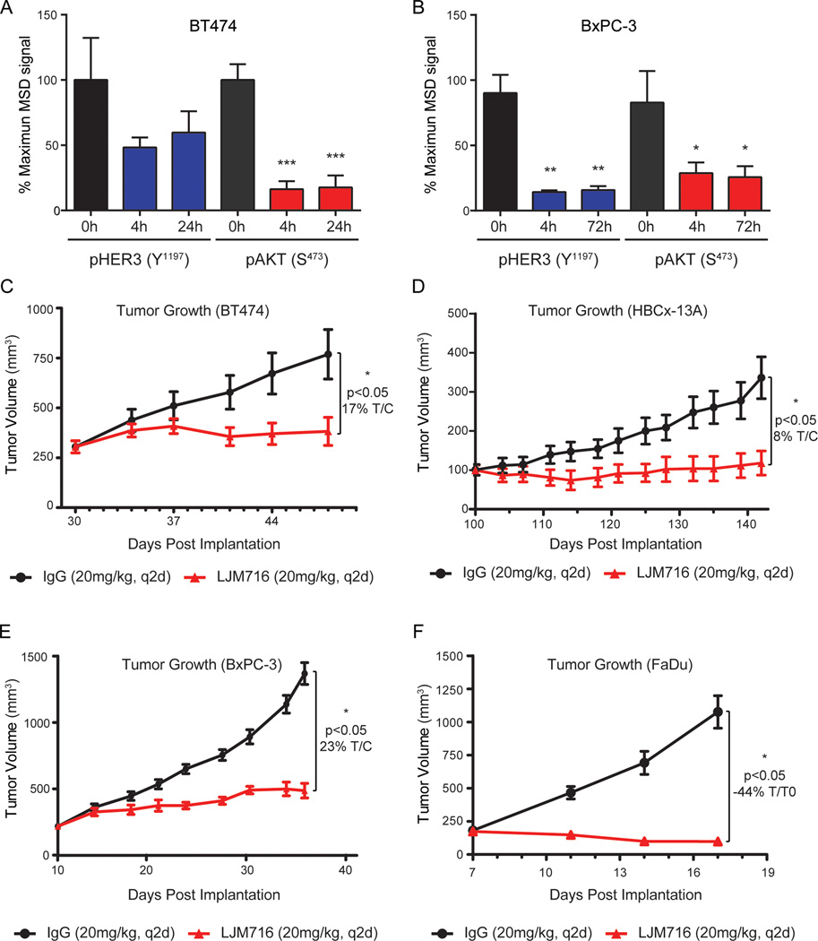
Having established that LJM716 inhibits multiple modes of HER3 activation in vitro, we next asked whether this activity was maintained in vivo. SCID mice bearing HER2 amplified BT474 breast cancer xenografts were treated with a single-dose of 20 mg/kg LJM716, resulting in maximal reduction of pHER3 by 52% and pAKT by 84% over a 24h period when compared to an equivalent dose of an isotype control antibody (Figure 2A). Similarly, in SCID mice bearing ligand-expressing BxPC-3 pancreatic xenografts, intravenous dosing of 20 mg/kg LJM716 resulted in 86% maximal inhibition of pHER3 and 74% inhibition of pAKT compared to isotype-matched treated controls (Figure 2B). These pharmacodynamic studies demonstrate that LJM716 is capable of inhibiting both HER2 and ligand mediated HER3 signaling in vivo as evidenced by decreased pHER3/ pAKT in both xenograft models.
Figure 2. LJM716 inhibits HER3 signaling in vivo and is efficacious in multiple ligand-dependent and independent xenograft tumor models.
(A) BT474 xenografts were dosed intravenously with a single 20 mg/kg dose of LJM716 and tumors were harvested at 0, 4 or 24 hours followed by analysis of lysates for pHER3 and pAKT levels by MSD assay.
(B) BxPC-3 xenografts were dosed intravenously with a single 20 mg/kg dose of LJM716 and tumors were harvested at 0, 4, or 72 hours followed by analysis of lysates for pHER3 and pAKT levels by MSD assay.
(C) BT474 xenografts were dosed intravenously every other day with 20 mg/kg LJM716 (red) or IgG (black).
(D) HBCx-13A primary human xenografts were dosed intravenously every other day with 20 mg/kg LJM716 (red) or IgG (black).
(E) BxPC-3 xenografts were dosed intravenously every other day with 20 mg/kg LJM716 (red) or IgG (black).
(F) FaDu xenografts were dosed intravenously every other day with 20 mg/kg LJM716 (red) or IgG (black).
Two-tailed non-paired t-test was performed for A+B. Data in C–F are presented as mean tumor volume ±SEM. All delta volumes subjected to One Way ANOVA and Tukeys post hoc analysis. * indicates p<0.05; ** indicates p<0.01; *** indicates p<0.001.
LJM716 displayed significant antitumor activity in BT474 (17% T/C) and HBCx-13A (7.7% T/C) HER2 amplified xenografts (Figure 2C & D). Treatment of SCID mice bearing NRG1 expressing xenografts with 20 mg/kg LJM716 induced tumor regression in FaDu (SCCHN), tumor stasis in Hara (Lung) and significant (T/C <25%) tumor growth inhibition in BxPC-3 and T3M4 (pancreatic) xenografts (Figure 2E & F, Table S5). LJM716 is therefore efficacious in HER2 amplified and NRG1 expressing tumor models in vivo.
LJM716 traps HER3 in the inactive conformation
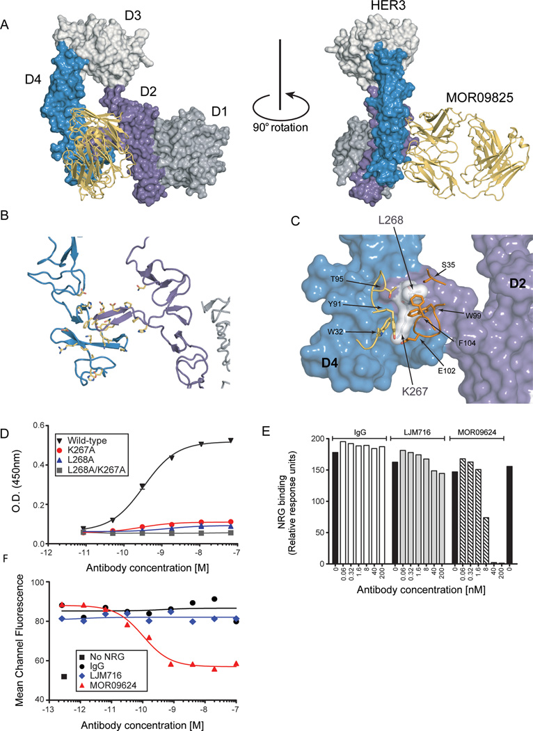
In an effort to understand the mechanism by which LJM716 inhibits HER3, the epitope was identified by determining the three-dimensional structure of the Fab fragment of MOR09825 bound to the HER3 extracellular domain. MOR09825, the parent Fab molecule of LJM716, has identical CDR regions and an identical biological profile to LJM716 (data not shown). The 3.4Å crystal structure reveals that MOR09825 binds to HER3 in the tethered (inactive) ErbB conformation and recognizes a non-linear epitope formed by two distinct domains D2 and D4 (Figure 3A). The epitope is centered over the D2/D4 interface with a similar occluded surface area for each domain (D2–706 Å2; D4–546 Å2) (Figure 3B). This unique binding mode has not been previously observed with other ErbB targeted antibodies and can only occur when domains 2 and 4 are juxtaposed in the tethered (inactive) HER3 conformation (27–29). The structure also reveals that the antibody heavy chain and light chain contribute approximately equally to the recognition of HER3 with the paratope comprising all three heavy chain CDRs and two light chain CDRs (Supplementary Figure S3B).
Figure 3. LJM716 locks HER3 in the inactive conformation and does not block ligand binding.
(A) Crystal structure of the HER3 extracellular domain in complex with MOR09825. Fab is in gold and the HER3 domains are individually colored: D1 (grey), D2 (purple), D3 (white) and D4 (blue). HER3 is in the inactive (tethered) conformation and the ligand binding site located in D1 and D3 is not occluded.
(B) HER3 residues comprising the MOR09825 epitope (yellow ball-and-sticks) are clustered in D2 and D4. MOR09825 is removed from the view to aid visualization.
(C) Close up of MOR09825 interactions with K267 and L268 located near the interface of the HER3 dimerization loop (D2) and auto-inhibitory domain (D4). MOR09825 light chain residues are highlighted in gold and MOR09825 heavy chain residues in orange.
(D) Binding of LJM716 to HER3 mutants K267A, L268A and K267A/ L268A expressed as recombinant proteins and determined by biochemical ELISA.
(E) Interaction analyses performed by capturing biotinylated NRG1 on the surface of a Bioacore™ sensor chip. Preformed HER3/ antibody complexes at the indication concentrations were injected over reference and active surfaces and their interactions with NRG1 observed.
(F) MCF7 cells were incubated with the indicated concentrations of antibodies prior to the addition of 10 nM NRG1-β1 EGF domain. Cell surface bound NRG1 was quantified by flow cytometry and is plotted as mean channel fluorescence on the Y axis.
The crystallographically-defined epitope is corroborated by biochemical characterization of the anti-HER3/HER3 interaction. ELISA characterization of MOR09825 and other Family 15 members binding to recombinantly-expressed HER3 domains and ECD shows that binding is only detected for the full ECD construct. No binding is observed for constructs containing isolated domains or domain pairs, D1-D2, D2, D3–4 or D4 (Supplementary Figure S3C). Additionally, inspection of the crystal structure indicated that HER3 residues Lys267 and Leu268 located within the D2 β-hairpin dimerization arm are involved in numerous interactions with both the heavy and light chains of the antibody (Figure 3C), suggesting they may be fundamentally important for binding. Mutation of Lys267 and/ or Leu268 to alanine abolishes LJM716 binding (Figure 3D), whereas binding of a D3-targeted antibody is largely unaffected by these mutations (Supplementary Figure S3D). These data demonstrate that residues within the β-hairpin dimerization arm are an integral part of the LJM716 epitope. We conclude that binding of LJM716 locks HER3 in the inactive conformation, thus preventing the large-scale structural rearrangements required for transition of HER3 to the extended conformation characteristic of activated ErbB receptors (30–32).
LJM716 does not prevent neuregulin binding to HER3
The crystal structure indicated that the ligand-binding site of HER3, which has been mapped to D1 and D3 by analogy to EGFR (31;32), was not occluded by Fab binding (Figures 3A and 3B). We explored the impact of LJM716 upon the HER3/NRG1 interaction. LJM716/HER3 complexes were pre-formed and passed over a surface plasmon resonance biosensor chip previously coated with NRG1. LJM716/ HER3 complexes were capable of binding immobilized NRG1 while a complex comprised of HER3 and a ligand blocking HER3 antibody (MOR09624) prevented binding to NRG1 (Figure 3E). Furthermore, the presence of LJM716 had no significant impact on the binding affinity of HER3 for NRG1 (Supplementary Figure S4) indicating that LJM716 does not influence NRG1 binding in biochemical assays. The affinity of NRG1 was consistent with that previously determined using a mutant form of HER3 that is in the locked conformation (33). To confirm these results in a cell-based system, we tested whether MCF7 cells pre-bound with LJM716 were capable of binding NRG1. Using flow cytometry we found that pre-binding of MCF7 with LJM716 had no impact on subsequent binding by NRG1 (Figure 3F). These findings strongly suggest that the LJM716 epitope is not essential for ligand binding and that LJM716 and NRG1 can bind concurrently.
LJM716 effectively combines with HER2 and EGFR inhibitors
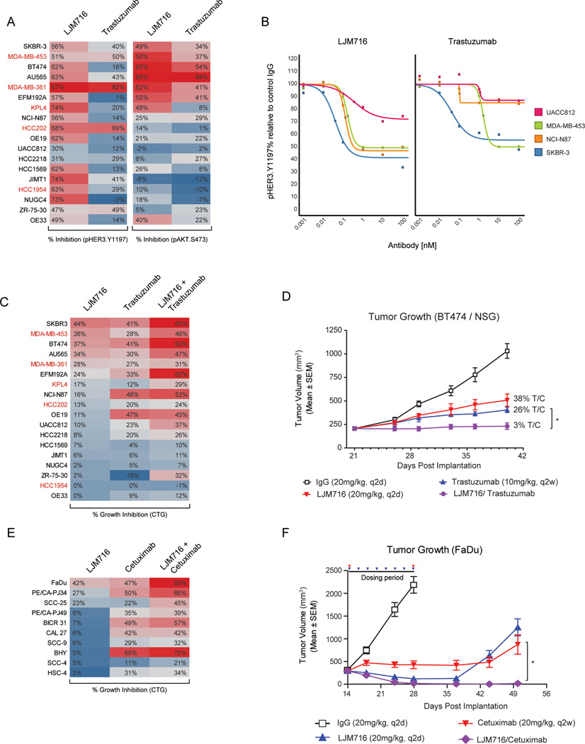
The inability of trastuzumab to fully inhibit HER2/3 complexes is thought to limit its therapeutic benefit, owing to incomplete PI3K pathway suppression (2;3). When we tested trastuzumab’s ability to inhibit pHER3 in a panel of HER2 amplified cell line models grown in full serum, robust (>50%) pHER3 inhibition was observed in only 3/19 cell lines (Figure 4A). In contrast, LJM716 treatment was more broadly active, lowering the IC50 of pHER3 inhibition (example MDA-MB-453) as well as enhancing maximal inhibition (example NCI-N87), so that 14/18 cell lines showed robust (>50%) pHER3 inhibition (Figure 4A & B). LJM716 induced inhibition of pAKT in a sub-set of cell lines (Figure 4A) all of which contained high levels of HER3 phosphorylation and in these cell lines LJM716 was capable of inducing growth inhibition (Figure 4C, Figure S1A & E). Trastuzumab similarly induced inhibition of pAKT (Figure 4A) and proliferation (Figure 4C) in an overlapping sub-set of cell lines despite its inability to effectively inhibit HER3. These data may indicate that HER2 can activate PI3K signaling in a HER3-dependent and independent manner, and that modulation of pAKT may be a key predictor of single-agent activity for both LJM716 and trastuzumab.
Figure 4. LJM716 displays in vitro and in vivo combination activity with trastuzumab and cetuximab.
(A) HER2 amplified cell lines grown in full-serum were treated with 10 nM LJM716 or trastuzumab for one hour and the impact on both pHER3 (Y1197) and pAKT (S473) determined by MSD assay. % inhibition relative to control IgG treated cells is visualized in the form of a heat map colored from blue (0% inhibition) to red (100% inhibition). Cell lines harboring hotspot PI3K mutations are highlighted in red.
(B) The HER2 amplified cell lines UACC812, MDA-MB-453, NCI-N87 and SKBR-3, grown in full-serum, were treated with increasing doses of LJM716 or trastuzumab for one hour and the impact on pHER3 (Y1197) was determined by MSD assay.
(C) HER2 amplified cell lines were dosed with LJM716 (33nM), trastuzumab (33nM) or LJM716 plus trastuzumab. Cells were grown for 5 days and cell viability determined by CTG. % inhibition relative to untreated cells is visualized in the form of a heat map.
(D) BT474 tumor xenografts were grown in NSG mice and treated with IgG (20 mg/kg, q2d), LJM716 (20 mg/kg, q2d), trastuzumab (10 mg/kg, q2w) or LJM716/ trastuzumab. Data are presented as mean tumor volume ±SEM. *p<0.05 by ANOVA post hoc Holm-Sidak test.
(E) SCCHN cell lines were treated with LJM716 (11nM), Cetuximab (11nM) or LJM716/ Cetuximab. Cells were grown for 5 days in full-serum and cell viability determined by CTG. % inhibition relative to untreated cells is visualized in the form of a heat map.
(F) FaDu tumor bearing mice were dosed for 14 days with IgG (20 mg/kg, q2d), LJM716 (20 mg/kg, q2d), Cetuximab (20 mg/kg, q2w) or LJM716/ Cetuximab. The re-growth of tumors was specifically monitored following cessation of dosing on day 28. *p<0.05 by Kruskal-Wallis ANOVA on ranks post hoc Dunn’s test.
To investigate the consequence of HER2 and HER3 dual inhibition we determined the growth inhibitory effect of simultaneously combining LJM716 with trastuzumab. In vitro, the addition of LJM716 to trastuzumab both increased the degree of growth inhibition obtained in sensitive cell lines (e.g. SKBR-3) and also induced a response in cell lines refractory to either agent alone (e.g. KPL4) (Figure 4C, Figure S7). The efficacy of LJM716/trastuzumab combination was tested in vivo using BT474 xenografts grown in NSG mice. BT474 xenografts grown in nude mice are exquisitely sensitive to trastuzumab, due in part to its potent ability to induce ADCC (34). Consequently, NSG mice were used since they lack functional NK cells in addition to B- and T-cells, thus potentially mimicking the impaired ability of trastuzumab to induce ADCC in those patients possessing low affinity Fcγ receptor polymorphisms (35). In NSG mice, both LJM716 and trastuzumab slowed tumor growth when dosed as single agents (Figure 4D). The combination of both agents was well tolerated and showed superior activity compared to either agent alone, inducing prolonged tumor stasis (Figure 4D, Figure S5A).
Since LJM716 also effectively inhibits NRG1-dependent HER3 activation we assessed its activity in models of SCCHN, a lineage that has recently been demonstrated to be driven by a NRG1/HER3 autocrine loop (22). LJM716 displayed single agent growth inhibitory activity in several of these cell lines (Figure 4E). Interestingly, the combination of LJM716 with the EGFR-targeted antibody cetuximab, which is approved for the treatment of metastatic SCCHN, was more potent in vitro compared to either single agent (Figure 4E). In vivo the LJM716/cetuximab combination was well-tolerated and capable of completely eradicating FaDu xenograft tumors (Figure 4F, Figure S5B) and in this regard was superior to either agent alone. These data indicate that LJM716 can inhibit multiple modes of HER3 activation and combination results in enhanced activity with both HER2 and EGFR targeted agents in indications driven by either HER2 or NRG1.
The HER2-targeted antibody pertuzumab, which blocks dimerization of HER2 with other ligand-activated ErbB members, was recently approved for treatment of metastatic breast cancer. We sought to compare the combination activity of trastuzumab/pertuzumab (T+P) with that of trastuzumab/LJM716 (T+L) in a set of HER2 amplified breast cancer cell lines. While T+P and T+L combinations were both highly potent at inhibiting colony, as well as 3D spheroid formation in the trastuzumab sensitive cell line BT474, the LJM716 containing combination was significantly more active in trastuzumab insensitive models like HR6 (36) and MDA-MB-453 cells (Figure 5A & B, Supplementary Figure S6). Increased activity of LJM716 containing treatments was accompanied by enhanced inhibition of HER3 and AKT (Figure 5C). In trastuzumab sensitive BT474 xenografts in vivo, T+P and T+L combinations are equally active at inducing tumor regressions (Figure 5D, S5C). When monitoring overall survival following a 35 day dosing period, both combinations equally prolong survival compared to either T or P alone (Figure 5E). These studies demonstrate that addition of LJM716 to trastuzumab is at least as active as T+P combination in trastuzumab sensitive settings, but enables enhanced inhibition of oncogenic signaling and proliferation in trastuzumab resistant settings.
Figure 5. The addition of LJM716 to trastuzumab enables more potent HER3/PI3K pathway signaling and growth inhibition than trastuzumab/pertuzumab combination, particularly in trastuzumab insensitive models.
(A) Cells were plated at 10,000 to 50,000 cells per well in 6-well plates and treated in triplicate with DMSO, 10 µg/ml LJM716, pertuzumab, and/or trastuzumab. Media containing antibodies was replenished every 3–4 days. Cells were stained with crystal violet when control treated cells were confluent, ranging from 14–21 days. Representative images and quantification of integrated intensity (% control) are shown. *, P < 0.05, t test.
(B) Cells were seeded in Matrigel and allowed to grow in the absence or presence of 10 µg/ml LJM716, pertuzumab, and/or trastuzumab as indicated. Medium was subsequently changed every 3 days. Images shown were recorded 15–19 days after cell seeding. Acini burden was quantified using the GelCount system. Each bar graph represents the mean + S.E.M. of triplicate samples. *, P < 0.05, t test.
(C) BT474 and MDA453 cells were treated with 10 µg/ml LJM 716, 10 µg/ml pertuzumab and/or 10 µg/ml trastuzumab for 1 or 24 hours. Whole cell lysates were prepared and separated in a 7% SDS gel followed by immunoblot analysis with antibodies directed against the indicated proteins.
(D) BT474 xenografts were treated with either IgG (20 mg/kg), trastuzumab (20 mg/kg), pertuzumab (20 mg/kg), LJM716 (20 mg/kg, q2d), trastuzumab+pertuzumab or trastuzumab+LJM716 for 35 days. Data are presented as mean tumor volume ±SEM.
(E) Kaplan-Meyer survival analysis following the end of 35 days of treatment. Mice were monitored for tumor re-growth and sacrificed once tumor burden was larger than 2000 mm3.
Discussion
Clinical and preclinical data suggest that resistance to ErbB targeted therapies occurs frequently and can arise through a variety of mechanisms. Mounting evidence has implicated HER3 in resistance to multiple targeted agents via both ligand-dependent and independent mechanisms (37;38). Through both structural and biochemical studies we have demonstrated that LJM716 binds a novel epitope that specifically locks HER3 in the inactive conformation. LJM716 binding ensures that the HER3 D2 dimerization arm remains inaccessible even in the presence of NRG1 or high-levels of HER2. Therefore, once bound to LJM716, HER3 is unable to transition to the active conformation and interact with partner receptors.
LJM716 inhibitory properties result in significant tumor growth inhibition in several different xenograft models that represent ligand-dependent and -independent modes of HER3 activation. The LJM716 mechanism of action offers several potential advantages when attempting to tackle clinical resistance to targeted agents. Inhibiting multiple modes of HER3 activation may prevent tumors from circumventing LJM716 activity by simply switching between ligand-dependent and ligand-independent HER3 activation as the tumor adapts in response to treatment. Evidence for an enrichment of HER2 amplification was recently shown in cetuximab refractory colorectal tumors (39), suggesting that plasticity in the expression of ErbB-family members known to activate HER3 is clinically relevant. The ability of LJM716 to lock HER3 in the inactive conformation may also prevent HER3 activation through the up-regulation of alternate heterodimer partners (40). A solely HER2 targeted therapy, like trastuzumab/pertuzumab combination is unable to suppress HER3-driven PI3K pathway signaling resulting from a switch from HER2/3 heterodimers to alternate signaling, such as EGFR/HER3 heterodimers (41). Our data demonstrate that NRG1_EGFR_HER3-driven cell lines are sensitive to LJM716 inhibition and that dual targeting of EGFR and HER3 can prevent the emergence of resistance in vivo (Figure 4E & F). One might speculate that consolidated HER family inhibition using a triple combination of HER2, LJM716 (HER3) and EGFR targeted agents, if tolerated, could provide additional benefit by restricting possible routes to resistance through adaptive switching between HER-family dimers.
HER3 is a central component of many interdependent signaling pathways activated in cancer. We hypothesize that combination of LJM716 with either HER2, EGFR or PI3K–targed agents may yield a more effective treatment strategy. Indeed, addition of LJM716 to trastuzumab resulted in increased inhibition of pAKT and improved in vitro efficacy exceeding that achieved by trastuzumab/pertuzumab combination (Figure 4B, Figure 5A & C). Interestingly, the presence of PIK3CA hotspot mutations, a clinically validated mechanism of trastuzumab resistance (2), appears to limit the added activity contributed by trastuzumab in LJM716/trastuzumab combinations (Figure S8). Furthermore, the solely HER2-targeted pertuzumab/trastuzumab combination was less effective than LJM716/trastuzumab in PIK3CA mutant cells, suggesting that PIK3CA mutations more dominantly impact sensitivity to HER2 inhibition compared to HER3 blockade.
The importance of having multiple methods of attacking oncogenic drivers is further underscored by the recent identification of cetuximab resistant EGFR mutants that remained sensitive to panitumumab (42). Since HER2 mutations located within the pertuzumab binding site have already been identified in multiple tumor types (43;44) it is possible that a similar resistance mechanism may arise in pertuzumab treated patients. In these cases LJM716 may present a complementary or superior approach for combination therapy with trastuzumab in both breast and gastric cancer.
In summary, we demonstrate that trapping HER3 in the inactive conformation is a highly effective therapeutic strategy that may be extended to other receptors, which are also activated in both a ligand-dependent and independent manner. Within the ErbB-family, both HER1 and HER4 can adopt a similar inactive conformation and thus may be targeted using an analogous approach. As a result of its novel mechanism LJM716 displays the unique potential to treat HER2/HER3 as well as NRG1_EGFR/ HER3 driven tumors either as a single agent or in combination with HER2, EGFR and PI3K targeted agents. LJM716 is currently undergoing clinical evaluation in HER2-positive breast and gastric cancer, as well as SCCHN.
Supplementary Material
Acknowledgments
The authors would like to thank the Novartis Protein Sciences group, specifically Thomas Pietzonka, Sabine Geiss, Rita Schmitz, Nadine Charara, and Mauro Zurini. In addition, we would like to thank the support of the MorphoSys team for SET affinity measurements. Use of the IMCA-CAT beamline 17-ID at the Advanced Photon Source was supported by the companies of the Industrial Macromolecular Crystallography Association through a contract with Hauptman-Woodward Medical Research Institute. Use of the Advanced Photon Source was supported by the U.S. Department of Energy, Office of Science, Office of Basic Energy Sciences, under Contract No. DE-AC02-06CH11357.
Grant support
This work was supported in part by ACS 118813-PF-10-070-01-TBG (JTG) and DOD BC093376 (JTG) postdoctoral fellowship awards, R01 Grant CA80195 (CLA), American Cancer Society (ACS) Clinical Research Professorship Grant CRP-07-234 (CLA), Breast Cancer Specialized Program of Research Excellence (SPORE) P50 CA98131, Stand Up to Cancer Dream Team Translational Research Grant, a Program of the Entertainment Industry Foundation (SU2C–AACR-DT0209) and Susan G. Komen for the Cure Foundation grant SAC100013 (CLA).
Reference List
- 1.Pohlmann PR, Mayer IA, Mernaugh R. Resistance to Trastuzumab in Breast Cancer. Clin Cancer Res. 2009;15:7479–7491. doi: 10.1158/1078-0432.CCR-09-0636. [DOI] [PMC free article] [PubMed] [Google Scholar]
- 2.Berns K, Horlings HM, Hennessy BT, Madiredjo M, Hijmans EM, Beelen K, et al. A functional genetic approach identifies the PI3K pathway as a major determinant of trastuzumab resistance in breast cancer. Cancer Cell. 2007;12:395–402. doi: 10.1016/j.ccr.2007.08.030. [DOI] [PubMed] [Google Scholar]
- 3.Junttila TT, Akita RW, Parsons K, Fields C, Lewis Phillips GD, Friedman LS, et al. Ligand-independent HER2/HER3/PI3K complex is disrupted by trastuzumab and is effectively inhibited by the PI3K inhibitor GDC-0941. Cancer Cell. 2009;15:429–440. doi: 10.1016/j.ccr.2009.03.020. [DOI] [PubMed] [Google Scholar]
- 4.Nagata Y, Lan KH, Zhou X, Tan M, Esteva FJ, Sahin AA, et al. PTEN activation contributes to tumor inhibition by trastuzumab, and loss of PTEN predicts trastuzumab resistance in patients. Cancer Cell. 2004;6:117–127. doi: 10.1016/j.ccr.2004.06.022. [DOI] [PubMed] [Google Scholar]
- 5.Dave B, Migliaccio I, Gutierrez MC, Wu MF, Chamness GC, Wong H, et al. Loss of phosphatase and tensin homolog or phosphoinositol-3 kinase activation and response to trastuzumab or lapatinib in human epidermal growth factor receptor 2-overexpressing locally advanced breast cancers. J Clin Oncol. 2011;29:166–173. doi: 10.1200/JCO.2009.27.7814. [DOI] [PMC free article] [PubMed] [Google Scholar]
- 6.Motoyama AB, Hynes NE, Lane HA. The efficacy of ErbB receptor-targeted anticancer therapeutics is influenced by the availability of epidermal growth factor-related peptides. Cancer Res. 2002;62:3151–3158. [PubMed] [Google Scholar]
- 7.Gijsen M, King P, Perera T, Parker PJ, Harris AL, Larijani B, et al. HER2 phosphorylation is maintained by a PKB negative feedback loop in response to anti-HER2 herceptin in breast cancer. PLoS Biol. 2010;8:e1000563. doi: 10.1371/journal.pbio.1000563. [DOI] [PMC free article] [PubMed] [Google Scholar]
- 8.Graus-Porta D, Beerli RR, Daly JM, Hynes NE. ErbB-2, the preferred heterodimerization partner of all ErbB receptors, is a mediator of lateral signaling. EMBO J. 1997;16:1647–1655. doi: 10.1093/emboj/16.7.1647. [DOI] [PMC free article] [PubMed] [Google Scholar]
- 9.Jura N, Shan Y, Cao X, Shaw DE, Kuriyan J. Structural analysis of the catalytically inactive kinase domain of the human EGF receptor 3. Proc Natl Acad Sci U S A. 2009;106:21608–21613. doi: 10.1073/pnas.0912101106. [DOI] [PMC free article] [PubMed] [Google Scholar]
- 10.Campbell MR, Amin D, Moasser MM. HER3 comes of age: new insights into its functions and role in signaling, tumor biology, and cancer therapy. Clin Cancer Res. 2010;16:1373–1383. doi: 10.1158/1078-0432.CCR-09-1218. [DOI] [PMC free article] [PubMed] [Google Scholar]
- 11.Mukherjee A, Badal Y, Nguyen XT, Miller J, Chenna A, Tahir H, et al. Profiling the HER3/PI3K pathway in breast tumors using proximity-directed assays identifies correlations between protein complexes and phosphoproteins. PLoS One. 2011;6:e16443. doi: 10.1371/journal.pone.0016443. [DOI] [PMC free article] [PubMed] [Google Scholar]
- 12.Cook RS, Garrett JT, Sanchez V, Stanford JC, Young C, Chakrabarty A, et al. ErbB3 ablation impairs PI3K/Akt-dependent mammary tumorigenesis. Cancer Res. 2011;71:3941–3951. doi: 10.1158/0008-5472.CAN-10-3775. [DOI] [PMC free article] [PubMed] [Google Scholar]
- 13.Holbro T, Beerli RR, Maurer F, Koziczak M, Barbas CF, III, Hynes NE. The ErbB2/ErbB3 heterodimer functions as an oncogenic unit: ErbB2 requires ErbB3 to drive breast tumor cell proliferation. Proc Natl Acad Sci U S A. 2003;100:8933–8938. doi: 10.1073/pnas.1537685100. [DOI] [PMC free article] [PubMed] [Google Scholar]
- 14.Lee-Hoeflich ST, Crocker L, Yao E, Pham T, Munroe X, Hoeflich KP, et al. A central role for HER3 in HER2-amplified breast cancer: implications for targeted therapy. Cancer Res. 2008;68:5878–5887. doi: 10.1158/0008-5472.CAN-08-0380. [DOI] [PubMed] [Google Scholar]
- 15.Chandarlapaty S, Sawai A, Scaltriti M, Rodrik-Outmezguine V, Grbovic-Huezo O, Serra V, et al. AKT inhibition relieves feedback suppression of receptor tyrosine kinase expression and activity. Cancer Cell. 2011;19:58–71. doi: 10.1016/j.ccr.2010.10.031. [DOI] [PMC free article] [PubMed] [Google Scholar]
- 16.Garrett JT, Olivares MG, Rinehart C, Granja-Ingram ND, Sanchez V, Chakrabarty A, et al. Transcriptional and posttranslational up-regulation of HER3 (ErbB3) compensates for inhibition of the HER2 tyrosine kinase. Proc Natl Acad Sci U S A. 2011;108:5021–5026. doi: 10.1073/pnas.1016140108. [DOI] [PMC free article] [PubMed] [Google Scholar]
- 17.Chakrabarty A, Sanchez V, Kuba MG, Rinehart C, Arteaga CL. Feedback upregulation of HER3 (ErbB3) expression and activity attenuates antitumor effect of PI3K inhibitors. Proc Natl Acad Sci U S A. 2011;109:2718–2723. doi: 10.1073/pnas.1018001108. [DOI] [PMC free article] [PubMed] [Google Scholar]
- 18.Narayan M, Wilken JA, Harris LN, Baron AT, Kimbler KD, Maihle NJ. Trastuzumab-induced HER reprogramming in "resistant" breast carcinoma cells. Cancer Res. 2009;69:2191–2194. doi: 10.1158/0008-5472.CAN-08-1056. [DOI] [PubMed] [Google Scholar]
- 19.Agus DB, Akita RW, Fox WD, Lewis GD, Higgins B, Pisacane PI, et al. Targeting ligand-activated ErbB2 signaling inhibits breast and prostate tumor growth. Cancer Cell. 2002;2:127–137. doi: 10.1016/s1535-6108(02)00097-1. [DOI] [PubMed] [Google Scholar]
- 20.Baselga J, Cortes J, Kim SB, Im SA, Hegg R, Im YH, et al. Pertuzumab plus trastuzumab plus docetaxel for metastatic breast cancer. N Engl J Med. 2012;366:109–119. doi: 10.1056/NEJMoa1113216. [DOI] [PMC free article] [PubMed] [Google Scholar]
- 21.Garrett JT, Sutton CR, Kuba MG, Cook RS, Arteaga CL. Dual blockade of HER2 in HER2-overexpressing tumor cells does not completely eliminate HER3 function. Clin Cancer Res. 2013;19:610–619. doi: 10.1158/1078-0432.CCR-12-2024. [DOI] [PMC free article] [PubMed] [Google Scholar]
- 22.Wilson TR, Lee DY, Berry L, Shames DS, Settleman J. Neuregulin-1-Mediated Autocrine Signaling Underlies Sensitivity to HER2 Kinase Inhibitors in a Subset of Human Cancers. Cancer Cell. 2011;20:158–172. doi: 10.1016/j.ccr.2011.07.011. [DOI] [PubMed] [Google Scholar]
- 23.Sheng Q, Liu X, Fleming E, Yuan K, Piao H, Chen J, et al. An activated ErbB3/NRG1 autocrine loop supports in vivo proliferation in ovarian cancer cells. Cancer Cell. 2010;17:298–310. doi: 10.1016/j.ccr.2009.12.047. [DOI] [PMC free article] [PubMed] [Google Scholar]
- 24.Prassler J, Steidl S, Urlinger S. In vitro affinity maturation of HuCAL antibodies: complementarity determining region exchange and RapMAT technology. Immunotherapy. 2009;1:571–583. doi: 10.2217/imt.09.23. [DOI] [PubMed] [Google Scholar]
- 25.Haenel C, Satzger M, Ducata DD, Ostendorp R, Brocks B. Characterization of high-affinity antibodies by electrochemiluminescence-based equilibrium titration. Anal Biochem. 2005;339:182–184. doi: 10.1016/j.ab.2004.12.032. [DOI] [PubMed] [Google Scholar]
- 26.Barretina J, Caponigro G, Stransky N, Venkatesan K, Margolin AA, Kim S, et al. The Cancer Cell Line Encyclopedia enables predictive modelling of anticancer drug sensitivity. Nature. 2012;483:603–607. doi: 10.1038/nature11003. [DOI] [PMC free article] [PubMed] [Google Scholar]
- 27.Cho HS, Leahy DJ. Structure of the extracellular region of HER3 reveals an interdomain tether. Science. 2002;297:1330–1333. doi: 10.1126/science.1074611. [DOI] [PubMed] [Google Scholar]
- 28.Bouyain S, Longo PA, Li S, Ferguson KM, Leahy DJ. The extracellular region of ErbB4 adopts a tethered conformation in the absence of ligand. Proc Natl Acad Sci U S A. 2005;102:15024–15029. doi: 10.1073/pnas.0507591102. [DOI] [PMC free article] [PubMed] [Google Scholar]
- 29.Ferguson KM, Berger MB, Mendrola JM, Cho HS, Leahy DJ, Lemmon MA. EGF activates its receptor by removing interactions that autoinhibit ectodomain dimerization. Mol Cell. 2003;11:507–517. doi: 10.1016/s1097-2765(03)00047-9. [DOI] [PubMed] [Google Scholar]
- 30.Cho HS, Mason K, Ramyar KX, Stanley AM, Gabelli SB, Denney DW, Jr, et al. Structure of the extracellular region of HER2 alone and in complex with the Herceptin Fab. Nature. 2003;421:756–760. doi: 10.1038/nature01392. [DOI] [PubMed] [Google Scholar]
- 31.Ogiso H, Ishitani R, Nureki O, Fukai S, Yamanaka M, Kim JH, et al. Crystal structure of the complex of human epidermal growth factor and receptor extracellular domains. Cell. 2002;110:775–787. doi: 10.1016/s0092-8674(02)00963-7. [DOI] [PubMed] [Google Scholar]
- 32.Garrett TP, McKern NM, Lou M, Elleman TC, Adams TE, Lovrecz GO, et al. Crystal structure of a truncated epidermal growth factor receptor extracellular domain bound to transforming growth factor alpha. Cell. 2002;110:763–773. doi: 10.1016/s0092-8674(02)00940-6. [DOI] [PubMed] [Google Scholar]
- 33.Kani K, Park E, Landgraf R. The extracellular domains of ErbB3 retain high ligand binding affinity at endosome pH and in the locked conformation. Biochemistry. 2005;44:15842–15857. doi: 10.1021/bi0515220. [DOI] [PubMed] [Google Scholar]
- 34.Clynes RA, Towers TL, Presta LG, Ravetch JV. Inhibitory Fc receptors modulate in vivo cytotoxicity against tumor targets. Nat Med. 2000;6:443–446. doi: 10.1038/74704. [DOI] [PubMed] [Google Scholar]
- 35.Musolino A, Naldi N, Bortesi B, Pezzuolo D, Capelletti M, Missale G, et al. Immunoglobulin G fragment C receptor polymorphisms and clinical efficacy of trastuzumab-based therapy in patients with HER-2/neu-positive metastatic breast cancer. J Clin Oncol. 2008;26:1789–1796. doi: 10.1200/JCO.2007.14.8957. [DOI] [PubMed] [Google Scholar]
- 36.Ritter CA, Perez-Torres M, Rinehart C, Guix M, Dugger T, Engelman JA, et al. Human breast cancer cells selected for resistance to trastuzumab in vivo overexpress epidermal growth factor receptor and ErbB ligands and remain dependent on the ErbB receptor network. Clin Cancer Res. 2007;13:4909–4919. doi: 10.1158/1078-0432.CCR-07-0701. [DOI] [PubMed] [Google Scholar]
- 37.Sergina NV, Rausch M, Wang D, Blair J, Hann B, Shokat KM, et al. Escape from HER-family tyrosine kinase inhibitor therapy by the kinase-inactive HER3. Nature. 2007;445:437–441. doi: 10.1038/nature05474. [DOI] [PMC free article] [PubMed] [Google Scholar]
- 38.Montero JC, Rodriguez-Barrueco R, Ocana A, Diaz-Rodriguez E, Esparis-Ogando A, Pandiella A. Neuregulins and cancer. Clin Cancer Res. 2008;14:3237–3241. doi: 10.1158/1078-0432.CCR-07-5133. [DOI] [PubMed] [Google Scholar]
- 39.Yonesaka K, Zejnullahu K, Okamoto I, Satoh T, Cappuzzo F, Souglakos J, et al. Activation of ERBB2 Signaling Causes Resistance to the EGFR-Directed Therapeutic Antibody Cetuximab. Sci Transl Med. 2011;3 doi: 10.1126/scitranslmed.3002442. 99ra86. [DOI] [PMC free article] [PubMed] [Google Scholar]
- 40.Engelman JA, Zejnullahu K, Mitsudomi T, Song Y, Hyland C, Park JO, et al. MET amplification leads to gefitinib resistance in lung cancer by activating ERBB3 signaling. Science. 2007;316:1039–1043. doi: 10.1126/science.1141478. [DOI] [PubMed] [Google Scholar]
- 41.Wheeler DL, Huang S, Kruser TJ, Nechrebecki MM, Armstrong EA, Benavente S, et al. Mechanisms of acquired resistance to cetuximab: role of HER (ErbB) family members. Oncogene. 2008;27:3944–3956. doi: 10.1038/onc.2008.19. [DOI] [PMC free article] [PubMed] [Google Scholar]
- 42.Montagut C, Dalmases A, Bellosillo B, Crespo M, Pairet S, Iglesias M, et al. Identification of a mutation in the extracellular domain of the Epidermal Growth Factor Receptor conferring cetuximab resistance in colorectal cancer. Nat Med. 2012;18:221–223. doi: 10.1038/nm.2609. [DOI] [PubMed] [Google Scholar]
- 43.Ding L, Getz G, Wheeler DA, Mardis ER, McLellan MD, Cibulskis K, et al. Somatic mutations affect key pathways in lung adenocarcinoma. Nature. 2008;455:1069–1075. doi: 10.1038/nature07423. [DOI] [PMC free article] [PubMed] [Google Scholar]
- 44.Kan Z, Jaiswal BS, Stinson J, Janakiraman V, Bhatt D, Stern HM, et al. Diverse somatic mutation patterns and pathway alterations in human cancers. Nature. 2010;466:869–873. doi: 10.1038/nature09208. [DOI] [PubMed] [Google Scholar]
Associated Data
This section collects any data citations, data availability statements, or supplementary materials included in this article.


