Abstract
The mechanical properties of enamel and dentin were studied using test specimens having the same shape and dimensions because these properties might vary with the experimental conditions and specimen shapes and dimensions. Healthy human teeth were used as specimens for mechanical tests. The stress (MPa), strain (%), and elastic modulus (E, MPa) of the specimens were obtained from compression tests. The maximum stresses of the enamel, dentin, and enamel–dentin specimens were 62.2 ± 23.8, 193.7 ± 30.6, and 126.1 ± 54.6 MPa, respectively. The maximum strains of the enamel, dentin, and enamel–dentin specimens were 4.5 ± 0.8%, 11.9 ± 0.1%, and 8.7 ± 2.7%, respectively. The elastic moduli of the enamel, dentin, and enamel–dentin specimens were 1338.2 ± 307.9, 1653.7 ± 277.9, and 1628.6 ± 482.7 MPa, respectively. The measured hardness value of enamel specimens (HV = 274.8 ± 18.1) was around 4.2 times higher than that of dentin specimens (HV = 65.6 ± 3.9). Judging from the measured values of the stress and strain of enamel specimens, enamel tended to fracture earlier than dentin; therefore, it was considered more brittle than dentin. However, judging from the measured hardness values, enamel was considered harder than dentin. Therefore, enamel has higher wear resistance, making it suitable for grinding and crushing foods, and dentin has higher force resistance, making it suitable for absorbing bite forces. The different mechanical roles of enamel and dentin may arise from their different compositions and internal structures, as revealed through scanning electron micrographs of enamel and dentin.
Keywords: Mechanical properties, compression tests, hardness tests, enamel, dentin
Introduction
Dental hard tissue comprises a combination of enamel and dentin, both of which have different compositions and structures. Enamel is mainly made of the mineral hydroxyapatite, which is crystalline calcium phosphate.1 It has a glossy surface and varies in color from light yellow to grayish white. It is the hardest tissue in the human body because it contains almost no water. Structurally, enamel covers the entire anatomic crown of the tooth above the gum and protects the dentin. During mastication, enamel comes in direct contact with food.2,3 Enamel comprises millions of trichome-like hexagonal-prism-shaped enamel rods having a diameter of 3–5 µm, which is 1/100th the thickness of hair. Tooth enamel does not regenerate once abraded or eroded.4 Enamel varies in thickness and density over the tooth surface, being thickest and hardest at the cusps or biting edges. The enamel of deciduous teeth is less hard than and half as thick as that of permanent teeth.5
Dentin consists of the mineral hydroxyapatite (70%), organic material (20%), and water (10%).6 Dentin is harder than bone but softer than enamel, and it is mostly made of phosphoric apatite crystallites.7 Tooth decay leads to the formation of cavities in the tooth. This process starts with a small hole on the surface of the hard enamel; however, once the decay passes through the enamel, it spreads wider and deeper because the softer dentin is generally easily decayed.2,3 Odontoblasts, which initiate dentin formation, are located around the pulp chamber as a single layer. The calcium ions of dentin and odontoblasts are connected by channels within dentinal tubules.6,7 Pain, pressure, and temperature are delivered to the nerves in the dentinal tubules via the channels of odontoblasts.8,9
Previous studies have elucidated the mechanical properties—specifically, the elastic modulus—of enamel and dentin through compression tests, nanoindentation tests, transmitted light microscopy (TLM) observation, and nanohardness tests (NHT).10–21 However, as enamel and dentin are not homogeneous and isotropic materials, they do not obey Hook’s law. Therefore, a comparison of mechanical properties based on the elastic modulus alone is not feasible; it is essential to also consider the stress and strain values. Furthermore, these studies did not investigate the mechanical properties of enamel and dentin using test specimens having the same shapes and dimensions.
Now, these properties may vary depending on the experimental conditions and specimen shapes and dimensions. Therefore, this study aims to determine the mechanical properties and primary roles of enamel and dentin by using test specimens having the same shapes and dimensions under the same experimental conditions. Specimens of dental hard tissues—enamel, dentin, and enamel–dentin (ED) composite—were used for compression tests, and the obtained data were analyzed to determine the mechanical properties with respect to the bite force. Furthermore, the hardness of enamel and dentin was tested and analyzed with respect to the wear resistance.
Materials and methods
Specimen preparation
Compression tests
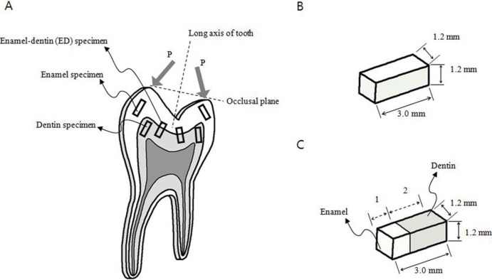
Five healthy human canines and 10 first premolars (age = 19.3 ± 4.1 years) were extracted and used as dental hard tissue specimens for the mechanical tests. Protocols were approved by the Institutional Review Board of the Samsung Medical Center for human research, and all individuals gave informed consent (IRB No. KBC12150). The extracted teeth were fixed in epoxy resin (ELR-3200; SPC Inc., Korea) moldings (epoxy resin:epoxy hardener = 40:10) and then cross-sectioned, as shown in Figure 1(a). The cross-sectioned teeth were machine-cut to obtain 10 dentin specimens, 10 enamel specimens, and 10 ED specimens having width, height, and length of 1.2, 1.2, and 3.0 mm, respectively, for the compression tests, as shown in Figure 1(b) and (c). Here, the geometric error was ±0.02 mm for the machine-cut process described below. Furthermore, the specimens were machine-cut along the direction of the force applied.
Figure 1.
Specimen preparation: (a) cross-sectioned after fixing a tooth within epoxy resin, (b) dimensions of enamel and dentin specimens, and (c) dimensions of ED specimens.
ED: enamel–dentin.
Here, P is load or force. Specimens were machine-cut and extracted along the direction of the force applied.
The teeth were cross-sectioned and machine-cut at a feeding speed of 0.2 mm/s and 1000 r/min by using a water-spray-cooled carborundum wheel (PSI-140-5L, Eagle Superabrasives, Inc, USA) in a micro diamond saw machine (RB216 Culux; R&B Inc., Korea) that can cut at a feeding speed of 0.005–5 mm/s and 200–5000 r/min. Each specimen was prepared for testing after it was wet-sand polished (METPOL2; R&B Inc., Korea) using a #3000 wet sanding polisher at 45 r/min, the speed of which ranged from 25 to 300 r/min. After polishing, each specimen was viewed at 40× magnification to check for the presence of cracks and to identify potential damages that may have occurred during polishing using a microscope (IX71; Olympus, Japan) with a 10× objective lens and 4×, 10×, 20×, and 40× eyepiece lenses. No cracks were found in most of the specimens. Finally, the specimens were kept in normal saline at room temperature (22°C) immediately before testing.
Here, the above machine-cut and polishing process can be new techniques to prepare the enamel and dentin specimens with the shape of a rectangular parallelepiped along the force direction which have a very small geometric error (±0.02 mm) without cracks.
Hardness test
After epoxy resin molding, the extracted mandibular canine (#43, Fédération dentaire internationale (FDI) tooth-numbering system) was cross-sectioned by using a micro diamond saw machine (RB216 Culux) at a feeding speed of 0.2 mm/s and 1000 r/min to maximize the square measurement area of the sectioned surface and to obtain dental hard tissue specimens for hardness tests (Figure 2). Two cross-sectioned specimens were prepared for the hardness test, and the sectioned surfaces were wet-sand polished with #400, #1200, and #3000 (in order) polishers (METPOL2) at 45 r/min.
Figure 2.
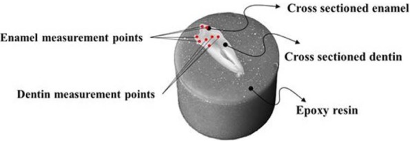
Cross-sectioned tooth specimen in epoxy resin for hardness test with measurement points.
Testing of mechanical properties
Compression test
A micro-load system (Universal Testing System; R&B Inc., Korea) was used for the compression tests (Figure 3(a)). Its maximum force capacity was 5 kN, its crosshead loading speed ranged from 0.0001 to 1600 mm/min, and it had ±0.5% operating accuracy. The volume of the load cell was decided by considering the failure loads of the specimens. The load cell was installed in the upper jig of the micro-load system to measure the load during the compression tests (Figure 3(b)). A 100-kgf load cell (SM603; MScell Inc., Korea) was used for the enamel, dentin, and ED specimens. A prepared specimen was placed on the lower jig for the compression test (Figure 3(c)). Subsequently, the upper jig was moved toward the lower jig, and then, the displacement and force were reset to zero when the force measured in the load cell was 0.05 N.
Figure 3.
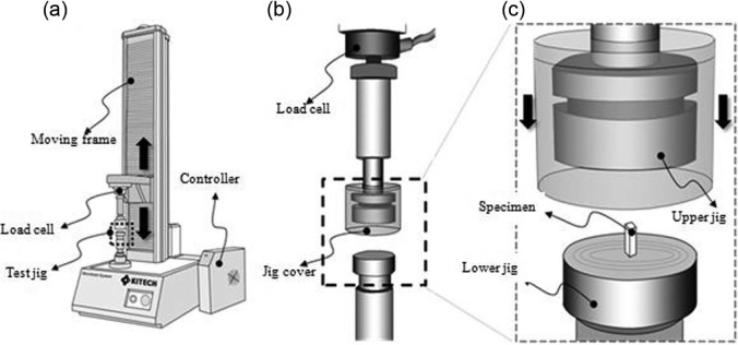
Micro-load system used in compression test: (a) selectable grip on lower part of the jig that is installed with a load cell on the upper part of the jig; (b) compression test jig—a jig cover was installed to prevent the broken pieces from popping when the specimens reached their breaking points; (c) compression test was performed after placing the specimens on the lower jig and lowering the upper jig.
In all compression tests, the loading speed was set at 0.1 mm/min. Compressive stresses (σ in MPa) and strains (ε in %) of the dental hard tissues and dental materials were respectively calculated using equations (1) and (2), and the elastic modulus was calculated using equation (3)
| (1) |
where σ = compressive stress, P = load or force, and A= cross-sectional area of specimen
| (2) |
where ε = strain, l = original length, and l′ = changed length, λ = l′−l)
| (3) |
where E = modulus of elasticity.
Hardness test
The hardness (Dimensionless Vickers Pyramid Number: HV) of the polished surfaces of the teeth was measured using a Vickers diamond indenter in a standard micro-hardness tester (HM; Mitutoyo, Japan) with 25 g to 1 kg loading control. An epoxy resin molding was fixed on the slide of the hardness tester and a 300-g indentation load was loaded to measure the hardness of the polished section. Data were obtained from four points on the enamel and four points on the dentin (Figure 2). After the hardness measurement, the indentation diagonal size and crack size were measured by optical microscopy in the micro-hardness tester.
Results
Compression test
The maximum force (N) and maximum displacement (mm) of each specimen were obtained from the compression test. The maximum stress (MPa) was obtained from equation (1), maximum strain (%) from equation (2), and modulus of elasticity (E, MPa) from equation (3).
Table 1 shows the mean values and standard deviations of the maximum stress (MPa), maximum strain (%), and E (MPa) of each material. A t-test was used to examine the significances of the maximum stress, maximum strain, and E values. The maximum stresses of the enamel, dentin, and ED specimens were 62.2 ± 23.8, 193.7 ± 30.6, and 126.1 ± 54.6 MPa, respectively. The maximum strains of the enamel, dentin, and ED specimens were 4.5 ± 0.8%, 11.9 ± 0.1%, and 8.7 ± 2.7%, respectively. The elastic modulus values of the enamel, dentin, and ED specimens were 1338.2 ± 307.9, 1653.7 ± 277.9, and 1628.6 ± 482.7 MPa, respectively; these values did not show a significant difference in the t-test (p > 0.1).
Table 1.
Mean values and standard deviations of mechanical properties of dental hard tissues from compression tests (n = 10/material).
| Compression test results | |||
|---|---|---|---|
| Specimen | Maximum stress (MPa) | Maximum strain (%) | E (MPa) |
| Enamel | 62.2 ± 23.8 | 4.5 ± 0.8 | 1338.2 ± 307.9† |
| Dentin | 193.7 ± 30.6 | 11.9 ± 0.1 | 1653.7 ± 277.9† |
| ED | 126.1 ± 54.6 | 8.7 ± 2.7 | 1628.6 ± 482.7† |
E: elastic modulus; ED: enamel–dentin.
Not significantly different (p > 0.1) in t-test.
Figure 4 shows a typical stress–strain curve of a specific test specimen, which represents a stress–strain relationship in the enamel, dentin, and ED specimens. The linear regression equation indicated that R2 for the enamel, dentin, and ED specimens was 0.9970, 0.9960, and 0.9988, respectively. The slope of the typical stress–strain curve of each specimen indicates the elastic modulus of that specimen. Judging from the slopes of all the materials in Figure 4 and Table 1, the elastic modulus of dentin was the highest, making it the stiffest material with a stiffness of 1653 ± 277.9 MPa. Furthermore, judging from the maximum stress and maximum strain of enamel, enamel tended to fracture earlier than the other specimens and was therefore considered a brittle material.
Figure 4.
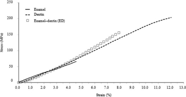
A typical stress–strain curve of the enamel, dentin, and ED specimens.
ED: enamel–dentin.
Hardness test
Table 2 shows the hardness values of the dental hard tissues. The hardness values of enamel and dentin were obtained through Vickers hardness tests and from Craig and Peyton.22 The hardness value of enamel (HV = 274.8 ± 18.1) was around 4.2 times higher than that of dentin (HV = 65.6 ± 3.9).
Table 2.
Vickers hardness (HV) of dental hard tissues.
| Dental hard tissue | Hardness (HV) from tests: mean (SD) | Hardness (HV) from reference |
|---|---|---|
| Enamel | 274.8 (18.1) | 283–37422 |
| Dentin | 65.6 (3.9) | 53–6322 |
SD: standard deviation.
Discussion and conclusion
The test results, listed in Table 1, showed that enamel and dentin, the basic components of a tooth, have different mechanical properties. The maximum stresses of enamel and dentin were 62.2 ± 23.8 and 193.7 ± 30.6 MPa, respectively, and that of dentin was around 3 times higher than that of enamel. Furthermore, the maximum strains of enamel and dentin were 4.5 ± 0.8% and 11.9 ± 0.1%, respectively, that is, that of dentin was around 2.5 times higher than that of enamel. The maximum stress and maximum strain of the ED specimens were 126.1 ± 54.6 MPa and 8.7 ± 2.7%, respectively; that is, the values of the mechanical properties of ED specimens were between those of enamel and dentin. These values were thought to be proportional to the amounts of enamel and dentin in the ED specimen (Table 1 and Figure 1(c)).
It was found that the differences were due to the differences in the composition and microstructure of the dental hard tissues. Enamel specimens comprise rows of hydroxyapatite (calcium and phosphorus salts) embedded in a protein matrix. Dentin specimens comprise mineralized connective tissue. ED specimens comprise both enamel and dentin, which are connected by the dentino-enamel junction (DEJ). As shown in Figure 5, enamel is rough and brittle, whereas dentin is smooth and solid.
Figure 5.
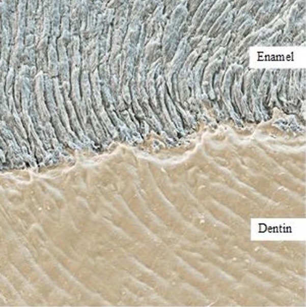
Dental hard tissues: colored scanning electron micrograph (SEM) of the boundary between tooth enamel (top) and dentin (bottom). Enamel is the outer covering of the crown (visible part) of the tooth. It is the hardest substance in the human body, comprising rows of hydroxyapatite (calcium and phosphorus salts) embedded in a protein matrix. Dentin makes up the majority of the tooth. It comprises mineralized connective tissue. Magnification: 500× when printed 10 cm wide (permitted by http://www.sciencephoto.com).
SEM: scanning electron micrograph.
Table 3 lists the elastic moduli of enamel and dentin obtained in this and previous studies. In the compression test, the elastic modulus of enamel was ~8.2–95.8 GPa and that of dentin was ~5.3 MPa to 13.3 GPa. In the nanoindentation test, the elastic modulus of enamel was ~3.2–98.3 GPa and that of dentin was 2–100 GPa. In TLM/NHT, the elastic modulus of dentin was ~30 GPa. Among previous studies, only Stanford et al.,10 and Fong et al.15reported the elastic moduli of both enamel and dentin. In this study, the elastic moduli of enamel, dentin, and ED specimens were found to be ~1.3 ± 0.3, 1.7 ± 0.3, and 1.6 ± 0.5 GPa, respectively. The value of the elastic modulus of enamel was around 1/6 to 1/65 lower than those in previous studies, and that of dentin was around 1/8 lower to 310 times higher than those in previous studies. However, as mentioned in the section “Introduction,” comparisons of the mechanical properties of dental hard tissues based on the elastic modulus alone are not feasible because they do not obey Hook’s law.
Table 3.
Elastic moduli of human enamel and dentin reported in this and previous studies.
| References | Test type | Shape | N | Dimensions (mm) | Tooth | E of enamel | E of dentin |
|---|---|---|---|---|---|---|---|
| This study | CMP |

|
Enamel 10 | 1.2 × 1.2 × 3.0 | Canines, premolars | 1338.2 ± 307.9 MPa | 1653.7 ± 277.9 MPa |
| Dentin 10 | |||||||
| Stanford et al.10 | CMP |

|
Enamel 14 | Diameter 0.9–1.57 | Unspecified | 8.2–37.2 GPa | 8.3–12.3 GPa |
| Dentin 8 | Length 0.7–2.2 | ||||||
| Craig et al.11 | CMP |

|
29 | Diameter 0.8 | Mandibular first molars | 62.7–95.8 GPa | – |
| Length 0.8–2.4 | |||||||
| Trengrove et al.12 | CMP |

|
68 | Diameter 4.0 | Central incisors; upper, lower canine | – | 5.3 ± 1.6 to 6.1 ± 1.6 MPa |
| Length 4.0 | |||||||
| Jantarat et al.13 | CMP |

|
9 | Outer diameter 3.5 | Maxillary incisors, canines | – | 13.3 ± 1.3 GPa |
| Internal diameter 1.5 | |||||||
| Length 6–10 | |||||||
| Haines14 | CMP | Cross-sectioned | – | Thickness 0.254 | Unspecified | – | 11.0 GPa |
| Fong et al.15 | N-I | Cross-sectioned | 6 | – | Incisor teeth | 98.3 ± 5.9 GPa | 24.8 ± 1.4 GPa |
| He et al.16 | N-I | Cross-sectioned | – | – | Premolar | 40–80 GPa | 60–100 GPa |
| Habelitz et al.17 | N-I | Cross-sectioned | 4 | – | Third molars | 75–90 GPa | – |
| Marshall et al.18 | N-I | Cross-sectioned | 5 | Thickness 1.0 | Third molars | – | 19.65 GPa |
| Arcís et al.19 | N-I | Cross-sectioned | 5 | Diameter 8.0, Thickness 2.0 | Unspecified | – | 2–8 GPa |
| Padmanabhan et al.20 | N-I | Cross-sectioned | 4 | – | Molar teeth | 3.22–3.51 GPa | – |
| Senawongse et al.21 | TLM, NHT | Cross-sectioned | 10 | Thickness 5 | Aged molars, young third molars | – | 29.9 ± 5.4 GPa |
| 10 | 30.9 ± 9.1 GPa |
E: elastic modulus, CMP: compression test, N-I: Nanoindentation, TLM: transmitted light microscopy, NHT: nanohardness test.
The stresses, strains, and elastic moduli of dental hard tissues as obtained from compression tests were compared with respect to the bite force. Furthermore, the hardness of dental hard tissues as obtained from Vickers hardness tests was compared and analyzed with respect to the wear resistance.
Judging from the stress and strain of the enamel specimens, enamel tended to fracture earlier than dentin and was therefore considered more brittle than dentin. However, judging from the measured hardness values, enamel was considered harder than dentin. Therefore, the mechanical role played by enamel is to grind (crush) food and protect dentin because of its higher wear resistance, and that played by dentin is to absorb bite forces because of its higher force resistance. The different mechanical roles of enamel and dentin may arise from their different compositions and internal microstructures, as revealed by the scanning electron micrographs (SEMs) shown in Figure 5 (top and bottom, respectively).
Future studies should address the mechanical properties of existing tooth reconstruction materials such as amalgam, dental ceramic, dental resin, gold alloy, zirconia, and titanium alloy using specimens having the same shapes and dimensions to determine the most suitable tooth reconstruction material for substituting the enamel and dentin parts of the tooth. Furthermore, the results of this and future studies can provide useful information for the development of a new tooth reconstruction material.
Footnotes
Declaration of conflicting interests: There is no conflict of interest regarding this manuscript.
Funding: This research was supported by KITECH, Republic of Korea.
References
- 1. Staines M, Robinson WH, Hood JAA. Spherical indentation of tooth enamel. J Mater Sci 1981; 16(9): 2551–2556 [Google Scholar]
- 2. Tillberg A, Järvholm B, Berglund A. Risks with dental materials. Dent Mater 2008; 24(7): 940–943 [DOI] [PubMed] [Google Scholar]
- 3. Vaderhobli RM. Advances in dental materials. Dent Clin North Am 2011; 55(3): 619–625 [DOI] [PubMed] [Google Scholar]
- 4. Cuy JL, Mann AB, Livi KJ, et al. Nanoindentation mapping of the mechanical properties of human molar tooth enamel. Arch Oral Biol 2002; 47: 281–291 [DOI] [PubMed] [Google Scholar]
- 5. Gašperšič D. Micromorphometric analysis of cervical enamel structure of human upper third molars. Arch Oral Biol 1995; 40(5): 453–457 [DOI] [PubMed] [Google Scholar]
- 6. Ten Cate AR. Oral histology: development, structure, and function. 5th ed. Toronto: Mosby, 1998, p. 150 [Google Scholar]
- 7. Marshall GW, Jr, Marshall SJ, Kinney JH, et al. The dentin substrate: structure and properties related to bonding. J Dent 1997; 25(6): 441–458 [DOI] [PubMed] [Google Scholar]
- 8. Arana-Chavez VE, Massa LF. Odontoblasts: the cells forming and maintaining dentine. Int J Biochem Cell Biol 2004; 36(8): 1367–1373 [DOI] [PubMed] [Google Scholar]
- 9. Murray PE, About I, Lumley PJ, et al. Odontoblast morphology and dental repair. J Dent 2003; 31(1): 75–82 [DOI] [PubMed] [Google Scholar]
- 10. Stanford WJ, Paffenbarger GC, Kumpula WJ, et al. Determination of some compressive properties of human enamel and dentin. J Am Dent Assoc 1958; 57(4): 487–495 [DOI] [PubMed] [Google Scholar]
- 11. Craig RG, Peyton FA, Johnson DW. Compressive properties of enamel, dental cements and gold. J Dent Res 1961; 40: 936–945 [Google Scholar]
- 12. Trengrove HG, Carter GM, Hood JAA. Stress relaxation properties of human dentin. Dent Mater 1995; 11(5–6): 305–310 [DOI] [PubMed] [Google Scholar]
- 13. Jantarat J, Palamara JEA, Lindner C, et al. Time-dependent properties of human root dentin. Dent Mater 2002; 18(6): 486–493 [DOI] [PubMed] [Google Scholar]
- 14. Haines DJ. Physical properties of human tooth enamel and enamel sheath material under load. J Biomech 1968; 1: 117–125 [DOI] [PubMed] [Google Scholar]
- 15. Fong H, Sarikaya M, White SN, et al. Nano-mechanical properties profiles across dentin–enamel junction of human incisor teeth. Mater Sci Eng C 2000; 7: 119–128 [Google Scholar]
- 16. He LH, Fujisawa N, Swain MV. Elastic modulus and stress–strain response of human enamel by nano-indentation. Biomaterials 2006; 27: 4388–4398 [DOI] [PubMed] [Google Scholar]
- 17. Habelitz S, Marshall SJ, Marshall GW, Jr, et al. Mechanical properties of human dental enamel on the nanometre scale. Arch Oral Biol 2001; 46: 173–183 [DOI] [PubMed] [Google Scholar]
- 18. Marshall GW, Jr, Balooch M, Gallagher RR, et al. Mechanical properties of the dentinoenamel junction: AFM studies of nanohardness, elastic modulus, and fracture. J Biomed Mater Res 2001; 54: 87–95 [DOI] [PubMed] [Google Scholar]
- 19. Arcís RW, López-Macipe A, Toledano M, et al. Mechanical properties of visible light-cured resins reinforced with hydroxyapatite for dental restoration. Dent Mater 2002; 18(1): 49–57 [DOI] [PubMed] [Google Scholar]
- 20. Padmanabhan SK, Balakrishnan A, Chu MC, et al. Micro-indentation fracture behavior of human enamel. Dent Mater 2010; 26: 100–104 [DOI] [PubMed] [Google Scholar]
- 21. Senawongse P, Otsuki M, Tagami J, et al. Age-related changes in hardness and modulus of elasticity of dentine. Arch Oral Biol 2006; 51(6): 457–463 [DOI] [PubMed] [Google Scholar]
- 22. Craig RG, Peyton FA. The microhardness of enamel and dentin. J Dent Res 1958; 37: 661–668 [DOI] [PubMed] [Google Scholar]


