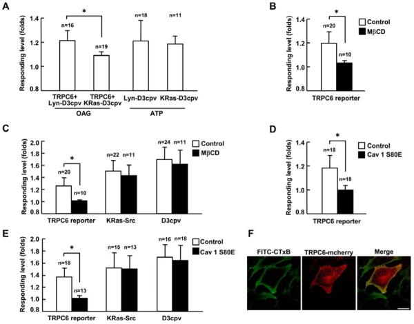
Fig. 3. The activation of TRPC6 depends on lipid rafts and caveolae.
(A) Responses of Lyn-D3cpv and KRas-D3cpv reporters upon 100 μM OAG or 10 nM ATP stimulation as indicated. Lyn-D3cpv or KRas-D3cpv reporter was transfected into HEK293T cells with (OAG stimulation) or without the co-transfection of wild type TRPC6 (ATP stimulation) as indicated. (B) and (D) Response of TRPC6 reporter to 100 μM OAG stimulation in HEK239T cells (B) with 10 μM MβCD pretreatment or (D) with the expression of dominate negative Caveolin-1 (Cav 1 S80E). MbCD was added into medium 1 hr before stimulation with OAG. (C) and (E) Responses of TRPC6, KRas-Src or D3cpv reporter to 10 ng/ml PDGF stimulation in MEFs (C) with 10 μM MβCD pretreatment or (E) with the expression of Cav1 S80E. MEFs transfected with TRPC6 reporter were pretreated with 1 μM TG for 1 hr before imaging. MEFs transfected with the D3cpv reporter were maintained in Ca2+ free HBSS buffer during imaging. (F) Confocal images of FITC-CTxB labeled ganglioside GM1 and TRPC6-mCherry in MEFs. “n” represents the cell number in each group. * indicates the significant difference between indicated groups (P < 0.01). Scale bar, 25 μm.

