Abstract
This study aims to investigate the neurostructural foundations of the human personality in young adults. High-resolution structural T1-weighted MR images of 71 healthy young individuals were processed using voxel-based morphometric (VBM) approach. Multiple regression analyses were performed to identify the associations between personality traits and gray matter volume (GMV). The Eysenck Personality Questionnaire-Revised, Short Scale for Chinese was chosen to assess the personality traits. This scale includes four dimensions, namely, extraversion, neuroticism, psychoticism, and lie. Particularly, we studied on two dimensions (extraversion and neuroticism) of Eysenck’s personality. Our results showed that extraversion was negatively correlated with GMV of the bilateral amygdala, the bilateral parahippocampal gyrus, the right middle temporal gyrus, and the left superior frontal gyrus, all of which are involved in emotional and social cognitive processes. These results might suggest an association between extraversion and affective processing. In addition, a positive correlation was detected between neuroticism and GMV of the right cerebellum, a key brain region for negative affect coordination. Meanwhile, a negative association was revealed between GMV of the left superior frontal gyrus and neuroticism. These results may prove that neuroticism is related to several brain regions involved in regulating negative emotions. Based on those findings, we concluded that brain regions involved in social cognition and affective process accounted for modulation and shaping of personality traits among young individuals. Results of this study may serve as a basis for elucidating the anatomical factors of personality.
Introduction
Extraversion and neuroticism are two important and frequently studied dimensions of human personality [1], [2]. According to Eysenck’s influential arousal theory [2], [3], extraversion is the result of individual differences in the level of activity in the cortico-reticular loop and other arousal systems (e.g., monoamine oxidase system and pituitary-adrenocortical system). Thus, individuals with low extraversion scores have higher baseline levels of cortical arousal and higher arousability of the cortex, i.e., greater changes in cortical activity in response to arousing stimuli, than individuals whose extraversion scores were high [4], [5]. Neuroticism, on the other hand, results from a lower threshold for activation in the limbic circuit, such that individuals with high neuroticism scores show great activation levels and low thresholds within subcortical structures [1], [2].
The aforementioned biological explanations of extraversion and neuroticism have attracted increasing interests in linking the two fundamental traits to functional and anatomical brain markers. On the functional level, many neuroimaging studies have examined extraversion and neuroticism using modern neuroimaging techniques, such as positron emission tomography, electroencephalogram, magnetic resonance perfusion imaging, and resting-state functional magnetic resonance imaging (fMRI) [6]–[11]. These studies have highlighted the functional neural correlations of extraversion and neuroticism, and further demonstrated the underlying neural mechanisms of personality dimensions, thereby providing neurobiological evidence for the hypothesized biological model of Eysenck’s personality.
Recent studies have used modern structural imaging techniques to detect whether or not personality traits are correlated with brain structures. Previous studies have suggested that personality may be associated with brain regions involved in emotional regulation and affective processing. For example, in an adolescent sample, Blankstein et al. [12] found that extraversion and neuroticism were negatively and positively correlated with the GMV of the medial frontal gyrus and subgenual anterior cingulate cortex in females, respectively. The opposite of this relationship between GMV and personality could be observed in males. Omura et al. [13] studied forty-one participants aged 19 to 29 years and found that extraversion was positively correlated with the left amygdala and orbitofrontal cortex but negatively correlated with the precentral cortex. DeYoung et al. [14] studied individuals aged 18 to 40 years and found that extraversion was positively correlated with GMV of the orbitofrontal cortex. Another VBM study [15] involving sixty-five participants aged 21 to 56 years revealed that extraversion was positively correlated with the anterior cingulate cortex volume, orbitofrontal volume, and right amygdala volume. This study also suggested that the correlation between extraversion and the volume of the anterior cingulate cortex varied in males and females. Obviously, the main results of these studies are inconsistent. Age effect is a dominant factor affecting changes in regional brain volume, and personality may interact with the effects of age on brain structures [16]. This inconsistency may be due in part to the age range of the recruited subjects [16]. To remove the potential effect of the wide age range, we concentrated on examining the relationships between personality traits and GMV of brain regions only in young adults.
Our aims in the present study were two-fold: first, we examined whether there are relationships between extraversion and neuroticism based on Eysenck’s model and regional brain GMV in young adults; second, we investigated whether the core brain structures involved in emotional processing are correlated with extraversion and neuroticism. Of note, since the definition and the neuropsychological mechanism of psychoticism, the third dimension in the Eysenck scale, are ambiguous [17], [18], we did not investigate the relationship between psychoticism and GMV.
Materials and Methods
Participants
Seventy-one right-handed healthy university students (34 males) underwent magnetic resonance imaging (MRI) scans at Jinling Hospital, Nanjing, China. The mean age of the participants was 22.35 years (standard deviation = 1.5, age range: 19–26 years). Informed written consents were obtained from all participants before any study procedure was initiated. Exclusion criteria included any history of psychiatric or neurological illness, brain injury, and alcohol or drug abuse. An informal interview prior to the MRI scanning was conducted to confirm that subjects did not use any psychotropic drugs. Left-handed individuals, who were assessed using Chinese revised version of the Edinburgh Handedness Inventory, were excluded [19]. The present study was approved by the local Medical Ethics Committee at Jinling Hospital, Nanjing University.
Personality Questionnaires
To assess personality traits, all participants completed the Eysenck Personality Questionnaire-Revised, Short Scale for Chinese (EPQ-RSC), a self-report questionnaire [20]–[23]. We chose the EPQ-RSC since it has been extensively used for clinical and research purposes in China. Furthermore, the validity and stability of this scale have been authenticated in Chinese subjects. The EPQ-RSC includes four dimensions: Extraversion (E), Neuroticism (N), Psychoticism (P), and Lie (L). Subjects responded to each item in the questionnaire with “yes” or “no” (coded as 1 or 0) depending on the applicability of the statement. The raw scores were translated into T-scores using the following formula [22]:
where mean is the mean value of the personality scores over all participants; SD is the standard deviation of the personality scores. Qian et al. [22] suggested that EPQ-RSC had satisfactory reliability and validity of extraversion, neuroticism and lie, whereas the reliability and validity of psychoticism were relatively lower. We focused our analyses on extraversion and neuroticism, two important and significant personality dimensions whose resultant T-scores were used for measuring correlations with GMV values in the present work.
MRI Data Acquisition
High-resolution 3D structural MRI scans of all subjects were performed on a 3T MRI scanner (Siemens-Trio, Erlangen, German) using a T1-weighted spoiled grass gradient recalled sequence at the Jinling Hospital, Nanjing, China. Tight but comfortable foam padding was used to minimize head motion, and earplugs were used to reduce scanner noise. The following parameters were used: repetition time (TR) = 2300 ms, echo time (TE) = 2.98 ms, flip angle = 9°, slice thickness = 1 mm, FOV = 24 cm×24 cm, matrix size = 512×512, and voxel size = 0.5 mm×0.5 mm×1 mm.
Data Preprocessing
Images were initially visually inspected for artifacts or structural abnormalities unrelated to healthy subjects. Subjects with general MRI contraindications were excluded in the following analyses. VBM analyses were performed using SPM8 (http://www.fil.ion.ucl.ac.uk/spm) as previously described [24]. The detailed procedures were as follows. First, the origin of each participant’s structural images was set to the anterior commissure manually. Second, all images were divided into gray matter, white matter, and cerebrospinal fluid, and then imported into a strictly aligned space [25]. Third, the segmented images were iteratively registered by the Diffeomorphic Anatomical Registration Through Exponentiated Lie Algebra toolbox [26]. This process created a template for a group of individuals. The resulting images were spatially normalized into the MNI space using an affine spatial normalization. An extra processing step was performed to multiply each spatially normalized image by its relative volume before and after normalization to maintain the total amount of each tissue. Finally, images were smoothed with an isotropic Gaussian kernel of 8 mm full width at half maximum.
Second-level Analyses
Voxel-based multiple regression analyses (based on general linear model) were performed by SPM8, with voxel-wise GMV value as dependent variable and N and E scores of personality traits as covariates of interest. In addition, sex, age, and total intracranial volume were used as external regressors to control their effects on both brain structure [27], [28] and personality [29]. We set the significance value at p<0.05 using the AlphaSim correction (combined height threshold of p<0.005 and a minimum cluster size of 172 voxels). This correction was conducted using the AlphaSim program embedded into the REST Software (http://www.restfmri.net/forum/REST_V1.8), which applied Monte Carlo simulation to calculate the probability of false positive detection by considering both the individual voxel probability threshold and cluster size [30].
Results
Sample Characteristics and Personality Scores
The descriptive statistics of sample characteristics and personality scores are shown in Table 1 . A significantly negative correlation was found between N and E (r = −0.4042, p<0.0001). The result was concordant with many previous studies, demonstrating an inverse correlation between neuroticism and extraversion [31]–[33].
Table 1. Descriptive statistics of sample characteristics and personality scores.
| Variable | Data | Range |
| Age (years) | 22.35±1.5 | 19–26 |
| Gender (male/female) | 34/37 | – |
| E scores | 54.68±8.39 | 35.75–73.05 |
| N scores | 44.42±11.95 | 24.98–65.70 |
Age and personality scores are displayed as mean±SD.
Personalities and GMV Values
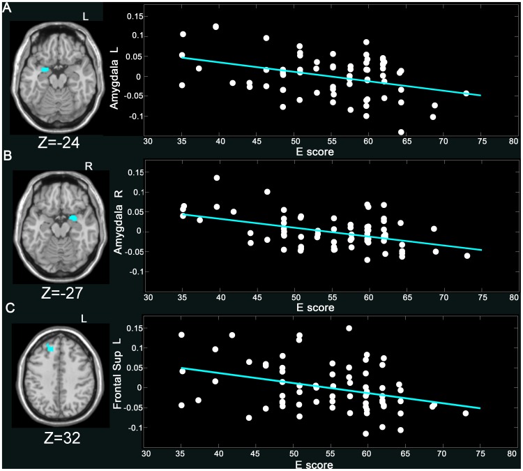
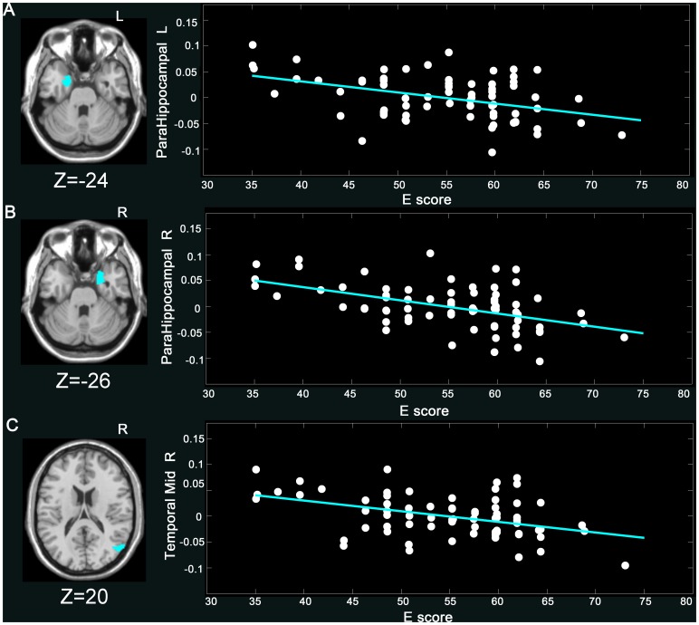
E scores were negatively correlated with GMV of the left amygdala (coordinates: x = −27, y = 3, z = −24), the right amygdala (coordinates: x = 27, y = 2, z = −27), the left parahippocampal gyrus (coordinates: x = −18, y = 3, z = −24), the right parahippocampal gyrus (coordinates: x = 27, y = 6, z = −26), the right middle temporal gyrus (coordinates: x = 54, y = −71, z = 20), and the left superior frontal gyrus (coordinates: x = −14, y = 29, z = 32) ( Figures 1 – 2 , and Table 2 ).
Figure 1. Negative correlation of E score and GMV (p<0.05, Alphasim corrected) in the (A) L amygdala (−27, 3, −24) (r = −0.43, p<0.001), (B) R amygdala (27, 2, −27) (r = −0.46, p<0.001), and (C) L superior frontal gyrus (−14, 29, 32) (r = −0.35, p<0.005).
The GMV values in the figure were extracted from the significant clusters after age, gender, and total intracranial volume of each subject were regressed out. More details of these regions are described in Table 2 .
Figure 2. Negative correlation of E score and GMV (p<0.05, Alphasim corrected) in the (A) L parahippocampal (−18, 3, −24) (r = −0.42, p<0.0001), (B) R parahippocampal (27, 6, −26) (r = −0.50, p<0.0001), and (C) R middle temporal gyrus (54, −71, 20) (r = −0.38, p<0.05).
The GMV values in the figure were extracted from the significant clusters after age, gender, and total intracranial volume of each subject were regressed out. More details of these regions are described in Table 2 .
Table 2. Brain regions in which GMV were significantly related with E and N.
| Personalities | Regions | BA | MNI coordinate | Direction of association | T value | ||
| x | y | z | |||||
| E | L AMYG | – | −27 | 3 | −24 | Negative | −4.22 |
| R AMYG | – | 27 | 2 | −27 | −5.11 | ||
| L ParaHG | 34 | −18 | 3 | −24 | −4.02 | ||
| R ParaHG | 28 | 27 | 6 | −26 | −5.10 | ||
| R MTG | 39 | 54 | −71 | 20 | −3.22 | ||
| L SFG | 8/9/32 | −14 | 29 | 32 | −3.63 | ||
| N | R CER | – | 8 | −41 | −14 | Positive | 3.36 |
| L SFG | 6/8 | −20 | 13 | 56 | Negative | −3.46 | |
MNI, Montreal Neurological Institute; BA, Brodmann’s area; E, extraversion; N, neurocitism; L, Left; R, right; AMYG, amygdala; ParaHG, parahippocampal gyrus; MTG, middle temporal gyrus; SFG, superior frontal gyrus; CER, cerebellum. T value represents the statistical value of peak voxel showing brain regions’ GMV correlated with E and N. Positive and negative T values indicate positive and negative correlations, respectively, between GMV and E/N scores.
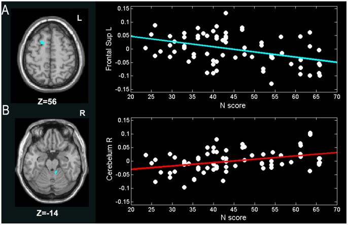
N scores were positively correlated with GMV of the right cerebellum (coordinates: x = 8, y = −41, z = −14) but negatively correlated with GMV of the left superior frontal gyrus (coordinates: x = −20, y = 13, z = 56) ( Figure 3 and Table 2 ).
Figure 3. Correlations of N score and GMV (p<0.05, Alphasim corrected) in the (A) L superior frontal gyrus (−20, 13, 56) (r = −0.3853, p<0.0001), and (B) R cerebellum (8, −41, −14) (r = 0.3720, p<0.05).
The GMV values in the figure were extracted from the significant clusters after age, gender, and total intracranial volume of each subject were regressed out. More details of these regions are described in Table 2 .
The results of our analyses are shown in Table 2 , which lists all clusters related to Eysenck’s personality of extraversion and neuroticism, controlling the effects of age, sex, and total intracranial volume.
Discussion
In the current study, we investigated two dimensions (extraversion and neuroticism) of Eysenck’s personality in young people. Extraversion and neuroticism are personality traits linked to emotional states. In general, extraverted individuals are susceptible to positive emotions, such as sociability, excitement, engagement, and enthusiasm, whereas neurotics are particularly susceptible to negative emotions, including fear, anxiety, and distress [32]. Controlling for age, sex, and total intracranial volume, we explored the correlations between extraversion and neuroticism and GMV values. The GMV of the bilateral amygdala, the bilateral parahippocampal gyrus, and right middle temporal gyrus were negatively correlated with extraversion. The GMV of the left superior frontal gyrus was negatively associated with both extraversion and neuroticism, whereas that of the right cerebellum was positively correlated with neuroticism. These results help to clarify the important relationships between the GMV of brain regions associated with emotional processing and personality traits.
GMV of Amygdala Negatively Associated with E
In the present study, the relationships between extraversion and GMV in several brain areas (the bilateral amygdala, the bilateral parahippocampal gyrus, the right middle temporal gyrus and the left superior frontal gyrus) were all found to be negative. This result indicates that individuals with high extraversion scores showed small GMV in the reported regions.
The direction of associations found in this study is consistent with previous structural studies that showed only negative correlations between extraversion and several frontal regions [31], [34], [35]. However, a positive relationship was also found between amygdala and extraversion in a biological model of five-factor traits [13], [36]–[38]. For example, Cremers et al. [38] observed that extraverts, compared with introverts, showed higher total GMV in the right amygdala. Omura et al. [13] discovered a positive association between extraversion and GMV in the left amygdala. Our inconsistent results may be partly attributed to many factors, including heterogeneity in personality measurements between the five-factor and Eysenck’s models. Some neurodevelopmental studies have reported that lower grey matter density can lead to better performance [39]–[42]. A smaller GMV may possibly indicate a higher efficiency in implementing specific functions associated with the amygdala. The association between extraversion and reduced amygdala GMV may have clinical significance since extraversion was found to be positively related to positive emotions after the induction of pleasant mood under Eysenck’s model [5]. Therefore, the relationship between GMV and extraversion in the amygdala, which is essential in emotional processing, supports Eysenck’s prediction for extraverts with positive effect.
GMV of the Parahippocampal Gyrus Negatively Associated with E
We also found an association between reduced GMV in the parahippocampal gyrus and extraverts. Jackson et al. [16] concluded that the interactive effects of personality with age in the parahippocampal gyrus were related to its function in emotional and memory processing. Moreover, Mobbs et al. [43] used event-related functional MRI to address the putative neural and behavioral associations between humor appreciation and extraversion. They found a positive correlation between humor-related activation and extraversion in the parahippocampal gyrus. Extraversion, according to Eysenck’s theory, results from the differences between the activity levels of an individual in the cortico-reticular loop and other arousal systems (e.g., monoamine oxidase and pituitary-adrenocortical systems) [3], [44], [45]. Those previous results suggest that the parahippocampal gyrus contributes to emotional processing, which is an important aspect in modulating or shaping personality. Our findings may sustain the idea that activation in the parahippocampal gyrus is involved in the positive effect. The current result showing larger GMV in introverts is also in line with the result of the previous studies, in which individuals who exhibited lower E scores showed greater neuronal coherence in the parahippocampal gyrus [9], [10].
GMV of the Middle Temporal Gyrus Negatively Associated with E
Negative correlation between extraversion and the GMV of the middle temporal gyrus was an additional interesting result. Previous studies demonstrated that the middle temporal gyrus is involved in several cognitive processes connected with personality traits [46]. Moreover, the middle temporal gyrus is involved in language and semantic memory processing [46]–[48], and multimodal sensory integration [49]. The relationship between GMV and extraversion detected in the right middle temporal gyrus suggested that extraverts are different from introverts in emotional processing. This suggestion was based on the assumption that extraverts are more behaviorally active, optimistic, and sociable than introverts [50], [51]. Lower grey matter density can lead to better performance [52]. Thus, we can conclude that extraverts perform working memory tasks more efficiently than introverts.
GMV of the Superior Frontal Gyrus Negatively Associated with E and N
A significantly negative relation was observed in the left superior frontal gyrus between GMV and both extraversion and neuroticism. The superior frontal gyrus was thought to contribute to higher cognitive functions and particularly to working memory [53]. In fMRI experiments, Goldberg et al. [54], [55] found that the superior frontal gyrus was involved in self-awareness, in coordination with the action of the sensory system. Kunisato et al. [56] have explored positive correlations between extraversion and fractional amplitude low-frequency fluctuations in the right superior frontal gyrus. Forsman et al. [57] also observed a negative relationship between the GMV of the left superior frontal gyrus and extraversion. Neuroimaging results indicated that the left superior frontal gyrus was significantly correlated with the regulation of negative emotions and partially significantly activated in regulating positive emotions [58], [59]. Personality traits were conjunct with emotions, in that the superior frontal gyrus involved in both positive and negative effects was also involved in extraversion and neuroticism.
GMV of Cerebellum Positively Associated with N
Our results showed that the GMV of the cerebellum was positively correlated with neuroticism. The cerebellum was involved in emotional processing, cognition, and regulation of mood [60]–[64]. Previous studies have demonstrated that the cerebellum was principally involved in motor and cognitive functions [65]–[69], less involved in emotional regulation and affective processing [70]–[72], and even less involved in personality individual differences [73]. A recent study in which cerebellar function was manipulated with transcranial direct current stimulation provided an additional evidence for cerebellar involvement during the processing of negatively valenced emotional stimuli [74]. Research has shown a positive relationship between neuroticism and high levels of the stress-related hormone cortisol [75]. Moreover, De Young et al. [14] found that the cerebellum was positively associated with neuroticism. Our results suggested that individuals with higher N scores had higher GMV in the cerebellum, which may agree with the theory on the involvement of the cerebellum in the vulnerability to experiencing negative effects and developing mood disorders [74], [75]. In conclusion, the GMV of the cerebellum is associated with neurotic personality traits, which are obviously related to negative emotions.
Limitations
Several limitations of the current study should be mentioned. First, our study was limited by a relatively small sample size. An expanded imaging dataset will be necessary to confirm our preliminary results in the future study. In addition, our study was an exploratory research. Thus, the AlphaSim correction was adopted for the second-level analyses. In the future, more conservative correction (such as family wise error (FWE) or false discovery rate (FDR) correction) should be applied to avoid false positives. Third, we examined the relationship between GMV and personality traits only in young adults sample in our study, future studies should include young, middle-aged, and elderly adults simultaneously in order to directly explore the effects of age on the relationship between personality and GMV. Finally, the obtained results were based on univariate statistical method. Previous studies have suggested that multivariate method is sensitive to the fine-grained spatial discriminative patterns and outperforms univariate analysis [76]–[78]. Future studies may be benefit from a multivariate approach to explore the neurostructural foundations of the human personality.
Conclusions
We used VBM, an objective structural analysis technique designed to evaluate brain structural features, to characterize the relationship between personality traits and GMV of brain regions in young adults. Extraversion was negatively correlated with GMV of the bilateral amygdala, the bilateral parahippocampal gyrus, the right middle temporal gyrus, and the left superior frontal gyrus. These results indicated that brain regions involved in the initial process of emotional information affected personality. In addition, a positive correlation was detected between neuroticism and GMV of the right cerebellum, whereas a negative association was determined between GMV of the left superior frontal gyrus and neuroticism. This result indicated that those regions contributed to negative emotional processing were associated with neuroticism. Overall, the present findings suggested that brain structures involved in social cognition and affective process accounted for modulation and shaping of personality in young people. We concluded that relationships do exist between GMV of brain regions and personality. Our conclusion was in accordance with the results of previous studies, which suggested that human personality traits are based on individual differences in brain structures.
Acknowledgments
We thank our volunteers for their participation in this study and thank the academic editor and four anonymous reviewers for their constructive comments that have improved the manuscript considerably.
Funding Statement
This work was supported by the 973 project 2012CB517901; and Natural Science Foundation of China (Grant Nos. 61035006, 61125304, 81171406 and 61273361), and the Specialized Research Fund for the Doctoral Program of Higher Education of China (Grant No. 20120185110028), and the Key Technology R&D Program of Sichuan Province (Grant No. 2012SZ0159). The funders had no role in study design, data collection and analysis, decision to publish, or preparation of the manuscript.
References
- 1.Eysenck HJ (1990) Biological dimensions of personality. 244–276.
- 2. Eysenck HJ (1967) The biological basis of personality. Transaction Pub 199: 1031–1034. [DOI] [PubMed] [Google Scholar]
- 3.Eysenck HJ, Eysenck M (1985) Personality and individual differences: A natural science approach. New York: Plenum Press.
- 4. Hagemann D, Hewig J, Walter C, Schankin A, Danner D, et al. (2009) Positive evidence for Eysenck’s arousal hypothesis:a combined EEG and MRI study with multiple measurement occasions. Personality and Individual Differences 47: 717–721. [Google Scholar]
- 5.Rusting C, Larsen R (1997) Extraversion, neuroticism, and susceptibility to positive and negative affect: A test of two theoretical models. Personality and Individual Differences.
- 6. Kim SH, Hwang JH, Park HS, Kim SE (2008) Resting brain metabolic correlates of neuroticism and extraversion in young men. Neuroreport 19: 883–886. [DOI] [PubMed] [Google Scholar]
- 7. O’Gorman RL, Kumari V, Williams SC, Zelaya FO, Connor SE, et al. (2006) Personality factors correlate with regional cerebral perfusion. Neuroimage 31: 489–495. [DOI] [PubMed] [Google Scholar]
- 8. Tran Y, Craig A, McIsaac P (2001) Extraversion-introversion and 8–13 Hz waves in frontal cortical regions. Personality Individual Diff 30: 205–215. [Google Scholar]
- 9. Wei L, Duan X, Yang Y, Liao W, Gao Q, et al. (2011) The synchronization of spontaneous BOLD activity predicts extraversion and neuroticism. Brain Res 1419: 68–75. [DOI] [PubMed] [Google Scholar]
- 10.Wei L, Duan X, Zheng C, Wang S, Gao Q, et al.. (2012) Specific frequency bands of amplitude low-frequency oscillation encodes personality. Hum Brain Mapp. [DOI] [PMC free article] [PubMed]
- 11. Adelstein JS, Shehzad Z, Mennes M, Deyoung CG, Zuo XN, et al. (2011) Personality is reflected in the brain’s intrinsic functional architecture. PLoS One 6: e27633. [DOI] [PMC free article] [PubMed] [Google Scholar]
- 12. Blankstein U, Chen JY, Mincic AM, McGrath PA, Davis KD (2009) The complex minds of teenagers: neuroanatomy of personality differs between sexes. Neuropsychologia 47: 599–603. [DOI] [PubMed] [Google Scholar]
- 13. Omura K, Todd Constable R, Canli T (2005) Amygdala gray matter concentration is associated with extraversion and neuroticism. Neuroreport 16: 1905–1908. [DOI] [PubMed] [Google Scholar]
- 14. DeYoung CG, Hirsh JB, Shane MS, Papademetris X, Rajeevan N, et al. (2010) Testing predictions from personality neuroscience. Brain structure and the big five. Psychol Sci 21: 820–828. [DOI] [PMC free article] [PubMed] [Google Scholar]
- 15. Cremers H, van Tol M-J, Roelofs K, Aleman A, Zitman FG, et al. (2011) Extraversion is linked to volume of the orbitofrontal cortex and amygdala. PloS one 6: e28421. [DOI] [PMC free article] [PubMed] [Google Scholar]
- 16. Jackson J, Balota DA, Head D (2011) Exploring the relationship between personality and regional brain volume in healthy aging. Neurobiol Aging 32: 2162–2171. [DOI] [PMC free article] [PubMed] [Google Scholar]
- 17. Eysenck HJ (1992) The definition and measurement of psychoticism. Personality and Individual Differences 13: 757–786. [Google Scholar]
- 18. Eysenck HJ (1997) Personality and experimental psychology: The unification of psychology and the possibility of a paradigm. Journal of Personality and Social Psychology 73: 1224–1237. [Google Scholar]
- 19. Oldfield RC (1971) The assessment and analysis of handedness: the Edinburgh inventory. Neuropsychologia 9: 97–113. [DOI] [PubMed] [Google Scholar]
- 20. Eysenck HJ (1946) The Measurement of Personality. [Resume]. Proc R Soc Med 40: 75–80. [DOI] [PMC free article] [PubMed] [Google Scholar]
- 21.Eysenck HJ (1991) Manual of the Eysenck personality scales (EPS Adult). London: Hodder & Stoughton.
- 22. Qian M, Wu G, Zhu R, Zhang S (2000) Development of the Revised Eysenck Personality Questionnaire Short Scale for Chinese (EPQ-RSC). Acta Psychol Sinica 32: 317–323. [Google Scholar]
- 23.Eysenck HJ, Eysenck SBG (1975) Manual of the Eysenck Personality Questionnaire. SanDiego, CA: Educational & Industrial Testing Service.
- 24. Liu F, Guo W, Yu D, Gao Q, Gao K, et al. (2012) Classification of Different Therapeutic Responses of Major Depressive Disorder with Multivariate Pattern Analysis Method Based on Structural MR Scans. PLoS One 7: e40968. [DOI] [PMC free article] [PubMed] [Google Scholar]
- 25. Ashburner J, Friston KJ (2000) Voxel-based morphometry–the methods. Neuroimage 11: 805–821. [DOI] [PubMed] [Google Scholar]
- 26. Ashburner J (2007) A fast diffeomorphic image registration algorithm. Neuroimage 38: 95–113. [DOI] [PubMed] [Google Scholar]
- 27. Good CD, Johnsrude IS, Ashburner J, Henson RN, Friston KJ, et al. (2001) A voxel-based morphometric study of ageing in 465 normal adult human brains. Neuroimage 14: 21–36. [DOI] [PubMed] [Google Scholar]
- 28. Sowell ER, Peterson BS, Thompson PM, Welcome SE, Henkenius AL, et al. (2003) Mapping cortical change across the human life span. Nat Neurosci 6: 309–315. [DOI] [PubMed] [Google Scholar]
- 29. Cloninger CR, Svrakic DM, Przybeck TR (1993) A psychobiological model of temperament and character. Arch Gen Psychiatry 50: 975–990. [DOI] [PubMed] [Google Scholar]
- 30.Chao-Gan Y, Yu-Feng Z (2010) DPARSF: a MATLAB toolbox for “pipeline” data analysis of resting-state fMRI. Frontiers in systems neuroscience 4. [DOI] [PMC free article] [PubMed]
- 31. Wright CI, Williams D, Feczko E, Barrett LF, Dickerson BC, et al. (2006) Neuroanatomical correlates of extraversion and neuroticism. Cereb Cortex 16: 1809–1819. [DOI] [PubMed] [Google Scholar]
- 32. Rusting CL, Larsen RJ (1997) Extraversion, neuroticism, and susceptibility to positive and negative affect: A test of two theoretical models. Personality and Individual Differences 22: 607–612. [Google Scholar]
- 33. Kim SH, Hwang JH, Park HS, Kim SE (2008) Resting brain metabolic correlates of neuroticism and extraversion in young men. Neuroreport 19: 883–886. [DOI] [PubMed] [Google Scholar]
- 34. Barrós-Loscertales A, Meseguer V, Sanjuán A, Belloch V, Parcet M, et al. (2006) Striatum gray matter reduction in males with an overactive behavioral activation system. European Journal of Neuroscience 24: 2071–2074. [DOI] [PubMed] [Google Scholar]
- 35.Coutinho JF, Sampaio A, Ferreira M, Soares JM, Gonçalves OF (2013) Brain correlates of pro-social personality traits: a voxel-based morphometry study. Brain imaging and behavior: 1–7. [DOI] [PubMed]
- 36. LeDoux JE (1995) Emotion: clues from the brain. Annu Rev Psychol 46: 209–235. [DOI] [PubMed] [Google Scholar]
- 37. Canli T, Sivers H, Whitfield SL, Gotlib IH, Gabrieli JD (2002) Amygdala response to happy faces as a function of extraversion. Science 296: 2191. [DOI] [PubMed] [Google Scholar]
- 38. Cremers H, van Tol MJ, Roelofs K, Aleman A, Zitman FG, et al. (2011) Extraversion is linked to volume of the orbitofrontal cortex and amygdala. PLoS One 6: e28421. [DOI] [PMC free article] [PubMed] [Google Scholar]
- 39. Sowell ER, Delis D, Stiles J, Jernigan TL (2001) Improved memory functioning and frontal lobe maturation between childhood and adolescence: a structural MRI study. Journal of the International Neuropsychological Society 7: 312–322. [DOI] [PubMed] [Google Scholar]
- 40. Sowell ER, Thompson PM, Leonard CM, Welcome SE, Kan E, et al. (2004) Longitudinal mapping of cortical thickness and brain growth in normal children. The Journal of Neuroscience 24: 8223–8231. [DOI] [PMC free article] [PubMed] [Google Scholar]
- 41. Sowell ER, Thompson PM, Holmes CJ, Jernigan TL, Toga AW (1999) In vivo evidence for post-adolescent brain maturation in frontal and striatal regions. Nature neuroscience 2: 859–861. [DOI] [PubMed] [Google Scholar]
- 42. Reiss AL, Abrams MT, Singer HS, Ross JL, Denckla MB (1996) Brain development, gender and IQ in children A volumetric imaging study. Brain 119: 1763–1774. [DOI] [PubMed] [Google Scholar]
- 43. Mobbs D, Hagan CC, Azim E, Menon V, Reiss AL (2005) Personality predicts activity in reward and emotional regions associated with humor. Proc Natl Acad Sci U S A 102: 16502–16506. [DOI] [PMC free article] [PubMed] [Google Scholar]
- 44. Rusting CL, Larsen RJ (1997) Extraversion, neuroticism, and susceptibility to positive and negative affect: A test of two theoretical models. Pers Indiv Differ 22: 607–612. [Google Scholar]
- 45. Canli T, Zhao Z, Desmond JE, Kang E, Gross J, et al. (2001) An fMRI study of personality influences on brain reactivity to emotional stimuli. Behav Neurosci 115: 33–42. [DOI] [PubMed] [Google Scholar]
- 46. Cabeza R, Nyberg L (2000) Imaging cognition II: An empirical review of 275 PET and fMRI studies. J Cogn Neurosci 12: 1–47. [DOI] [PubMed] [Google Scholar]
- 47. Chao LL, Haxby JV, Martin A (1999) Attribute-based neural substrates in temporal cortex for perceiving and knowing about objects. Nat Neurosci 2: 913–919. [DOI] [PubMed] [Google Scholar]
- 48. Tranel D, Damasio H, Damasio AR (1997) A neural basis for the retrieval of conceptual knowledge. Neuropsychologia 35: 1319–1327. [DOI] [PubMed] [Google Scholar]
- 49. Mesulam MM (1998) From sensation to cognition. Brain 121 (Pt 6): 1013–1052. [DOI] [PubMed] [Google Scholar]
- 50. Depue RA, Collins PF (1999) Neurobiology of the structure of personality: dopamine, facilitation of incentive motivation, and extraversion. Behav Brain Sci 22: 491–517 discussion 518–469. [DOI] [PubMed] [Google Scholar]
- 51. Costa PT Jr, McCrae RR (1988) Personality in adulthood: a six-year longitudinal study of self-reports and spouse ratings on the NEO Personality Inventory. J Pers Soc Psychol 54: 853–863. [DOI] [PubMed] [Google Scholar]
- 52. Lieberman MD, Rosenthal R (2001) Why introverts can’t always tell who likes them: multitasking and nonverbal decoding. Journal of personality and social psychology 80: 294. [DOI] [PubMed] [Google Scholar]
- 53. du Boisgueheneuc F, Levy R, Volle E, Seassau M, Duffau H, et al. (2006) Functions of the left superior frontal gyrus in humans: a lesion study. Brain 129: 3315–3328. [DOI] [PubMed] [Google Scholar]
- 54. Goldberg II, Harel M, Malach R (2006) When the brain loses its self: prefrontal inactivation during sensorimotor processing. Neuron 50: 329–339. [DOI] [PubMed] [Google Scholar]
- 55. Vince G (2006) Watching the brain ‘switch off’ self-awareness. Dana Foundation Brain in the News 13: 2. [Google Scholar]
- 56. Kunisato Y, Okamoto Y, Okada G, Aoyama S, Nishiyama Y, et al. (2011) Personality traits and the amplitude of spontaneous low-frequency oscillations during resting state. Neurosci Lett 492: 109–113. [DOI] [PubMed] [Google Scholar]
- 57. Forsman LJ, de Manzano O, Karabanov A, Madison G, Ullen F (2012) Differences in regional brain volume related to the extraversion-introversion dimension–a voxel based morphometry study. Neurosci Res 72: 59–67. [DOI] [PubMed] [Google Scholar]
- 58. Mak AK, Hu ZG, Zhang JX, Xiao ZW, Lee TM (2009) Neural correlates of regulation of positive and negative emotions: an fmri study. Neurosci Lett 457: 101–106. [DOI] [PubMed] [Google Scholar]
- 59. Shaywitz SE, Shaywitz BA, Pugh KR, Fulbright RK, Skudlarski P, et al. (1999) Effect of estrogen on brain activation patterns in postmenopausal women during working memory tasks. JAMA 281: 1197–1202. [DOI] [PubMed] [Google Scholar]
- 60. Snider RS, Maiti A (1976) Cerebellar contributions to the Papez circuit. J Neurosci Res 2: 133–146. [DOI] [PubMed] [Google Scholar]
- 61. Heath RG, Dempesy CW, Fontana CJ, Myers WA (1978) Cerebellar stimulation: effects on septal region, hippocampus, and amygdala of cats and rats. Biol Psychiatry 13: 501–529. [PubMed] [Google Scholar]
- 62. Schutter DJ, van Honk J (2005) The cerebellum on the rise in human emotion. Cerebellum 4: 290–294. [DOI] [PubMed] [Google Scholar]
- 63.Liu F, Guo W, Liu L, Long Z, Ma C, et al.. (2012) Abnormal amplitude low-frequency oscillations in medication-naive, first-episode patients with major depressive disorder: A resting-state fMRI study. Journal of affective disorders. [DOI] [PubMed]
- 64.Liu F, Hu M, Wang S, Guo W, Zhao J, et al.. (2012) Abnormal regional spontaneous neural activity in first-episode, treatment-naive patients with late-life depression: A resting-state fMRI study. Progress in Neuro-Psychopharmacology and Biological Psychiatry. [DOI] [PubMed]
- 65. Oliveri M, Torriero S, Koch G, Salerno S, Petrosini L, et al. (2007) The role of transcranial magnetic stimulation in the study of cerebellar cognitive function. The Cerebellum 6: 95–101. [DOI] [PubMed] [Google Scholar]
- 66. Foti F, Mandolesi L, Cutuli D, Laricchiuta D, De Bartolo P, et al. (2010) Cerebellar damage loosens the strategic use of the spatial structure of the search space. The Cerebellum 9: 29–41. [DOI] [PubMed] [Google Scholar]
- 67. Oliveri M, Bonnì S, Turriziani P, Koch G, Gerfo EL, et al. (2009) Motor and linguistic linking of space and time in the cerebellum. PloS one 4: e7933. [DOI] [PMC free article] [PubMed] [Google Scholar]
- 68.Laricchiuta D, Petrosini L, Piras F, Macci E, Cutuli D, et al.. (2012) Linking novelty seeking and harm avoidance personality traits to cerebellar volumes. Human Brain Mapping. [DOI] [PMC free article] [PubMed]
- 69.Picerni E, Petrosini L, Piras F, Laricchiuta D, Cutuli D, et al.. (2013) New evidence for the cerebellar involvement in personality traits. Frontiers in behavioral neuroscience 7. [DOI] [PMC free article] [PubMed]
- 70. Schmahmann JD, Sherman JC (1998) The cerebellar cognitive affective syndrome. Brain 121: 561–579. [DOI] [PubMed] [Google Scholar]
- 71. Schmahmann JD, Weilburg JB, Sherman JC (2007) The neuropsychiatry of the cerebellum–insights from the clinic. The Cerebellum 6: 254–267. [DOI] [PubMed] [Google Scholar]
- 72. Turner BM, Paradiso S, Marvel CL, Pierson R, Boles Ponto LL, et al. (2007) The cerebellum and emotional experience. Neuropsychologia 45: 1331–1341. [DOI] [PMC free article] [PubMed] [Google Scholar]
- 73. O’Gorman R, Kumari V, Williams S, Zelaya F, Connor S, et al. (2006) Personality factors correlate with regional cerebral perfusion. Neuroimage 31: 489–495. [DOI] [PubMed] [Google Scholar]
- 74. Ferrucci R, Giannicola G, Rosa M, Fumagalli M, Boggio PS, et al. (2012) Cerebellum and processing of negative facial emotions: cerebellar transcranial DC stimulation specifically enhances the emotional recognition of facial anger and sadness. Cogn Emot 26: 786–799. [DOI] [PMC free article] [PubMed] [Google Scholar]
- 75. Nater UM, Hoppmann C, Klumb PL (2010) Neuroticism and conscientiousness are associated with cortisol diurnal profiles in adults–role of positive and negative affect. Psychoneuroendocrinology 35: 1573–1577. [DOI] [PubMed] [Google Scholar]
- 76. Norman KA, Polyn SM, Detre GJ, Haxby JV (2006) Beyond mind-reading: multi-voxel pattern analysis of fMRI data. Trends in cognitive sciences 10: 424–430. [DOI] [PubMed] [Google Scholar]
- 77.Liu F, Guo W, Fouche J-P, Wang Y, Wang W, et al.. (2013) Multivariate classification of social anxiety disorder using whole brain functional connectivity. Brain Structure and Function: 1–15. [DOI] [PubMed]
- 78. Liu F, Wee C-Y, Chen H, Shen D (2014) Inter-modality relationship constrained multi-modality multi-task feature selection for Alzheimer’s Disease and mild cognitive impairment identification. NeuroImage 84: 466–475. [DOI] [PMC free article] [PubMed] [Google Scholar]


