

Abstract
When the Mn2+ and Ca2+ ions normally present in concanavalin A are removed, the protein becomes incapable of binding saccharides. To explore the structural differences between the native and demetallized forms and their effects on the saccharide-binding properties of the protein, we have refined and compared the crystal structures of both forms. Refinement, carried out by automated difference Fourier methods, has revealed a number of differences between the two structures as well as minor differences between the two crystallographically independent monomers in the demetallized structure. Significant differences between the holo- and apoproteins are confined to the region where the metals are bound. These differences include a reorganization and disordering of the loop, consisting of residues 7-25, that contains all of the direct metal ligands of the protein. In some molecules, the side chain of arginine-228 appears to move into the metal-binding region, possibly compensating in part for the absence of the metal's positive charge. The cis peptide observed in the native protein at alanine-207 is apparently not present in the demetallized protein. The conformational differences affect many of the residues currently thought to be involved in the specific binding of saccharides.
Keywords: x-ray crystallography, molecular replacement, refinement, metal-protein interactions, lectins
Full text
PDF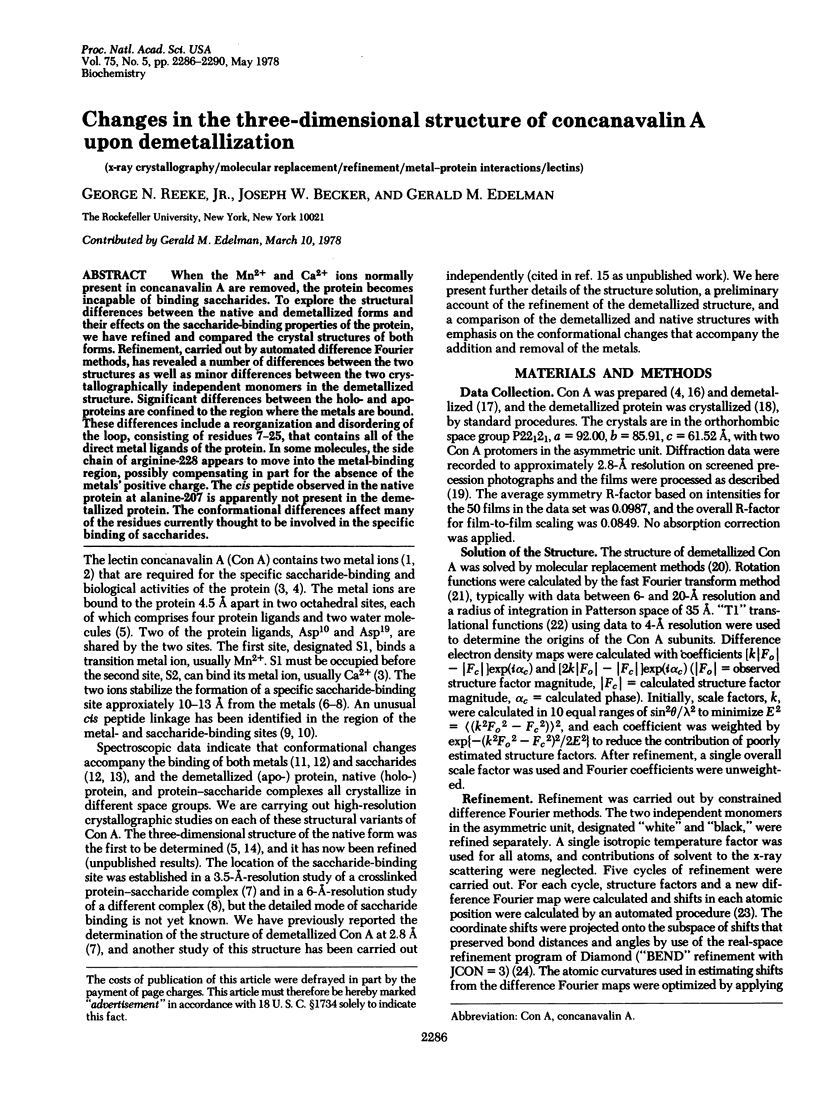
Images in this article
Selected References
These references are in PubMed. This may not be the complete list of references from this article.
- Agrawal B. B., Goldstein I. J. Protein-carbohydrate interactin. XV. The role of bivalent cations in concanavalin A-polysaccharide interaction. Can J Biochem. 1968 Sep;46(9):1147–1150. doi: 10.1139/o68-170. [DOI] [PubMed] [Google Scholar]
- Agrawal B. B., Goldstein I. J. Protein-carbohydrate interaction. VI. Isolation of concanavalin A by specific adsorption on cross-linked dextran gels. Biochim Biophys Acta. 1967 Oct 23;147(2):262–271. [PubMed] [Google Scholar]
- Becker J. W., Reeke G. N., Jr, Cunningham B. A., Edelman G. M. New evidence on the location of the saccharide-binding site of concanavalin A. Nature. 1976 Feb 5;259(5542):406–409. doi: 10.1038/259406a0. [DOI] [PubMed] [Google Scholar]
- Becker J. W., Reeke G. N., Jr, Wang J. L., Cunningham B. A., Edelman G. M. The covalent and three-dimensional structure of concanavalin A. III. Structure of the monomer and its interactions with metals and saccharides. J Biol Chem. 1975 Feb 25;250(4):1513–1524. [PubMed] [Google Scholar]
- Brewer C. F., Sternlicht H., Marcus D. M., Grollman A. P. Binding of 13 C-enriched -methyl-D-glucopyranoside to concanavalin A as studied by carbon magnetic resonance. Proc Natl Acad Sci U S A. 1973 Apr;70(4):1007–1011. doi: 10.1073/pnas.70.4.1007. [DOI] [PMC free article] [PubMed] [Google Scholar]
- Brown R. D., 3rd, Brewer C. F., Koenig S. H. Conformation states of concanavalin A: kinetics of transitions induced by interaction with Mn2+ and Ca2+ ions. Biochemistry. 1977 Aug 23;16(17):3883–3896. doi: 10.1021/bi00636a026. [DOI] [PubMed] [Google Scholar]
- Cunningham B. A., Wang J. L., Pflumm M. N., Edelman G. M. Isolation and proteolytic cleavage of the intact subunit of concanavalin A. Biochemistry. 1972 Aug 15;11(17):3233–3239. doi: 10.1021/bi00767a016. [DOI] [PubMed] [Google Scholar]
- Edelman G. M., Cunningham B. A., Reeke G. N., Jr, Becker J. W., Waxdal M. J., Wang J. L. The covalent and three-dimensional structure of concanavalin A. Proc Natl Acad Sci U S A. 1972 Sep;69(9):2580–2584. doi: 10.1073/pnas.69.9.2580. [DOI] [PMC free article] [PubMed] [Google Scholar]
- Freer S. T., Alden R. A., Carter C. W., Jr, Kraut J. Crystallographic structure refinement of Chromatium high potential iron protein at two Angstroms resolution. J Biol Chem. 1975 Jan 10;250(1):46–54. [PubMed] [Google Scholar]
- Grimaldi J. J., Sykes B. D. Concanavalin A: a stopped flow nuclear magnetic resonance study of conformational changes induced by Mn++, Ca++, and alpha-methyl-D-mannoside. J Biol Chem. 1975 Mar 10;250(5):1618–1624. [PubMed] [Google Scholar]
- Hardman K. D., Ainsworth C. F. Structure of concanavalin A at 2.4-A resolution. Biochemistry. 1972 Dec 19;11(26):4910–4919. doi: 10.1021/bi00776a006. [DOI] [PubMed] [Google Scholar]
- Hardman K. D., Ainsworth C. F. Structure of the concanavalin A-methyl alpha-D-mannopyranoside complex at 6-A resolution. Biochemistry. 1976 Mar 9;15(5):1120–1128. doi: 10.1021/bi00650a026. [DOI] [PubMed] [Google Scholar]
- Hardman K. D. Crystallography of a metal-containing protein, concanavalin A. Adv Exp Med Biol. 1973;40:103–123. doi: 10.1007/978-1-4684-3240-4_6. [DOI] [PubMed] [Google Scholar]
- Jack A., Weinierl J., Kalb A. J. An x-ray crystallographic study of demetallized concanavalin A. J Mol Biol. 1971 May 28;58(1):389–395. doi: 10.1016/0022-2836(71)90254-3. [DOI] [PubMed] [Google Scholar]
- Kalb A. J., Levitzki A. Metal-binding sites of concanavalin A and their role in the binding of alpha-methyl d-glucopyranoside. Biochem J. 1968 Oct;109(4):669–672. doi: 10.1042/bj1090669. [DOI] [PMC free article] [PubMed] [Google Scholar]
- McCubbin W. D., Oikawa K., Kay C. M. Circular dichroism studies on concanavalin A. Biochem Biophys Res Commun. 1971 May 7;43(3):666–674. doi: 10.1016/0006-291x(71)90666-8. [DOI] [PubMed] [Google Scholar]
- Pflumm M. N., Wang J. L., Edelman G. M. Conformational changes in concanavalin A. J Biol Chem. 1971 Jul 10;246(13):4369–4370. [PubMed] [Google Scholar]
- Reeke G. N., Jr, Becker J. W., Edelman G. M. The covalent and three-dimensional structure of concanavalin A. IV. Atomic coordinates, hydrogen bonding, and quaternary structure. J Biol Chem. 1975 Feb 25;250(4):1525–1547. [PubMed] [Google Scholar]
- Yariv J., Kalb A. J., Levitzki A. The interaction of concanavalin A with methyl alpha-D-glucopyranoside. Biochim Biophys Acta. 1968 Sep 3;165(2):303–305. doi: 10.1016/0304-4165(68)90063-9. [DOI] [PubMed] [Google Scholar]