Abstract
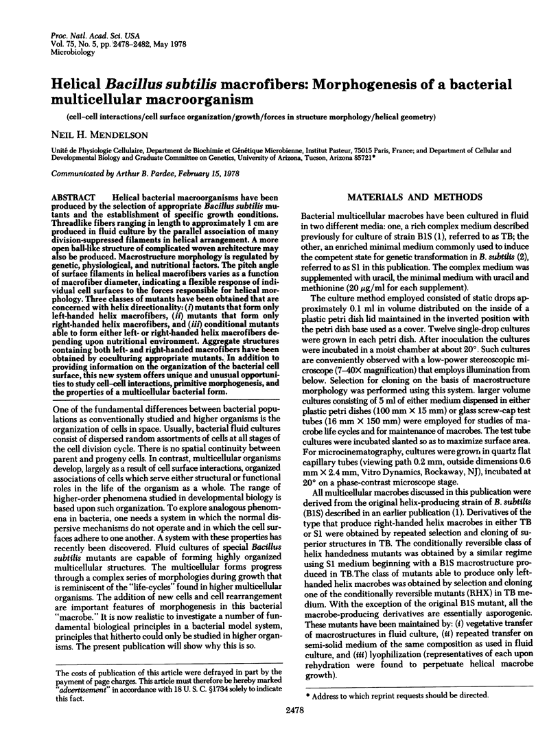
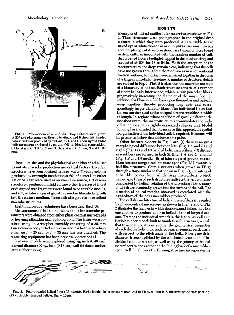
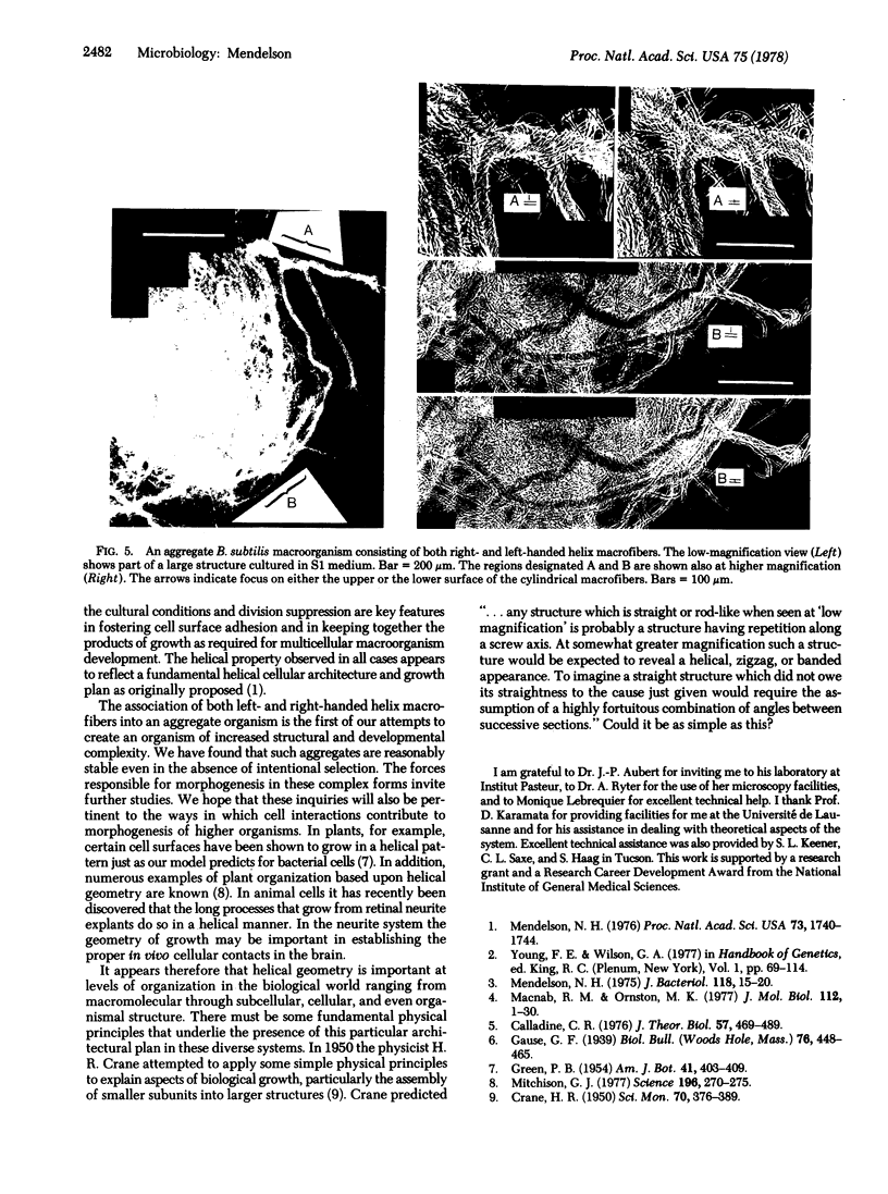
Helical bacterial macroorganisms have been produced by the selection of appropriate Bacillus subtilis mutants and the establishment of specific growth conditions. Threadlike fibers ranging in length to approximately 1 cm are produced in fluid culture by the parallel association of many division-suppressed filaments in helical arrangement. A more open ball-like structure of complicated woven architecture may also be produced. Macrostructure morphology is regulated by genetic, physiological, and nutritional factors. The pitch angle of surface filaments in helical macrofibers varies as a function of macrofiber diameter, indicating a flexible response of individual cell surfaces to the forces responsible for helical morphology. Three classes of mutants have been obtained that are concerned with helix directionality: (i) mutants that form only left-handed helix macrofibers, (ii) mutants that form only right-handed helix macrofibers, and (iii) conditional mutants able to form either left- or right-handed helix macrofibers depending upon nutritional environment. Aggregate structures containing both left- and right-handed macrofibers have been obtained by coculturing appropriate mutants. In addition to providing information on the organization of the bacterial cell surface, this new system offers unique and unusual opportunities to study cell-cell interactions, primitive morphogenesis, and the properties of a multicellular bacterial form.
Keywords: cell-cell interactions, cell surface organization, growth, forces in structure morphology, helical geometry
Full text
PDF


Images in this article
Selected References
These references are in PubMed. This may not be the complete list of references from this article.
- Calladine C. R. Design requirements for the construction of bacterial flagella. J Theor Biol. 1976 Apr;57(2):469–489. doi: 10.1016/0022-5193(76)90016-3. [DOI] [PubMed] [Google Scholar]
- Coyne S. I., Mendelson N. H. Clonal analysis of cell division in the Bacillus subtilis div IV-B1 minicell-producing mutant. J Bacteriol. 1974 Apr;118(1):15–20. doi: 10.1128/jb.118.1.15-20.1974. [DOI] [PMC free article] [PubMed] [Google Scholar]
- Macnab R. M., Ornston M. K. Normal-to-curly flagellar transitions and their role in bacterial tumbling. Stabilization of an alternative quaternary structure by mechanical force. J Mol Biol. 1977 May 5;112(1):1–30. doi: 10.1016/s0022-2836(77)80153-8. [DOI] [PubMed] [Google Scholar]
- Mendelson N. H. Helical growth of Bacillus subtilis: a new model of cell growth. Proc Natl Acad Sci U S A. 1976 May;73(5):1740–1744. doi: 10.1073/pnas.73.5.1740. [DOI] [PMC free article] [PubMed] [Google Scholar]
- Mitchison G. J. Phyllotaxis and the fibonacci series. Science. 1977 Apr 15;196(4287):270–275. doi: 10.1126/science.196.4287.270. [DOI] [PubMed] [Google Scholar]


