

Abstract
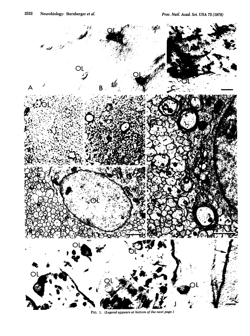
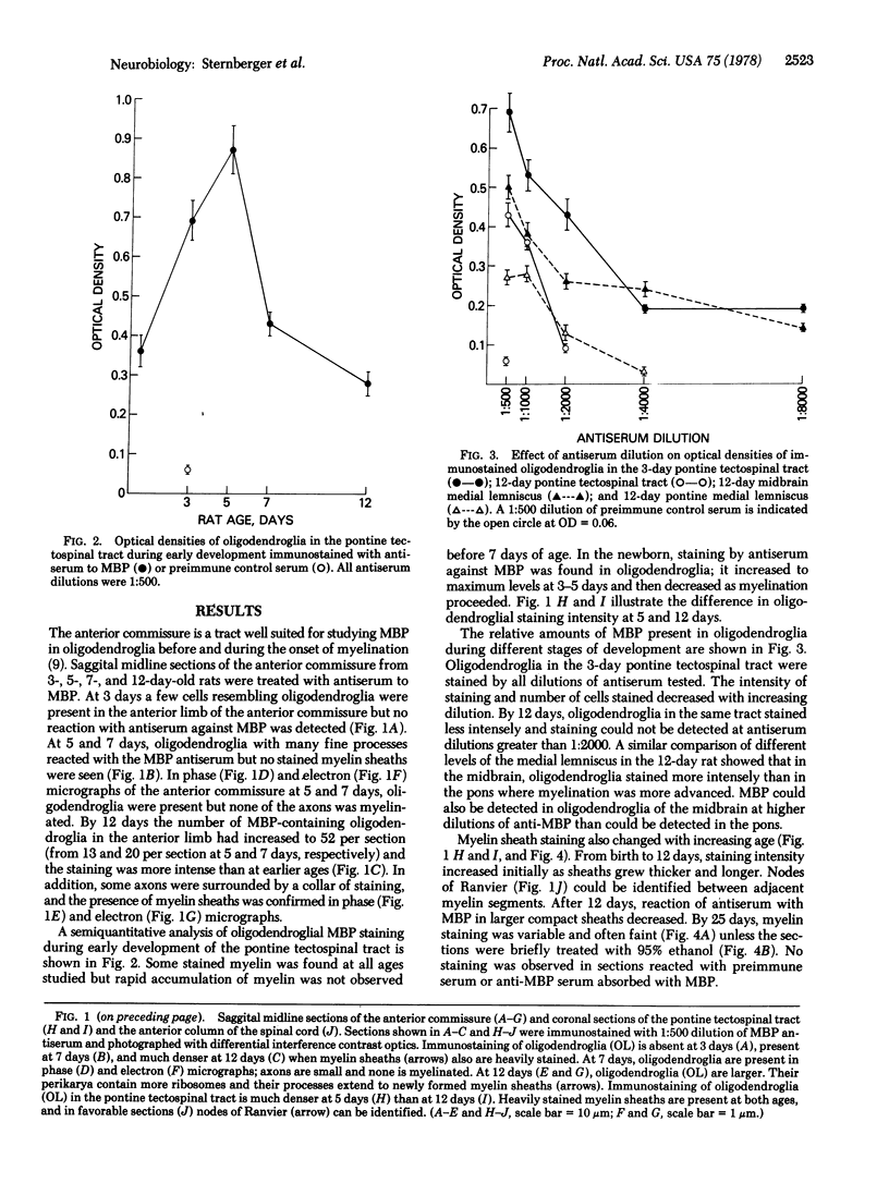
A specific antibody to myelin basic protein has been used to localize the protein in developing rat oligodendroglia and myelin. Basic protein is found in the oligodendroglial cytoplasm of anterior commissures of 5- and 7-day old rats before the beginning of myelination. Staining of basic protein in oligodendroglia increases, becoming most intense during early myelination; it decreases during rapid myelination. Staining intensity of oligodendroglia is dependent upon age, brain region, and nervous tract studied. In myelin, reaction of basic protein with antibody decreases when large compact sheaths are present, unless tissue sections are first treated with alcohol.
Full text
PDF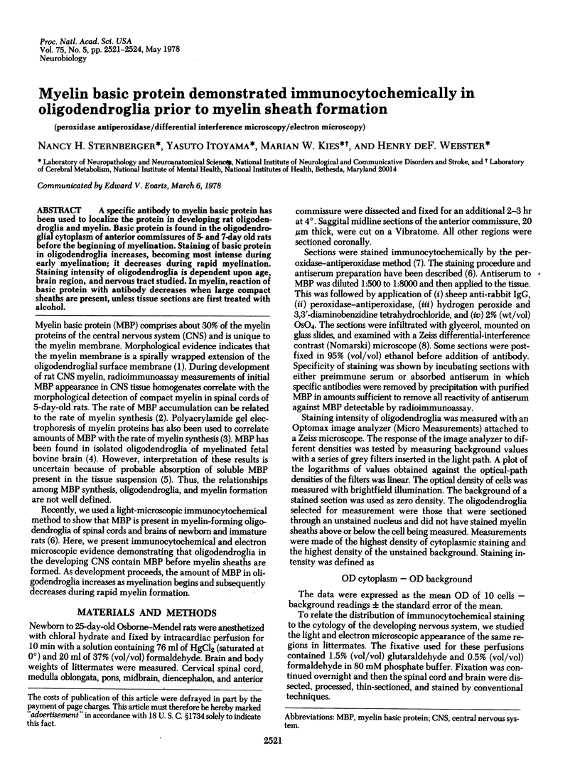
Images in this article
Selected References
These references are in PubMed. This may not be the complete list of references from this article.
- Banik N. L., Smith M. E. Protein determinants of myelination in different regions of developing rat central nervous system. Biochem J. 1977 Feb 15;162(2):247–255. doi: 10.1042/bj1620247. [DOI] [PMC free article] [PubMed] [Google Scholar]
- Cohen S. R., Guarnieri M. Immunochemical measurement of myelin basic protein in developing rat brain: an index of myelin synthesis. Dev Biol. 1976 Mar;49(1):294–299. doi: 10.1016/0012-1606(76)90276-1. [DOI] [PubMed] [Google Scholar]
- Fewster M. E., Einstein E. R., Csejtey J., Blackstone S. C. Proteins in the bovine oligodendroglia cells at various stages of brain development. Neurobiology. 1974;4(6):388–401. [PubMed] [Google Scholar]
- McDermott J. R., Iqbal K., Wisniewski H. M. The encephalitogenic activity and myelin basic protein content of isolated oligodendroglia. J Neurochem. 1977 May;28(5):1081–1088. doi: 10.1111/j.1471-4159.1977.tb10672.x. [DOI] [PubMed] [Google Scholar]
- Sternberger L. A., Hardy P. H., Jr, Cuculis J. J., Meyer H. G. The unlabeled antibody enzyme method of immunohistochemistry: preparation and properties of soluble antigen-antibody complex (horseradish peroxidase-antihorseradish peroxidase) and its use in identification of spirochetes. J Histochem Cytochem. 1970 May;18(5):315–333. doi: 10.1177/18.5.315. [DOI] [PubMed] [Google Scholar]
- Sternberger N. H., Itoyama Y., Kies M. W., Webster H deF Immunocytochemical method to identify basic protein in myelin-forming oligodendrocytes of newborn rat C.N.S. J Neurocytol. 1978 Apr;7(2):251–263. doi: 10.1007/BF01217922. [DOI] [PubMed] [Google Scholar]
- Sturrock R. R. Development of the mouse anterior commissure. III. Changes in total number of neuroglia, mitotic cells and dead cells in the anterior and posterior limbs with age. Zentralbl Veterinarmed C. 1976 Sep;5(3):244–252. doi: 10.1111/j.1439-0264.1976.tb00772.x. [DOI] [PubMed] [Google Scholar]
- Webster H. de F., Reier P. J., Kies M. W., O'Connell M. F. A simple method for quantitative morphological studies of CNS demyelination: whole mounts of tadpole optic nerves examined by differential-interference microscopy. Brain Res. 1974 Oct 11;79(1):132–138. doi: 10.1016/0006-8993(74)90572-1. [DOI] [PubMed] [Google Scholar]
- Whitaker J. N. The antigenicity of myelin encephalitogenic protein: production of antibodies to encephalitogenic protein with deoxyribonucleic acid--encephalitogenic protein complexes. J Immunol. 1975 Feb;114(2 Pt 2):823–828. [PubMed] [Google Scholar]