Abstract
Recent evidence indicates that inflammation plays a critical role in the initiation and progression of hypertensive kidney disease. However, the signaling mechanisms underlying the induction of inflammation are poorly understood. We found that CXCL16 was induced in renal tubular epithelial cells in response to angiotensin II in a NF-κB-dependent manner. To determine if CXCL16 plays a role in angiotensin II-induced renal inflammation and fibrosis, wild-type and CXCL16 knockout mice were infused with angiotensin II at 1500 ng/kg/min for up to 4 weeks. Wild-type and CXCL16 knockout mice had comparable blood pressure at baseline. Angiotensin II treatment led to an increase in blood pressure that was similar between WT and CXCL16 knockout mice. CXCL16 knockout mice were protected from angiotensin II-induced renal dysfunction, proteinuria, and fibrosis. CXCL16 deficiency suppressed bone marrow-derived fibroblast accumulation and myofibroblast formation in the kidneys of angiotensin II-treated mice, which was associated with less expression of extracellular matrix proteins. Furthermore, CXCL16 deficiency inhibited infiltration of F4/80+ macrophages and CD3+ T cells in the kidneys of angiotensin II-treated mice compared with wild-type mice. Finally, CXCL16 deficiency reduced angiotensin II-induced proinflammatory cytokine expressions in the kidneys. Taken together, our results indicate that CXCL16 plays a pivotal role in the pathogenesis of angiotensin II-induced renal injury and fibrosis through regulation of macrophage and T cell infiltration and bone marrow-derived fibroblast accumulation.
Keywords: Angiotensin II, Renal fibrosis, Inflammation, Chemokine, NF-κB
Introduction
Hypertension is a major cause of chronic kidney disease (CKD). A prominent pathological feature in patients with hypertensive kidney disease is inflammation, tubular atrophy and renal fibrosis. The degree of renal fibrosis correlates well with the prognosis of chronic kidney disease1. Renal interstitial fibrosis is characterized by substantial fibroblast activation and excessive production and deposition of extracellular matrix (ECM), which leads to the destruction of renal parenchyma and progressive loss of kidney function. The current therapeutic options in the clinical setting for this devastating condition are limited and often ineffective except for dialysis or kidney transplantation, thus making chronic kidney failure one of the most expensive diseases to treat on a per patient basis2. Despite improvement in the knowledge of diverse aspects of CKD, the initial molecular events leading to chronic renal fibrosis and eventually chronic renal failure remain elusive. Therefore, a better understanding of the cellular and molecular mechanisms underlying the initiation and progression of chronic kidney disease is essential for developing effective strategies to treat this devastating disorder and prevent its progression.
A large body of evidence indicates that activation of the renin-angiotensin system (RAS) plays a central role in the pathogenesis of chronic kidney disease3. The underlying mechanisms involved in angiotensin II (Ang II)-induced hypertensive kidney disease are incompletely understood. Recent studies have shown that inflammatory and immune cell infiltration and altered chemokine and cytokine production are characteristic for hypertensive kidney damage4, 5. The infiltration of circulating cells into sites of injury is mediated by locally produced chemokines through interaction with their respective receptors6.
Chemokines are divided into four subfamilies – CXC, CC, C, and CX3C – according to the number and spacing of conserved cysteine residues in their sequences6. Chemokine (C-X-C motif) ligand 16 (CXCL16) is a recently discovered cytokine belonging to the CXC chemokine subfamily7. There are two forms of CXCL16. The transmembrane form of CXCL16 functions as an adhesion molecule for CXCR6 expressing cells while the soluble form of CXCL16 mediates infiltration of circulating cells into sites of injury8, 9. In this study, we investigated the role of CXCL16 in renal injury and fibrosis in Ang II-induced hypertensive kidney disease using CXCL16 knockout mice.
Materials and Methods
A full description of animals and methods can be found in the online-only Data Supplement.
Results
Ang II Induces CXCL16 Expression
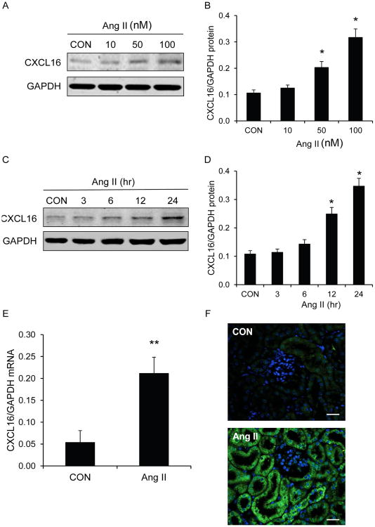
To determine if Ang II regulates CXCL16 gene expression, mouse kidney tubular epithelial cells were treated with various concentrations of Ang II for different periods of time. Western blot analysis showed that Ang II treatment significantly induced CXCL16 protein expression in vitro in a dose- and time- dependent manner (Figure 1A-D).
Figure 1. Ang II induces CXCL16 expression.
Ang II time-dependently increases CXCL16 protein expression in kidney tubular epithelial cells (A and B). Ang II dose-dependently increases CXCL16 protein expression in kidney tubular epithelial cells (C and D). * P<0.05 vs vehicle controls. n=4 per group for B and D. Ang II induces CXCL16 mRNA in the kidneys (E). ** P<0.01 vs vehicle-treated controls. n=6 per group. Representative photomicrographs of kidney sections stained for CXCL16 (green) and DAPI (blue) (F). Scale bar, 25μm.
We then determined if CXCL16 was induced in the kidney in response to Ang II infusion. Quantitative real time RT-PCR showed that mRNA level of CXCL16 was upregulated in the kidneys after Ang II treatment compared with that of control kidneys (Figure 1E). To identify the cell type responsible for CXCL16 production in the kidney, serial kidney sections were stained with an anti-CXCL16 antibody. The results revealed that CXCL16 protein was mainly induced in tubular epithelial cells of Ang II-treated kidneys (Figure 1F).
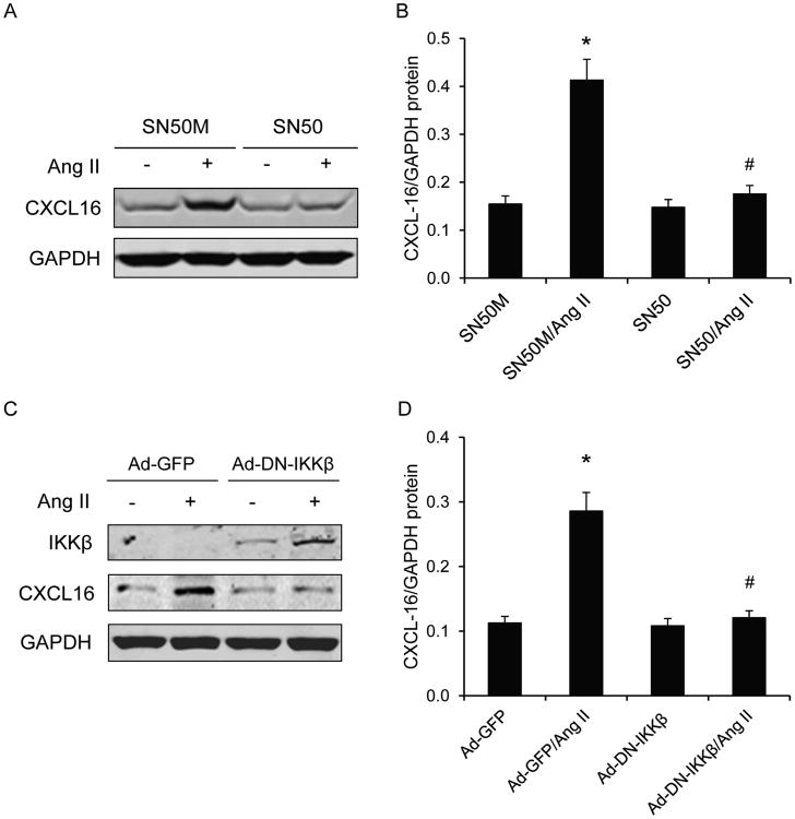
Because NF-κB is a master regulator of inflammation, we next evaluated if NF-κB mediates Ang II-induced CXCL16 gene expression in vitro. Mouse kidney tubular epithelial cells were pretreated with NF-κB inhibitor SN50 or its inactive control peptide SN50M for 30 min and then treated with Ang II at 100 nM or vehicle for 24 hr. Western blot analysis showed that SN50 abolished Ang II-induced CXCL16 gene expression (Figure 2 A and B). These data indicate that NF-κB plays an essential role in Ang II-induced CXCL16 expression in kidney tubular epithelial cells.
Figure 2. Inhibition of NF-κB blocks Ang II-induced CXCL16 protein expression in kidney tubular epithelial cells.
SN50 abolishes Ang II-induced CXCL16 protein expression (A and B). * P<0.05 vs vehicle controls; # P<0.05 vs Ang II. n=4 per group. Ectopic expression of DN-IKKβ blocks Ang II-induced CXCL16 protein expression (C and D). Cell lysates were immunoblotted with specific antibodies as indicated. * P<0.05 vs Ad-GFP controls; # P<0.05 vs Ad-GFP-Ang II. n=4 per group.
To verify that NF-κB mediates Ang II-induced CXCL16 gene expression, mouse tubular epithelial cells were transduced with a recombinant adenovirus expressing a dominant negative form of IKKβ, an upstream signaling of NF-κB. Cells transduced with a recombinant adenovirus expressing eGFP were used as controls. Western blot analysis showed that ectopic expression of dominant negative IKKβ completely blocked Ang II-induced CXCL16 expression (Figure 2 C and D).
Blood Pressure
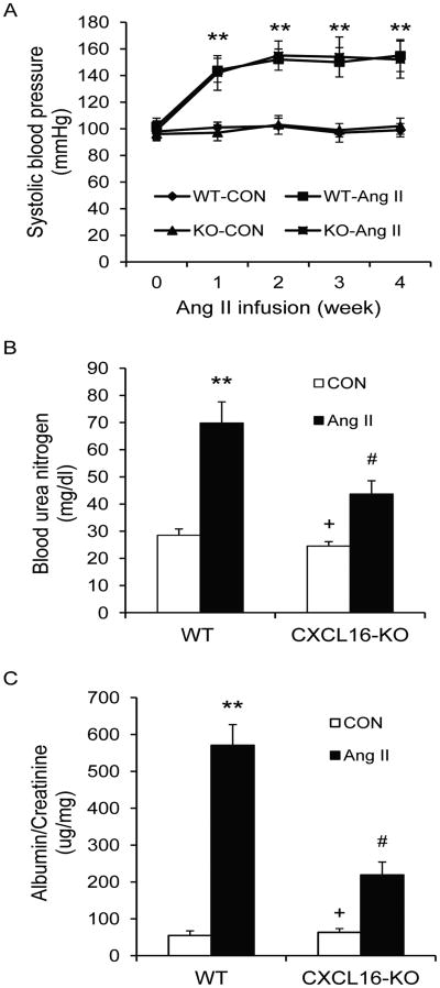
To determine the functional significance of CXCL16 induction in the kidney, WT and CXCL16 KO mice were treated with vehicle or Ang II for 4 weeks. There were no significant differences in systolic blood pressure among the four groups at baseline. Ang II treatment led to an increase in systolic blood pressure in both WT and CXCL16 KO mice that was comparable between the two treatment groups (Figure 3A).
Figure 3. Effect of CXCL16 deficiency on blood pressure, renal function and proteinuria.
CXCL16 deficiency has no effect on Ang II-induced elevation of blood pressure (A). ** P<0.01 between Ang II-treated groups and vehicle-treated control groups. n=6 per group. CXCL16 deficiency significantly prevents Ang II-induced renal dysfunction (B). ** P<0.01 vs WT controls; + P < 0.05 vs KO Ang II; and # P < 0.05 vs WT Ang II. n=6 per group. CXCL16 deficiency reduces Ang II-induced albuminuria (C). ** P<0.01 vs WT controls; + P < 0.05 vs KO Ang II; and # P < 0.05 vs WT Ang II. n=6 per group.
Renal Function
Treatment with Ang II for 4 weeks caused kidney dysfunction in WT mice as reflected by significant elevation of blood urea nitrogen (BUN). Kidney function was preserved in CXCL16 KO mice with BUN markedly lower than WT mice (Figure 3B).
Albuminuria
WT mice developed massive albuminuria after Ang II treatment for 4 weeks, whereas Ang II-treated CXCL16 KO mice produced significantly less albuminuria (Figure 3C).
Kidney Injury and Fibrosis
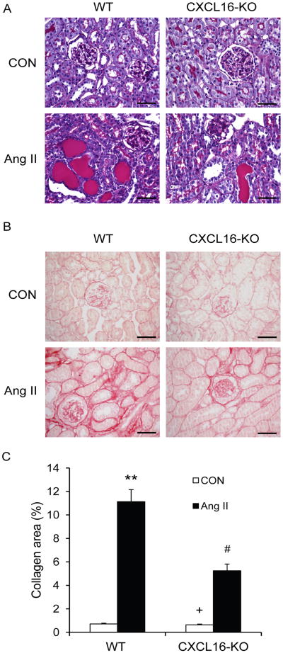
To assess the effect of CXCL16 deficiency on Ang II-induced hypertensive kidney damage, kidney sections were stained with PAS and scored for histological injury after 4 weeks of saline or Ang II infusion. On a semiquantitative scale that includes glomerulosclerosis, interstitial disease, fibrosis, and vascular injury, the 2 saline-infused groups had minimal kidney damage (Figure 4A and Table 1), Ang II caused a marked increase in the severity of kidney injury in the WT mice, which was substantially reduced in CXCL16 KO mice (Figure 4A and Table 1). Sirius red staining showed that Ang II-treated WT mice developed significant collagen deposition in the kidney compared with saline-treated WT mice (Figure 4 B and C). These fibrotic responses were significantly reduced in CXCL16 KO mice after 4 weeks of Ang II infusion (Figure 4 B and C).
Figure 4. CXCL16 deficiency suppresses renal injury and fibrosis.
A. Representative photomicrographs of PAS stained sections showing Ang II-induced kidney damage in WT and CXCL16 KO mice. Scale bar, 50μm. B. Representative photomicrographs of kidney sections from WT and CXCL16 KO mice 4 weeks after Ang II or saline treatment stained with Sirius red for assessment of total collagen deposition. Scale bar, 50μm. C. Quantitative analysis of interstitial collagen content in the kidneys of WT and CXCL16 KO mice. ** P < 0.01 vs WT controls; + P < 0.05 vs KO Ang II; and # P < 0.05 vs WT Ang II. n= 6 per group.
Table 1. CXCL16 Deficiency Prevents Ang II-induced Kidney Injury.
| Experimental Group | Glomerular Injury | Tubulointerstitial Disease | Vascular Damage | Total Injury |
|---|---|---|---|---|
| WT-CON | 0.21±0.03 | 0.23±0.03 | 0.15±0.02 | 0.59±0.07 |
| WT-Ang II | 2.53±0.42* | 4.55±0.54* | 1.14±0.16* | 8.22±0.95* |
| KO-CON | 0.19±0.03† | 0.22±0.03† | 0.14±0.02‡ | 0.55±0.07‡ |
| KO-Ang II | 1.25±0.17§ | 2.37±0.05§ | 0.56±0.31§ | 4.18±0.52§ |
p<0.01 versus WT-CON;
p<0.01 versus KO-Ang II;
p<0.05 versus KO-Ang II;
p<0.05 versus WT-Ang II. n=6 per group.
CXCL16 Deficiency Attenuates ECM Protein Expression
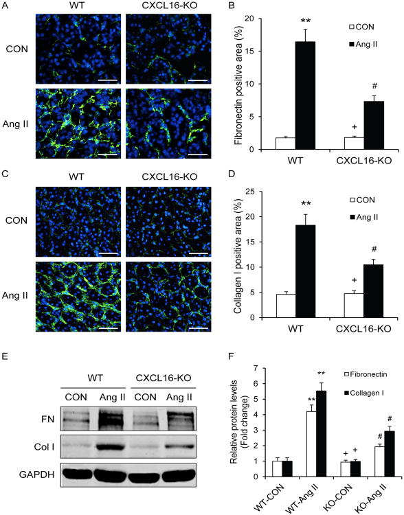
We next examined the effect of CXCL16 deficiency on the expression and accumulation of collagen I and fibronectin, two major components of ECM. Immunofluorescence and Western blot analysis demonstrated that CXCL16 deficiency attenuated the upregulation of collagen I and fibronectin in the kidneys after 4 weeks of Ang II infusion (Figure 5). These data indicate that CXCL16 deficiency inhibits Ang II-induced ECM protein expression.
Figure 5. CXCL16 deficiency reduces fibronectin and collagen I expression.
A. Representative photomicrographs of fibronectin immunofluorescence staining in the kidneys of WT and CXCL16 KO mice 4 weeks after Ang II or saline treatment. Scale bar, 50μm. B. Quantitative analysis of fibronectin positive area in the kidneys of WT and CXCL16 KO mice. ** P <0.01 vs WT controls, + P <0.05 vs KO Ang II, and # P <0.05 vs WT Ang II. n=6 per group. C. Representative photomicrographs of collagen I immunofluorescence staining in the kidneys of WT and CXCL16 KO mice 4 weeks after Ang II or saline treatment. Scale bar, 50μm. D. Quantitative analysis of collagen I positive area in the kidneys of WT and CXCL16 KO mice 4 weeks after Ang II or saline treatment. ** P <0.01 vs WT controls; + P <0.05 vs KO Ang II; and # P <0.05 vs WT Ang II. n=6 per group. E. Representative Western blots show the protein levels of fibronectin and collagen I in the kidneys of WT and CXCL16 KO mice 4 weeks after Ang II or saline treatment. F. Quantitative analysis of fibronectin and collagen I protein expression in the kidneys of WT and CXCL16 KO mice. ** P < 0.01 vs WT controls; + P < 0.05 vs KO Ang II; and # P < 0.05 vs WT Ang II. n=6 per group.
Myeloid Fibroblasts Accumulation
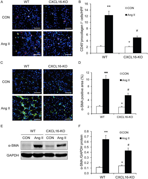
Recent evidence indicates that myeloid fibroblasts contribute significantly to the pathogenesis of renal fibrosis10-14. To examine the effect of CXCL16 deficiency on the accumulation of myeloid fibroblasts in the kidney, WT and CXCL16 KO mice were infused with vehicle or Ang II for 2 weeks. Kidney sections were stained for CD45 and procollagen I and examined with a fluorescence microscope. The results showed that the number of bone marrow-derived fibroblasts dual positive for CD45 and procollagen I was significantly reduced in the kidneys of Ang II-treated CXCL16 KO mice compared with WT mice (Figure 6 A and B). These data indicate that CXCL16 has an important role in recruiting bone marrow-derived fibroblasts into the kidney in response to Ang II.
Figure 6. CXCL16 deficiency suppresses bone marrow-derived fibroblast accumulation and myofibroblast formation in the kidney.
A. Representative photomicrographs of kidney sections from WT and CXCL16 KO mice 2 weeks after Ang II or saline treatment stained for CD45 (red), procollagen I (green), and DAPI (blue). Scale bar, 50μm. B. Quantitative analysis of CD45+ and procollagen I+ fibroblasts in kidneys of WT and CXCL16 KO mice 2 weeks after Ang II or saline treatment. ** P < 0.01 vs WT controls; + P < 0.05 vs KO Ang II; and # P < 0.05 vs WT Ang II. n=6 per group. C. Representative photomicrographs of α-SMA immunofluorescent staining in kidneys of WT and CXCL16 KO mice 4 weeks after Ang II or saline treatment. Scale bar, 50μm. D. Quantitative analysis of α-SMA protein expression in kidneys of WT and CXCL16 KO mice. ** P <0.01 vs WT controls; + P <0.05 vs KO Ang II; and # P <0.05 vs WT Ang II. n=6 per group. E. Representative Western blots show the levels of α-SMA protein expression in the kidneys of WT and CXCL16 KO mice 4 weeks after Ang II or saline treatment. F. Quantitative analysis of α-SMA protein expression in the kidneys of WT and CXCL16 KO mice. ** P <0.01 vs WT controls; + P <0.05 vs KO Ang II; and # P <0.05 vs WT Ang II. n=5 per group.
To determine if CXCL16 deficiency influences myofibroblast population, kidney sections were stained for α-SMA, a marker of myofibroblasts, and examined with a fluorescence microscope. The results revealed that Ang II-treated CXCL16 KO mice exhibited a significant reduction in the number of α-SMA+ myofibroblasts in the kidneys compared with Ang II-treated WT mice (Figure 6 C and D). Consistent with these findings, Western blot analysis showed that CXCL16 deficiency significantly reduced the protein expression levels of α-SMA in the kidneys after Ang II treatment compared with WT mice (Figure 6 E and F). These results indicate that CXCL16 deficiency reduces the population of myofibroblasts in the kidney.
Macrophage and T cell Infiltration
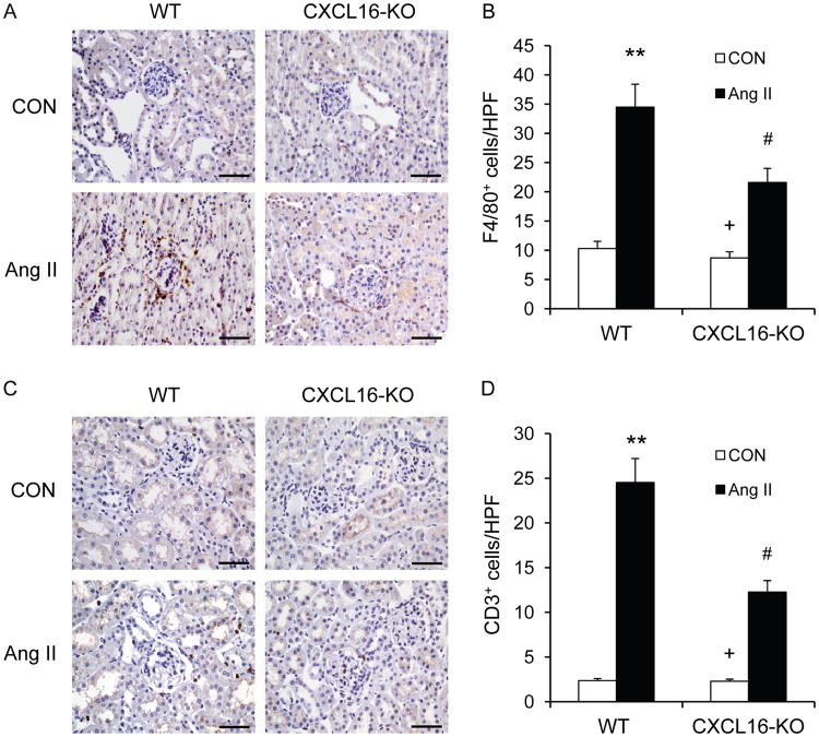
To examine if CXCL16 plays a role in the regulation of inflammatory cell infiltration into the kidney, WT and CXCL16 KO mice were infused with vehicle or Ang II for 2 weeks. Kidney sections were stained for F4/80 and CD3. Significant infiltration of macrophages and T cells was observed in the kidneys of WT mice after Ang II treatment compared with the vehicle-treated control group (Figure 7). In comparison, CXCL16 deficiency significantly inhibited macrophage and T cell infiltration into the kidneys after Ang II treatment (Figure 7). These results indicate that CXCL16 plays a critical role in recruiting inflammatory cell into the kidney in Ang II-induced hypertensive kidney disease.
Figure 7. CXCL16 deficiency reduces macrophage and T cell infiltration.
A. Representative photomicrographs of kidney sections stained for F4/80 (a macrophage marker) (brown) and counterstained with hematoxylin (blue) in WT and CXCL16 KO mice 2 weeks after Ang II or saline treatment. Scale bar, 50μm. B. Quantitative analysis of F4/80+ macrophages in the kidneys of WT and CXCL16 KO mice. **P < 0.01 vs WT controls; # P < 0.05 vs WT Ang II; + P < 0.05 vs KO Ang II. n=6 in each group. C. Representative photomicrographs of kidney sections stained for CD3 (a T lymphocyte marker) (brown) and counterstained with hematoxylin (blue) in WT and CXCL16 KO mice 2 weeks after Ang II or saline treatment. Scale bar, 50μm. D. Quantitative analysis of CD3+ T cells in the kidneys of WT and CXCL16 KO mice. **P < 0.01 vs WT controls; # P < 0.01 vs WT Ang II; + P < 0.05 vs KO Ang II. n=6 in each group.
Proinflammatory Cytokine Production
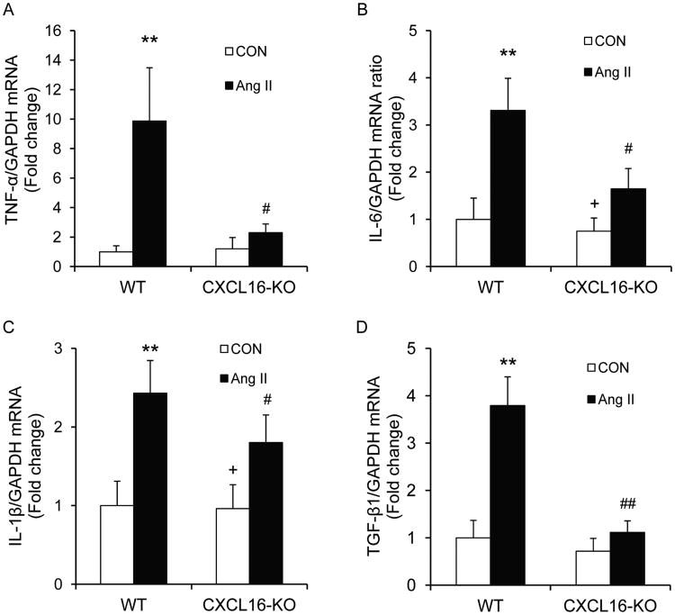
We next examined the effect of CXCL16 deficiency on the expression of known proinflammatory cytokines that are involved in the pathogenesis of kidney injury15. The mRNA levels of IL-6, TNF-α, IL-1β, and TGF-β1 were increased significantly in the kidneys of WT mice after Ang II treatment (Figure 8). In contrast, the upregulation of IL-6, TNF-α, IL-1β, and TGF-β1 was greatly attenuated in the kidneys of CXCL16 KO mice after Ang II treatment (Figure 8). These results suggest that CXCL16 regulates proinflammatory cytokine expression in the kidney in response to Ang II.
Figure 8. CXCL16 deficiency inhibits proinflammatory cytokine expression.
A. Quantitative analysis of TNF-α mRNA expression in the kidneys of WT and CXCL16 KO mice 2 weeks after Ang II or saline treatment. **P < 0.01 vs WT controls; # P < 0.05 vs WT Ang II. n=4 in each group. B. Quantitative analysis of IL-6 mRNA expression in the kidneys of WT and CXCL16 KO mice 2 weeks after Ang II or saline treatment. **P < 0.01 vs WT control; # P < 0.05 vs WT Ang II; + P < 0.05 vs KO Ang II. n=4 in each group. C. Quantitative analysis of IL-1β mRNA expression in the kidneys of WT and CXCL16 KO mice 2 weeks after Ang II or saline treatment. **P < 0.01 vs WT control; # P < 0.05 vs WT Ang II; + P < 0.05 vs KO Ang II. n=4 in each group. D. Quantitative analysis of TGF-β1 mRNA expression in the kidneys of WT and CXCL16 KO mice 2 weeks after Ang II or saline treatment. **P < 0.01 vs WT controls; ## P < 0.01 vs WT Ang II. n=4 in each group.
Discussion
In this study, we have demonstrated that Ang II induces CXCL16 gene expression in kidney tubular epithelial cells via activation of NF-κB and deletion of CXCL16 led to renal protection in an experimental model of chronic progressive kidney disease induced by Ang II infusion. In the Ang II-induced hypertension model, genetic deletion of CXCL16 preserves renal function, reduces urinary albumin excretion, and diminishes tubulointerstitial disease, glomerular injury, and vascular damage. It is noteworthy that genetic deletion of CXCL16 has no effect on arterial blood pressure. These findings are significant because the renal protection is achieved even in the setting of sustained hypertension.
Mounting evidence indicates that activation of RAS plays a central role in the development of chronic kidney disease. The underlying mechanisms involved in Ang II-induced hypertensive kidney disease are incompletely understood. Recent studies have shown that inflammatory and immune cell infiltration and altered chemokine production are characteristic for hypertensive kidney disease4, 5. The infiltration of circulating cells into sites in injury is mediated by locally produced chemokines through interaction with their respective receptors.
Chemokines are a group of structurally related cytokines that are classified into four major subfamilies: CC, CXC, C, and CX3C based on amino-terminal cysteine motif6. CXCL16 is a newly discovered chemokine that belongs to the CXC chemokine subfamily7. There are two forms of CXCL16. The transmembrane form of CXCL16 is composed of a CXC chemokine domain, a mucin-like stalk, a transmembrane domain and a cytoplasmic tail. The soluble form of CXCL16 resulting from cleavage by ADAM10 at the cell surface is composed of the extracellular stalk and the chemokine domain. The transmembrane form of CXCL16 functions as an adhesion molecule for CXCR6 (the only known receptor for CXCL16) expressing cells while the soluble form of CXCL16 functions as a chemoattractant to promote migration of CXCR6 expressing cells including T cells, monocytes, and myeloid fibroblasts14, 16, 17. CXCL16 has been reported to be expressed in the kidney8, 14, 18. However, its role in the pathogenesis of Ang II-induced renal injury is not known. In the present study, we demonstrate that Ang II induces CXCL16 gene expression in tubular epithelial cells in a time- and dose-dependent fashion. Furthermore, inhibition of NF-κB signaling with a pharmacological inhibitor or dominant negative IKKβ abolishes Ang II-induced CXCL16 gene expression. These data indicate that NF-κB signaling mediates Ang II-induced CXCL16 gene expression in kidney tubular epithelial cells. We provide experimental evidence that CXCL16 is pathologically important in the development of renal injury and fibrosis because deletion of CXCL16 inhibits the recruitment of bone marrow-derived fibroblasts, macrophages, and T cells into the kidney and the development of renal interstitial fibrosis and proteinuria in response to Ang II treatment. These data indicate that CXCL16 signaling plays an important role in the recruitment of bone marrow-derived fibroblasts, macrophages, and T cells into the kidney and pathogenesis of renal injury.
A significant finding of this study is the marked reduction in Ang II-induced renal interstitial fibrosis in CXCL16 KO mice. Renal interstitial fibrosis is a hallmark of chronic kidney disease and the degree of interstitial fibrosis strongly predicts the prognosis of chronic kidney disease1. Renal interstitial fibrosis is characterized by substantial fibroblast activation and excessive production and deposition of extracellular matrix (ECM), which leads to the destruction of renal parenchyma and progressive loss of kidney function. Because activated fibroblasts are the principal cells responsible for ECM production in the fibrotic kidney, their activation is regarded as a key event in the pathogenesis of renal fibrosis19, 20. However, the origin of these fibroblasts remains controversial. They are traditionally thought to arise from resident renal fibroblasts21-23. Recent evidence suggests they may originate from bone marrow-derived cells10-14, 24. Bone marrow-derived fibroblast precursors termed fibrocytes are derived from a subpopulation of circulating mononuclear cells25-29. These cells express hematopoietic markers such as CD45 and CD11b and mesenchymal markers such as collagen I and vimentin. We and others have shown that these cells migrate into the kidney in response to obstructive injury and contribute significantly to the development of renal fibrosis11, 14, 30, 31. However, the signaling mechanisms underlying the recruitment of these bone marrow-derived fibroblast precursors into the kidney are incompletely understood. In the present study, we showed that CXCL16 is pathologically important in recruiting myeloid fibroblasts into the kidney and the development of renal fibrosis because deletion of CXCL16 inhibits bone marrow-derived fibroblast accumulation and myofibroblast formation in the kidney and the development of renal interstitial fibrosis in response to chronic Ang II treatment. These data indicate that CXCL16 signaling plays a key role in the recruitment of bone marrow-derived fibroblasts into the kidney and the development of renal fibrosis in Ang II-induced hypertensive nephropathy.
Macrophages and T cells have been shown to play an important role in the development of hypertensive kidney disease32. Ang II is a key factor in the regulation of inflammatory response in hypertensive end organ damage3, 33. CXCR6, the only known receptor for CXCL16, is expressed in various leukocyte subsets including monocytes and T cells16, 17. In the present study, our results demonstrate that Ang II-induced interstitial infiltration of macrophages and T cells into the kidney is significantly attenuated in CXCL16 KO mice. These data indicate that CXCL16 signaling mediates Ang II-induced macrophage and T cell infiltration into the kidney.
In addition to establishing a critical role of CXCL16 in recruiting inflammatory cells into the kidney, our results indicate that absence of CXCL16 influences proinflammatory cytokine expression in the kidney. This is relevant because proinflammatory cytokines – TNF-α, IL-6, and IL-1β have been implicated in the pathogenesis of Ang II-induced target organ damage34. A recent experimental study has shown that IL-6 contributes to Ang II-induced hypertension and renal damage35. Population based clinical studies have demonstrated that a significant association between inflammatory markers and renal function, supporting a link between inflammation and the development of CKD36-38.
TGF-β1 is a key profibrotic cytokine that contributes to renal damage and tubulointerstitial fibrosis39. Studies have demonstrated that TGF-β1 functions as a downstream mediator of Ang II-induced renal fibrosis and both factors share several intracellular mechanisms involved in the regulation of ECM protein expression3, 40, 41. Although studies have shown that bone marrow-derived fibroblasts significantly contribute to the pathogenesis of renal fibrosis, the molecular mechanisms underlying the differentiation of these cells into activated fibroblasts are not fully understood. Recent studies have demonstrated that TGF-β1 is a critical factor for the migration and differentiation of these myeloid fibroblasts precursors42, 43. In the present study, our results show that Ang II induces TGF-β1 gene expression in the kidney of WT mice and CXCL16 deficiency significantly attenuates Ang II-induced TGF-β1 gene expression. The decrease in TGF-β1 could be another mechanism by which CXCL16 deficiencysuppresses Ang II-induced renal fibrosis.
Perspectives
Our studies identify CXCL16 as a critical factor that regulates Ang II-induced renal injury and fibrosis. In response to Ang II, CXCL16 recruits macrophages, T cells, and myeloid fibroblasts into the kidney leading to renal injury and fibrosis. These findings suggest that inhibition of CXCL16 signaling could constitute a novel therapeutic strategy for hypertensive kidney disease.
Novelty and Significance.
What Is New?
We report that CXCL16 is a critical factor that regulates Ang II-induced renal injury and fibrosis.
What Is Relevant?
The role of CXCL16 in the development of hypertensive kidney disease is not known. Out study shows that CXCL16 plays a critical role in the development of hypertensive kidney disease, suggesting that inhibition of CXCL16 signaling represents a novel therapy for hypertensive kidney disease.
Summary
This study demonstrates that CXCL16 regualtes hypertensive renal injury and fibrosis via recruitment of macrophages, T cells, and myeloid fibroblasts into the kidney and suggests that inhibition of CXCL16 signaling could be a novel therapeutic target for hypertensive kidney disease.
Acknowledgments
We thank Dr. William E. Mitch for critical reading of the manuscript. We also thank Dr. Shuhua Han at Baylor College of Medicine for providing CXCL16 knockout mice.
Sources of Funding: This work was supported in part by the National Institutes of Health grants K08HL92958, R01DK95835 and an American Heart Association grant 11BGIA7840054 to Y.W. ML Entman was supported by the National Institutes of Health grant R01HL89792.
Footnotes
Disclosures: None
References
- 1.Nath KA. The tubulointerstitium in progressive renal disease. Kidney Int. 1998;54:992–994. doi: 10.1046/j.1523-1755.1998.00079.x. [DOI] [PubMed] [Google Scholar]
- 2.Bidani AK, Griffin KA. Pathophysiology of hypertensive renal damage: Implications for therapy. Hypertension. 2004;44:595–601. doi: 10.1161/01.HYP.0000145180.38707.84. [DOI] [PubMed] [Google Scholar]
- 3.Ruiz-Ortega M, Ruperez M, Esteban V, Rodriguez-Vita J, Sanchez-Lopez E, Carvajal G, Egido J. Angiotensin ii: A key factor in the inflammatory and fibrotic response in kidney diseases. Nephrol Dial Transplant. 2006;21:16–20. doi: 10.1093/ndt/gfi265. [DOI] [PubMed] [Google Scholar]
- 4.Crowley SD, Frey CW, Gould SK, Griffiths R, Ruiz P, Burchette JL, Howell DN, Makhanova N, Yan M, Kim HS, Tharaux PL, Coffman TM. Stimulation of lymphocyte responses by angiotensin ii promotes kidney injury in hypertension. Am J Physiol Renal Physiol. 2008;295:F515–524. doi: 10.1152/ajprenal.00527.2007. [DOI] [PMC free article] [PubMed] [Google Scholar]
- 5.Guzik TJ, Hoch NE, Brown KA, McCann LA, Rahman A, Dikalov S, Goronzy J, Weyand C, Harrison DG. Role of the t cell in the genesis of angiotensin ii induced hypertension and vascular dysfunction. J Exp Med. 2007;204:2449–2460. doi: 10.1084/jem.20070657. [DOI] [PMC free article] [PubMed] [Google Scholar]
- 6.Fernandez EJ, Lolis E. Structure, function, and inhibition of chemokines. Annu Rev Pharmacol Toxicol. 2002;42:469–499. doi: 10.1146/annurev.pharmtox.42.091901.115838. [DOI] [PubMed] [Google Scholar]
- 7.Matloubian M, David A, Engel S, Ryan JE, Cyster JG. A transmembrane cxc chemokine is a ligand for hiv-coreceptor bonzo. Nat Immunol. 2000;1:298–304. doi: 10.1038/79738. [DOI] [PubMed] [Google Scholar]
- 8.Garcia GE, Truong LD, Li P, Zhang P, Johnson RJ, Wilson CB, Feng L. Inhibition of cxcl16 attenuates inflammatory and progressive phases of anti-glomerular basement membrane antibody-associated glomerulonephritis. Am J Pathol. 2007;170:1485–1496. doi: 10.2353/ajpath.2007.060065. [DOI] [PMC free article] [PubMed] [Google Scholar]
- 9.Zhang L, Ran L, Garcia GE, Wang XH, Han S, Du J, Mitch WE. Chemokine cxcl16 regulates neutrophil and macrophage infiltration into injured muscle, promoting muscle regeneration. Am J Pathol. 2009;175:2518–2527. doi: 10.2353/ajpath.2009.090275. [DOI] [PMC free article] [PubMed] [Google Scholar]
- 10.Li J, Deane JA, Campanale NV, Bertram JF, Ricardo SD. The contribution of bone marrow-derived cells to the development of renal interstitial fibrosis. Stem Cells. 2007;25:697–706. doi: 10.1634/stemcells.2006-0133. [DOI] [PubMed] [Google Scholar]
- 11.Sakai N, Wada T, Yokoyama H, Lipp M, Ueha S, Matsushima K, Kaneko S. Secondary lymphoid tissue chemokine (slc/ccl21)/ccr7 signaling regulates fibrocytes in renal fibrosis. Proc Natl Acad Sci U S A. 2006;103:14098–14103. doi: 10.1073/pnas.0511200103. [DOI] [PMC free article] [PubMed] [Google Scholar]
- 12.Grimm PC, Nickerson P, Jeffery J, Savani RC, Gough J, McKenna RM, Stern E, Rush DN. Neointimal and tubulointerstitial infiltration by recipient mesenchymal cells in chronic renal-allograft rejection. N Engl J Med. 2001;345:93–97. doi: 10.1056/NEJM200107123450203. [DOI] [PubMed] [Google Scholar]
- 13.Broekema M, Harmsen MC, van Luyn MJ, Koerts JA, Petersen AH, van Kooten TG, van Goor H, Navis G, Popa ER. Bone marrow-derived myofibroblasts contribute to the renal interstitial myofibroblast population and produce procollagen i after ischemia/reperfusion in rats. J Am Soc Nephrol. 2007;18:165–175. doi: 10.1681/ASN.2005070730. [DOI] [PubMed] [Google Scholar]
- 14.Chen G, Lin SC, Chen J, He L, Dong F, Xu J, Han S, Du J, Entman ML, Wang Y. Cxcl16 recruits bone marrow-derived fibroblast precursors in renal fibrosis. J Am Soc Nephrol. 2011;22:1876–1886. doi: 10.1681/ASN.2010080881. [DOI] [PMC free article] [PubMed] [Google Scholar]
- 15.Chung AC, Lan HY. Chemokines in renal injury. J Am Soc Nephrol. 2011;22:802–809. doi: 10.1681/ASN.2010050510. [DOI] [PubMed] [Google Scholar]
- 16.Kim CH, Kunkel EJ, Boisvert J, Johnston B, Campbell JJ, Genovese MC, Greenberg HB, Butcher EC. Bonzo/cxcr6 expression defines type 1-polarized t-cell subsets with extralymphoid tissue homing potential. J Clin Invest. 2001;107:595–601. doi: 10.1172/JCI11902. [DOI] [PMC free article] [PubMed] [Google Scholar]
- 17.Sato T, Thorlacius H, Johnston B, Staton TL, Xiang W, Littman DR, Butcher EC. Role for cxcr6 in recruitment of activated cd8+ lymphocytes to inflamed liver. J Immunol. 2005;174:277–283. doi: 10.4049/jimmunol.174.1.277. [DOI] [PubMed] [Google Scholar]
- 18.Okamura DM, Lopez-Guisa JM, Koelsch K, Collins S, Eddy AA. Atherogenic scavenger receptor modulation in the tubulointerstitium in response to chronic renal injury. Am J Physiol Renal Physiol. 2007;293:F575–585. doi: 10.1152/ajprenal.00063.2007. [DOI] [PubMed] [Google Scholar]
- 19.Neilson EG. Mechanisms of disease: Fibroblasts--a new look at an old problem. Nat Clin Pract Nephrol. 2006;2:101–108. doi: 10.1038/ncpneph0093. [DOI] [PubMed] [Google Scholar]
- 20.Strutz F, Muller GA. Renal fibrosis and the origin of the renal fibroblast. Nephrol Dial Transplant. 2006;21:3368–3370. doi: 10.1093/ndt/gfl199. [DOI] [PubMed] [Google Scholar]
- 21.Picard N, Baum O, Vogetseder A, Kaissling B, Le Hir M. Origin of renal myofibroblasts in the model of unilateral ureter obstruction in the rat. Histochem Cell Biol. 2008;130:141–155. doi: 10.1007/s00418-008-0433-8. [DOI] [PMC free article] [PubMed] [Google Scholar]
- 22.Qi W, Chen X, Poronnik P, Pollock CA. The renal cortical fibroblast in renal tubulointerstitial fibrosis. Int J Biochem Cell Biol. 2006;38:1–5. doi: 10.1016/j.biocel.2005.09.005. [DOI] [PubMed] [Google Scholar]
- 23.Powell DW, Mifflin RC, Valentich JD, Crowe SE, Saada JI, West AB. Myofibroblasts. I. Paracrine cells important in health and disease. Am J Physiol. 1999;277:C1–9. doi: 10.1152/ajpcell.1999.277.1.C1. [DOI] [PubMed] [Google Scholar]
- 24.Iwano M, Plieth D, Danoff TM, Xue C, Okada H, Neilson EG. Evidence that fibroblasts derive from epithelium during tissue fibrosis. J Clin Invest. 2002;110:341–350. doi: 10.1172/JCI15518. [DOI] [PMC free article] [PubMed] [Google Scholar]
- 25.Chesney J, Bacher M, Bender A, Bucala R. The peripheral blood fibrocyte is a potent antigen-presenting cell capable of priming naive t cells in situ. Proc Natl Acad Sci U S A. 1997;94:6307–6312. doi: 10.1073/pnas.94.12.6307. [DOI] [PMC free article] [PubMed] [Google Scholar]
- 26.Chesney J, Bucala R. Peripheral blood fibrocytes: Novel fibroblast-like cells that present antigen and mediate tissue repair. Biochem Soc Trans. 1997;25:520–524. doi: 10.1042/bst0250520. [DOI] [PubMed] [Google Scholar]
- 27.Metz CN. Fibrocytes: A unique cell population implicated in wound healing. Cell Mol Life Sci. 2003;60:1342–1350. doi: 10.1007/s00018-003-2328-0. [DOI] [PMC free article] [PubMed] [Google Scholar]
- 28.Quan TE, Cowper S, Wu SP, Bockenstedt LK, Bucala R. Circulating fibrocytes: Collagen-secreting cells of the peripheral blood. Int J Biochem Cell Biol. 2004;36:598–606. doi: 10.1016/j.biocel.2003.10.005. [DOI] [PubMed] [Google Scholar]
- 29.Bucala R, Spiegel LA, Chesney J, Hogan M, Cerami A. Circulating fibrocytes define a new leukocyte subpopulation that mediates tissue repair. Mol Med. 1994;1:71–81. [PMC free article] [PubMed] [Google Scholar]
- 30.Niedermeier M, Reich B, Rodriguez Gomez M, Denzel A, Schmidbauer K, Gobel N, Talke Y, Schweda F, Mack M. Cd4+ t cells control the differentiation of gr1+ monocytes into fibrocytes. Proc Natl Acad Sci U S A. 2009;106:17892–17897. doi: 10.1073/pnas.0906070106. [DOI] [PMC free article] [PubMed] [Google Scholar]
- 31.Yang J, Chen J, Yan J, Zhang L, Chen G, He L, Wang Y. Effect of interleukin 6 deficiency on renal interstitial fibrosis. PLoS One. 2012;7:e52415. doi: 10.1371/journal.pone.0052415. [DOI] [PMC free article] [PubMed] [Google Scholar]
- 32.Luft FC, Dechend R, Muller DN. Immune mechanisms in angiotensin ii-induced target-organ damage. Ann Med. 2012;44(1):S49–54. doi: 10.3109/07853890.2011.653396. [DOI] [PubMed] [Google Scholar]
- 33.Mann DL. Angiotensin ii as an inflammatory mediator: Evolving concepts in the role of the renin angiotensin system in the failing heart. Cardiovasc Drugs Ther. 2002;16:7–9. doi: 10.1023/a:1015355112501. [DOI] [PubMed] [Google Scholar]
- 34.Mehta PK, Griendling KK. Angiotensin ii cell signaling: Physiological and pathological effects in the cardiovascular system. Am J Physiol Cell Physiol. 2007;292:C82–97. doi: 10.1152/ajpcell.00287.2006. [DOI] [PubMed] [Google Scholar]
- 35.Zhang W, Wang W, Yu H, Zhang Y, Dai Y, Ning C, Tao L, Sun H, Kellems RE, Blackburn MR, Xia Y. Interleukin 6 underlies angiotensin ii-induced hypertension and chronic renal damage. Hypertension. 2012;59:136–144. doi: 10.1161/HYPERTENSIONAHA.111.173328. [DOI] [PMC free article] [PubMed] [Google Scholar]
- 36.Pruijm M, Ponte B, Vollenweider P, Mooser V, Paccaud F, Waeber G, Marques-Vidal P, Burnier M, Bochud M. Not all inflammatory markers are linked to kidney function: Results from a population-based study. Am J Nephrol. 2012;35:288–294. doi: 10.1159/000335934. [DOI] [PubMed] [Google Scholar]
- 37.Keller C, Katz R, Cushman M, Fried LF, Shlipak M. Association of kidney function with inflammatory and procoagulant markers in a diverse cohort: A cross-sectional analysis from the multi-ethnic study of atherosclerosis (mesa) BMC Nephrol. 2008;9:9. doi: 10.1186/1471-2369-9-9. [DOI] [PMC free article] [PubMed] [Google Scholar]
- 38.Upadhyay A, Larson MG, Guo CY, Vasan RS, Lipinska I, O'Donnell CJ, Kathiresan S, Meigs JB, Keaney JF, Jr, Rong J, Benjamin EJ, Fox CS. Inflammation, kidney function and albuminuria in the framingham offspring cohort. Nephrol Dial Transplant. 2011;26:920–926. doi: 10.1093/ndt/gfq471. [DOI] [PMC free article] [PubMed] [Google Scholar]
- 39.Zeisberg M, Neilson EG. Mechanisms of tubulointerstitial fibrosis. J Am Soc Nephrol. 2010;21:1819–1834. doi: 10.1681/ASN.2010080793. [DOI] [PubMed] [Google Scholar]
- 40.Wolf G. Renal injury due to renin-angiotensin-aldosterone system activation of the transforming growth factor-beta pathway. Kidney Int. 2006;70:1914–1919. doi: 10.1038/sj.ki.5001846. [DOI] [PubMed] [Google Scholar]
- 41.Lan HY. Diverse roles of tgf-beta/smads in renal fibrosis and inflammation. Int J Biol Sci. 2011;7:1056–1067. doi: 10.7150/ijbs.7.1056. [DOI] [PMC free article] [PubMed] [Google Scholar]
- 42.Abe R, Donnelly SC, Peng T, Bucala R, Metz CN. Peripheral blood fibrocytes: Differentiation pathway and migration to wound sites. J Immunol. 2001;166:7556–7562. doi: 10.4049/jimmunol.166.12.7556. [DOI] [PubMed] [Google Scholar]
- 43.Scholten D, Reichart D, Paik YH, Lindert J, Bhattacharya J, Glass CK, Brenner DA, Kisseleva T. Migration of fibrocytes in fibrogenic liver injury. The American journal of pathology. 2011;179:189–198. doi: 10.1016/j.ajpath.2011.03.049. [DOI] [PMC free article] [PubMed] [Google Scholar]


