Abstract
Achondroplasia is considered as a form of skeletal dysplasia/dwarfism that manifests with stunted stature and disproportionate limb shortening. Achondroplasia is of special interest in the field of dentistry because of its characteristic craniofacial features. It has been considered as the most common short-limbed dwarfism syndrome. Very few authors have reported the presence of oligodontia in achondroplastic patients. The present paper reports a rare case of oligodontia in a young, female, achondroplastic patient.
Keywords: Achondroplasia, dwarfism, skeletal dysplasia
INTRODUCTION
Achondroplasia is a non-lethal form of chondrodysplasia.[1,2] It is the most common form of skeletal dysplasia and is characterized by short limb dwarfism, affecting the growth of the tubular bones, spine and skull. Prevalence is estimated to be 1 in 15,000. It is inherited as autosomal dominant trait with complete penetrance.[3] Approximately 80% of cases are due to de novo dominant mutations. Its clinical phenotype has been recognized for centuries and the notion that it reflects a disturbance of cartilage-mediated (endochondral) bone growth has existed for about 100 years. It occurs as a result of mutation in one copy of the fibroblast growth factor receptor 3 genes (FGFR3) [Online Mendelian Inheritance in Man database (OMIM) - Entry ID: 134934] of which more than 95% of patients have the same point mutation in FGFR3.[4]
Characteristic phenotypic features include disproportionate short stature, megalencephaly, a prominent forehead (frontal bossing), midface hypoplasia, rhizomelic shortening of the arms and legs, a normal trunk length, prominent lumbar lordosis, genu varum and a trident hand configuration.[5] The dentition typically is normal; however, delayed eruption of teeth with malocclusion is reported in some cases. We report a rare case of achondroplasia with oligodontia in a young female patient.
CASE REPORT
A 16-year-old female reported to Department of Oral Medicine and Radiology with the chief complaint of multiple missing teeth. The patient had short stature and walked with an abnormal gait. The patient gave familial history of her mother and sister affected in similar manner. The patient was born to non-consanguineous parents, delivered normally at 8.5 months of gestation. The patient's father reported that she had large and dysmorphic head at the time of birth. The gross motor developmental milestones were delayed significantly, a finding often attributed to the large head of these patients, but fine motor developmental milestones, social and adaptive milestones and language milestones were reportedly normal according to her father.
The patient appeared to be well-adjusted, healthy and intelligent. General physical examination showed short stature, with normal trunk length and rhizomelic shortening of the arms and legs [Figure 1]. Lumbar lordosis; prominent buttocks; and short, stubby fingers with trident hand configuration were also evident. Anthropometry (at age 16 years) revealed a height of 124 cm (midparental target height 143.6 cm by Tanner's formula), weight of 42 kg, mesomelic dwarfism with upper limb to lower limb ratio of >1 and an arm span of 100 cm (expected to be 124 cm). The head circumference was 58 cm. Her vital signs were within the normal limits. Systemic examination did not reveal any other abnormality. Extraoral examination revealed facial features such as brachycephaly, midfacial hypoplasia, flat nasal bridge, frontal bossing and competent lips. A concave facial profile was also noticed; however, the mandible appeared normal and the chin was not prominent [Figure 2].
Figure 1.
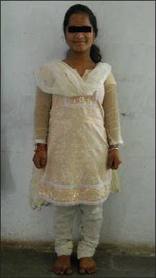
Clinical picture of patient showing short stature and rhizomelic shortening of arms and legs
Figure 2.
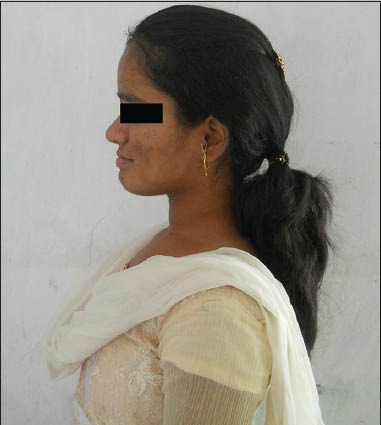
Clinical picture showing concave profile
Intraoral examination revealed the presence of eight teeth (11, 15, 17, 21, 25, 27, 36 and 46). No abnormality was seen with intraoral soft tissues except for hypertonic lips [Figure 3].
Figure 3.
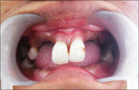
Intraoral picture showing multiple missing teeth and hypertonic lips
On radiographic examination, her orthopantomogram showed the presence of impacted maxillary canines (13 and 23) and the follicles of all third molars (18, 28, 38 and 48) [Figure 4]. The lateral cephalogram revealed an enlarged calvarium with increased transverse diameter, shortening of the skull base with frontal bossing and a retruded maxilla. The mandible appeared normal however [Figure 5]. The hand-wrist radiograph showed a trident hand configuration [Figure 6]. Based on the history, clinical examination and radiological investigations; a final diagnosis of achondroplasia was made.
Figure 4.
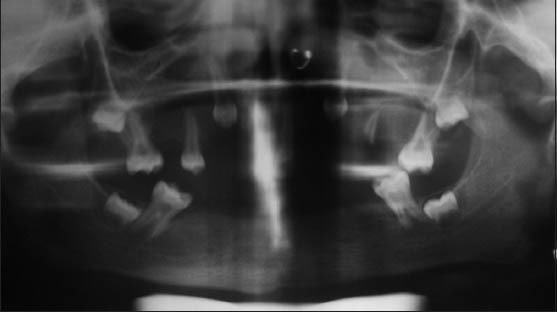
Orthopantomogram showing presence of impacted maxillary canines and follicles of third molars
Figure 5.
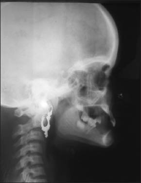
Lateral cephalogram showing maxillary retrusion, frontal bossing, large calvarium and shortened skull base
Figure 6.
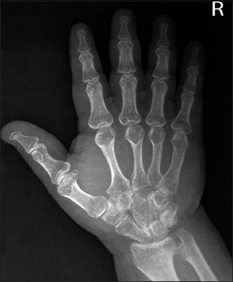
Hand-wrist radiograph showing trident hand configuration
The patient and her parents were provided with genetic counseling and the generally good prognosis of the condition was emphasized. The patient was then referred to Department of Prosthodontics for further treatment.
DISCUSSION
Achondroplasia is an autosomal dominant disorder with complete penetrance. The common mutation causes a gain of function of the FGFR3 gene, resulting in decreased endochondral ossification, inhibited proliferation of chondrocytes in growth plate cartilage, decreased cellular hypertrophy and decreased cartilage matrix production; leading to a variety of manifestations and complications. The gene has been mapped to band 4p16.3. In the heterozygous state, achondroplasia is non-lethal, with most patients having a relatively normal life span and normal intelligence. They are at risk, however, for conditions such as cervicomedullary compression, spinal stenosis, obesity or obstructive sleep apnea.[6]
Affected babies are short at birth and grow slowly throughout childhood; the average final height for women is 123 cm and 130 cm for men. Characteristic clinical features are typically present at birth; with affected patients having short limbs, a long narrow trunk and a large head with midfacial hypoplasia and a prominent forehead. The limb shortening is greatest in the proximal segments (rhizomelic limb shortening) and the fingers often display a trident configuration. Most joints are hyperextensible, but extension is restricted at the elbow.[7] In our patient all of these findings were present, including a height of 124 cm, rhizomelic limb shortening, large head, midfacial hypoplasia and trident hand configuration. Although the majority of affected children will be healthy, approximately 10% can develop significant complications.
The radiographic features of achondroplasia exhibit large calvarial bones with a small cranial base and facial bones. The vertebral pedicles are short throughout the spine as noted on a lateral radiograph. The interpedicular distance, which normally increases from the first to the fifth lumbar vertebrae, decreases in achondroplasia. The iliac bones are short and round and the acetabular roofs are flat. The tubular bones are short with mild irregular and flared metaphyses. The fibula is disproportionately long compared with the tibia. Hand radiographs show shortening of the metacarpals and phalanges with trident configuration.[7]
The dentition of achondroplastic dwarfs is usually normal, although there may be delayed eruption. Developmentally absent teeth have been reported rarely.[2,8] Stafne (1950) reported retarded eruption of many permanent teeth in a 30-year-old affected male.[8] Several odonto-stomatologic manifestations of achondroplasia have been previously reported; including skeletal and dental class III malocclusion, a narrow maxilla, macroglossia and an open bite between the posterior teeth.[8] The previous cases reported in literature have showed presence of posterior cross bite, Class III malocclusion and abnormally large tongue in all the cases.[9,10,11] Incidence of periodontal disease, benign migratory glossitis and trigeminal neuralgia in achondroplastic patients was also noted.[8,9,11] There was no available data regarding the incidence of oligodontia in achondroplastic children. Our patient had midfacial hypoplasia with retruded maxilla, however, marked mandibular prognathism was not observed. In our case the finding of oligodontia was more pronounced than any other cases reported in literature.
The differential diagnosis of achondroplasia includes other chondrodystrophies including hypochondroplasia and chondroectodermal dysplasia. Although hypochondroplasia is associated with dwarfism, it can be differentiated from achondroplasia by milder clinical features. Brachycephaly is much less pronounced in hypochondroplasia and also the trident hand configuration is not seen in hypochondroplasia.[4]
Dealing with achondroplastic children may require special psychological management during dental treatment, as the presence of disproportionate short stature can cause a number of psychosocial and social problems. Furthermore, special precautions in head control during dental intervention are essential, due to the possible presence of craniocervical instability, foramen magnum stenosis and limited neck extension; as these may contribute to respiratory complications.[6]
CONCLUSION
A patient with achondroplasia not only requires specific medical management, but also requires special attention with definitive dental management along with psychological support to help him lead a normal life and cope up with the medical as well as social challenges of life.
Footnotes
Source of Support: Nil.
Conflict of Interest: None declared.
REFERENCES
- 1.Hennekam R, Allanson J, Krantz I. 5th ed. New York: Oxford University Press; 2010. Gorlin's Syndromes of the Head and Neck. [Google Scholar]
- 2.Shivapathasundharam RR. 6th ed. New Delhi (India): Elsevier; 2011. Shafer's Textbook of Oral Pathology. [Google Scholar]
- 3.Bellu AG, Heffron WT, Ortiz de Luna RI, Hecht JT, Horton WA, Machado M, et al. Achondroplasia is defined by recurrent G380R mutations of FGFR3. Am J Hum Genet. 1995;56:368–73. [PMC free article] [PubMed] [Google Scholar]
- 4.Chitayat D, Fermandez B, Gardener A, Moore L, Glance P, Dunn M, et al. Compound heterozygosity for the achondroplasia-hypochondroplasia FGFR3 mutations: Prenatal diagnosis and post-natal outcome. Am J Med Genet. 1999;84:401–5. [PubMed] [Google Scholar]
- 5.Cohen MM., Jr Some achondroplasia with short limbs: Molecular perspectives. Am J Med Genet. 2002;112:304–13. doi: 10.1002/ajmg.10780. [DOI] [PubMed] [Google Scholar]
- 6.Rohilla S, Kaushik A, Vinod V, Tanwar R, Kumar M. Orofacial manifestations of achondroplasia. EXCLI J. 2012;11:538–54. [PMC free article] [PubMed] [Google Scholar]
- 7.Sethi RS, Kumar L, Chaurasiya OS, Gorakh R. Achondroplasia: A case report. Curr Pediatr Res. 2011;15:137–42. [Google Scholar]
- 8.Chawla K, Lamba AK, Faraz F, Tandon S. Achondroplasia and periodontal disease. J Indian Soc Periodontol. 2012;16:138–40. doi: 10.4103/0972-124X.94624. [DOI] [PMC free article] [PubMed] [Google Scholar]
- 9.Al-Saleem A, Al-Jobair A. Achondroplasia: Craniofacial manifestations and considerations in dental management. Saudi Dent J. 2010;22:195–9. doi: 10.1016/j.sdentj.2010.07.001. [DOI] [PMC free article] [PubMed] [Google Scholar]
- 10.Celenk P, Arici S, Celenk C. Oral findings in a typical case of achondroplasia. J Int Med Res. 2003;31:236–8. doi: 10.1177/147323000303100311. [DOI] [PubMed] [Google Scholar]
- 11.Takada Y, Morimoto T, Sugawara T, Ohno K. Trigeminal neuralgia associated with achondroplasia. Case report with literature review. Acta Neurochir (Wien) 2001;143:1173–6. doi: 10.1007/s007010100010. [DOI] [PubMed] [Google Scholar]


