Abstract
Carcinoma of the uterine cervix is a common neoplasm among Indian women; in fact, it is the commonest malignancy among rural Indian women. Uterine cervical cancer spreads mainly to the regional lymph nodes, with distant metastasis rarely occurring. Major sites of distant metastasis are lung, bone, and liver. Skin metastasis from carcinoma of the uterine cervix is a very rare event. The reported incidence ranges from 0.1 to 2%. Here we describe a 60-year-old woman with cervical cancer who developed metastatic lesions on the lower abdominal wall and also over the inner aspects of thigh.
Keywords: Skin metastasis, cancer, cervix, cutaneous metastasis in gynaecological cancer
Özet
Uterin serviks kanseri Hintli kadınlarda yaygın görülen bir neoplazmdır; kırsal kesimdeki Hintli kadınlarda gerçekten en yaygın görülen malignitedir. Uterin serviks kanseri çoğunlukla bölgesel lenf bezlerine yayılmakta olup uzak metastazlar nadiren ortaya çıkmaktadır. Başlıca uzak metastaz bölgeleri akciğer, kemik ve karaciğerdir. Uterin serviks kanserinin cilt metastazı oldukça nadir bir durumdur. Bildirilen insidans %0.1 ila %2 arasında değişmektedir. Biz burada servikal kanseri olan, alt karın duvarında ve ayrıca uyluğun iç kısımlarında metastatik lezyonlar gelişen 60 yaşında bir kadın hastayı tanımlamaktayız.
Introduction
Cancer of the uterine cervix, in addition to local contiguous invasion, spreads mainly through local lymphatics. Local lymph nodes are commonly involved, meaning that distant metastasis is rare. Skin metastasis from cervical cancer is a very unusual event; particularly, skin metastasis as a primary or recurrent presentation (as in the below-described case) of cervical cancer is very unusual indeed. Although cervical cancer is one of the commonest malignant neoplasms among Indian women-in fact, it is the commonest malignancy among women in rural parts of India (1)-skin metastasis from cervical cancer is extremely rare.
Case Report
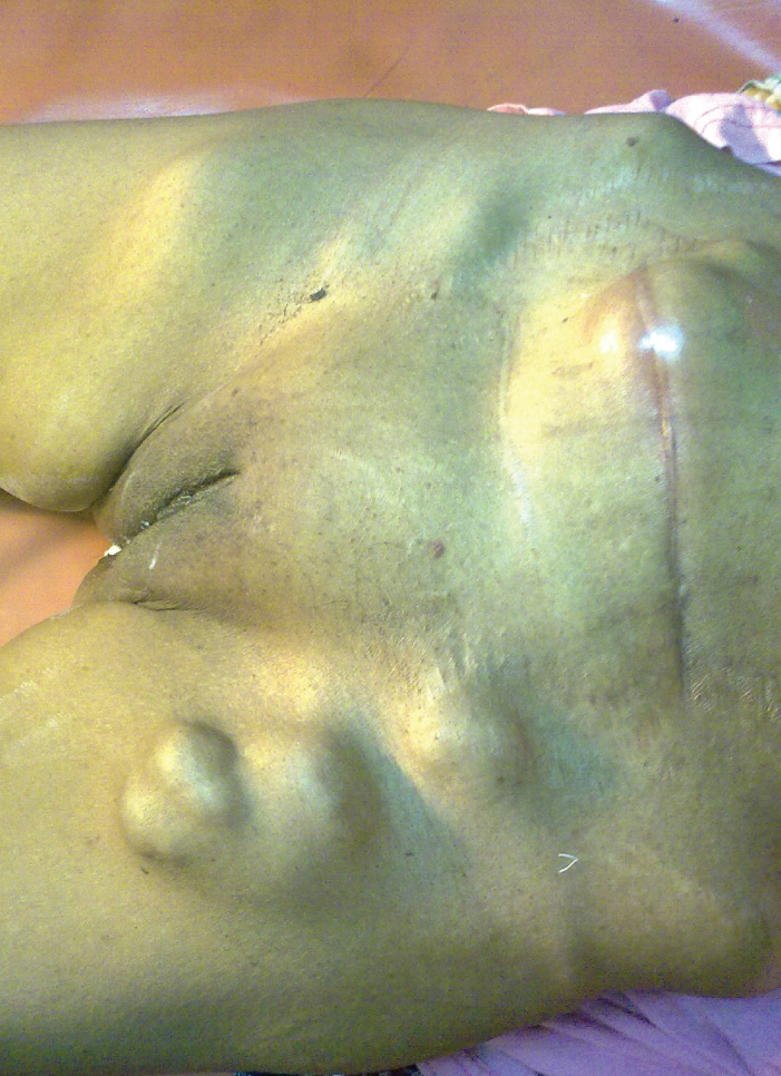
A 60-year-old female was admitted to our Medical College with multiple swellings on the lower abdomen and inguinal region. This was associated with vague abdominal discomfort. On general examination she was cachectic; abdominal examination revealed a suprapubic scar and a nodular swelling over it. There were three swellings on the left upper thigh and inguinal region (Figure 1). Per vaginal examination showed a healthy vault.
Figure 1.

Skin nodules in upper thigh along with the inguinal & abdominal swellings
Previous records revealed that she had been diagnosed with non-keratinising moderately differentiated squamous cell carcinoma of the cervix. One year previously she was admitted with a cervical growth which proved to be malignant and was in FIGO Stage IIa. The initial tumour size was approximately 4cm at the largest diameter without any parametrial involvement. Routine work-up including chest X-ray and abdominal ultrasound did not reveal any para-aortic nodal or distant spread. She underwent Modified Radical Hysterectomy (Wertheim’s Operation). Histopathological examination from the post-operative specimen showed involvement of the bilateral internal iliac nodes. However, she did not receive any adjuvant therapy, although she was advised to do so, and she did not return for follow-up.
On this occasion, biopsy was taken from the skin nodules over the left upper thigh, which showed cutaneous deposits of metastatic squamous cell carcinoma. Excision biopsy from the swollen inguinal nodes also revealed deposits of squamous cell carcinoma in the lymph nodes. She was diagnosed as a case of metastatic uterine cervical carcinoma, i.e. cervical cancer spreading to the skin and inguinal lymph nodes. Clinical and colposcopic examination revealed that the vaginal vault was free from tumour. Radiological investigations did not show the metastatic involvement of any other organ.
The patient was treated with six cycles of chemotherapy with Cisplatin (80 mg/m2) and Paclitaxel (225mg/m2; infusion over 3 hours) on day 1, every 3 weeks. She received the treatment according to the schedule. At the end of the chemotherapy course there was a 50% reduction in the size of the lesion and the enlarged lymph nodes disappeared. She received palliative radiotherapy of 3000-CGy external beam irradiation in 10 fractions over 2 weeks according to a conventional fractionation schedule.
The lesions responded well to radiation therapy; however, four months later, she developed bilateral lung metastasis and died within three months. The period between the date of her presentation at our OPD with skin metastasis to the date of her death was seven month long.
Discussion
Metastatic carcinoma of the skin is an uncommon occurrence. Breast cancer is one of the most common primary tumours to metastasise to skin; gynaecological malignancies rarely give rise to metastatic deposits on the skin. Samaila et al. (2), from a search of twenty years institutional data from Nigeria, could find only four instances of cutaneous deposits from gynaecological malignancies, and in only one of these cases was the primary malignancy in the uterine cervix. Skin metastasis from uterine cervical carcinoma is a rare event. The reported incidence ranges from 0.1 to 2% (3).
To search PubMed for previously reported cases, we used the keywords “cancer cervix” and “skin metastasis”. All of the relevant literatures were thoroughly reviewed. Relevant cases are presented here in Table 1 (4–31). Studies dealing with cutaneous metastasis from all primary malignancies, among which some are from cancer of the cervix, are not included in Table 1 (3, 32, 33). Fifteen cervical cancer patients (out of 1190 patients) with skin metastasis from the study by Imchi et al. (34) are also not presented in the Table.
Table 1.
Summary of results within the melatonin group and control groups
| Case | Author | Sites of Metastasis | Previous Treatment Received | Histology |
|---|---|---|---|---|
| 1 | Daw & Riley (4) | Umbilicus | - | SCC |
| 2 | Tharakaram et al. (5) | Thigh | - | SCC |
| 3 | Chapman et al. (6) | Umbilicus | Initial Presentation | SCC |
| 4. | Bohme et al. (7) | Two cases: One at Scalp, another in inguinal region | - | - |
| 5. | Hayes et al. (8) | Upper back | - | SCC |
| 6. | Lane et al. (9) | Laparoscopic port site | - | ASC |
| 7. | Selo-Ojeme et al. (10) | Hysterectomy incision site | Radical Hysterectomy | AC |
| 8. | Pertzborn et al. (11) | Hand | On Initial Presentation | - |
| 9. | Maheswari et al. (12) | Scalp | Radiotherapy | SCC |
| 10. | Tjalma et al. (13) | Laparoscopic port site | Radiotherapy | SCC |
| 11. | Behtash et al. (14) | Abdominal wall (drain site) | Radical Surgery & Radiotherapy | SCC |
| 12. | Liro et al. (15) | Abdominal wall | Radical Surgery & Radiotherapy | SCC |
| 13. | Agarwal et al. (16) | Scalp | Radiotherapy | SCC |
| 14. | Park et al. (17) | Scalp | - | SCC |
| 15. | Srivastava et al. (18) | Incisional site | Radical Surgery & Radiotherapy | SCC |
| 16. | Chung et al. (19) | Scalp presenting as alopecia neoplastica | Radical Surgery | SCC |
| 17. | Sachdev & Jain (20) | Incisional site | Surgery | SCC |
| 18. | Chen et al. (21) | Extremities, trunk, scalp | Chemo-radiotherapy | SCC |
| 19. | Abhishek et al. (22) | Scalp | AC | |
| 20. | Behtash et al. (23) | Umbilicus | Chemo-radiotherapy | SCC |
| 21 | Ozdemir & Tuncbilek (24) | Nasal dorsum | - | SCC |
| 22. | Deka et al. (Two Cases) (25) | Surgical scar | - | SCC |
| 23. | Takagi et al. (26) | Scalp | Surgery & Chemoradiation | SCC |
| 24. | Elamurugan et al. (27) | Palm (hand) | Radiotherapy | SCC |
| 25. | Agrawal et al. (28) | Abdominal wall | Chemoradiotherapy | SCC |
| 26. | Fogaca et al. (29) | All over | Surgery | Neuroendocrine Cancer |
| 27. | Lee et al. (30) | All over | - | |
| 28. | Chung et al. (31) | - | - |
The most common sites of cutaneous metastases in cervical carcinoma are the abdominal wall and lower extremities (8). This is consistent with other carcinomas, in that metastatic spread to the skin is commonly located near the site of the primary tumour (33). The usual mode of spread has been suggested to be the lymphatic system (34). Imchi et al. (34), after reviewing 1190 patients with cancer of the cervix, including 15 of whom developed skin metastasis, observed that the incidence of skin metastasis was 0.8% in stage I, 1.2% in stage II, 1.2% in stage III, and 4.8% in stage IV. The incidence of skin metastasis seemed to be higher in patients with adenocarcinoma and undifferentiated carcinoma than in patients with squamous cell carcinoma. Macroscopically, three common patterns of skin metastases, such as nodules, plaques, and inflammatory telangiectasic lesions, have been recognised (33). Skin metastases from cervical carcinoma occur predominantly in cases of tumour recurrences, with metastases developing up to 10 years after the initial diagnosis and averaging less than 1 year (35). The prognosis associated with cutaneous metastasis of cervical carcinoma is poor. Usually, metastases to the skin occurring in patients with carcinoma of the cervix represent a pre-terminal event with a time from diagnosis to death of 3 months (3). Systemic treatment in patients with advanced disease is palliative. Cisplatin is the single most active agent for the treatment of cervical cancer; however, recent evidence suggests that combination chemotherapy with cisplatin and paclitaxel may improve progression-free survival over the use of cisplatin alone (36). Palliative radiation is useful for controlling symptoms (37).
The most important prognostic factor, in cases like this, is the time interval between the initial diagnosis of the primary genital malignancy and the appearance of skin metastasis, whether metastasis is isolated or as a part of more widespread systemic recurrence. The earlier the metastasis occurs, the worse the prognosis for the patient is. The intent of treatment remains palliation, either by radiation, chemotherapy or surgery (alone or in combination). Most of the previously reported cases had undergone some kind of surgical intervention for their disease, either radical surgical intervention or lymphadenectomy (in some cases laparoscopic lymph node dissection), before the disease was treated by radiation therapy or chemoradiotherapy. Careful handling of tissue during operation, extension of radiation therapy portals to include the surgical scar and uncompromised primary treatment might prevent the development of skin metastases. Research targeted on the mechanism of local cancer spread and the interaction of cancer cells with the surgical wound environment may improve the knowledge regarding the pathogenesis of skin metastases and its clinical prognosis.
Footnotes
Ethics Committee Approval: Ethics committee approval was received for this study.
Informed Consent: Written informed consent was obtained from patients who participated in this study.
Peer-review: Externally peer-reviewed.
Author contributions: Concept – B.B., S.M.; Design - B.B., S.M.; Supervision - B.B., S.M.; Resource - B.B., S.M.; Materials - B.B., S.M.; Data Collection&/or Processing - B.B., S.M.; Analysis&/or Interpretation - B.B., S.M.; Literature Search - B.B., S.M.; Writing - B.B., S.M.; Critical Reviews - B.B., S.M.
Conflict of Interest: No conflict of interest was declared by the authors.
Financial Disclosure: No financial disclosure was declared by the authors.
References
- 1.National Cancer Registry Programme. Consolidated Report of Population Based Cancer Registries 2001 – 2004 incidence and distribution of cancer. 2006 Dec; [Google Scholar]
- 2.Samaila MO, Adesiyun AG, Waziri GD, Koledade KA, Kolawole AO. Cutaneous umbilical metastases in post-menopausal females with gynaecological malignancies. J Turkish-German Gynecol Assoc. 2012;13:204–7. doi: 10.5152/jtgga.2012.29. [DOI] [PMC free article] [PubMed] [Google Scholar]
- 3.Brady LW, O’Neil EA, Farber SH. Unusual sites of metastases. Semin Oncol. 1977;4:59–64. [PubMed] [Google Scholar]
- 4.Daw E, Riley S. Umbilical metastasis from squamous carcinoma of the cervix. Case report. Br J Obstet Gynaecol. 1982;89:1066. doi: 10.1111/j.1471-0528.1982.tb04669.x. [DOI] [PubMed] [Google Scholar]
- 5.Tharakaram S, Rajendran SS, Premalatha S, Yesudian P, Zahara A. Cutaneous metastasis from carcinoma cervix. Int J Dermatol. 1985;24:598–9. doi: 10.1111/j.1365-4362.1985.tb05860.x. [DOI] [PubMed] [Google Scholar]
- 6.Chapman GW, Jr, Abreo F, Thompson H. Squamous cell carcinoma of the cervix metastatic to the umbilicus. J Natl Med Assoc. 1987;79:1293, 1296–7. [PMC free article] [PubMed] [Google Scholar]
- 7.Böhme M, Baumann D, Donat H, Lenz E, Röder K. Rare type of metastases in progressive cervix carcinoma. Zentralbl Gynakol. 1990;112:1357–62. [PubMed] [Google Scholar]
- 8.Hayes AG, Berry AD., 3rd Cutaneous metastasis from squamous cell carcinoma of the cervix. J Am Acad Dermatol. 1992;26:846–50. doi: 10.1016/0190-9622(92)70119-z. [DOI] [PubMed] [Google Scholar]
- 9.Lane G, Tay J. Port-site metastasis following laparoscopic lymphadenectomy for adenosquamous carcinoma of the cervix. Gynecol Oncol. 1999;74:130–3. doi: 10.1006/gyno.1999.5379. [DOI] [PubMed] [Google Scholar]
- 10.Selo-Ojeme DO, Bhide M, Aggarwal VP. Skin incision recurrence of adenocarcinoma of the cervix five years after radical surgery for stage 1A disease. Int J Clin Pract. 1998;52:519. [PubMed] [Google Scholar]
- 11.Pertzborn S, Buekers TE, Sood AK. Hematogenous skin metastases from cervical cancer at primary presentation. Gynecol Oncol. 2000;76:416–7. doi: 10.1006/gyno.1999.5704. [DOI] [PubMed] [Google Scholar]
- 12.Maheshwari GK, Baboo HA, Ashwathkumar R, Dave KS, Wadhwa MK. Scalp metastasis from squamous cell carcinoma of the cervix. Int J Gynecol Cancer. 2001;11:244–6. doi: 10.1046/j.1525-1438.2001.00074.x. [DOI] [PubMed] [Google Scholar]
- 13.Tjalma WA, Winter-Roach BA, Rowlands P, De Barros Lopes A. Port-site recurrence following laparoscopic surgery in cervical cancer. Int J Gynecol Cancer. 2001;11:409–12. doi: 10.1046/j.1525-1438.2001.01049.x. [DOI] [PubMed] [Google Scholar]
- 14.Behtash N, Ghaemmaghami F, Yarandi F, Ardalan FA, Khanafshar N. Cutaneous metastasis from carcinoma of the cervix at the drain site. Gynecol Oncol. 2002;85:209–11. doi: 10.1006/gyno.2001.6559. [DOI] [PubMed] [Google Scholar]
- 15.Liro M, Kobierski J, Brzóska B. Isolated metastases of cervical cancer to the abdominal wall--a case report. Ginekol Pol. 2002;73:704–8. [PubMed] [Google Scholar]
- 16.Agarwal U, Dahiya P, Chauhan A, Sangwan K, Purwar P. Scalp metastasis in carcinoma of the uterine cervix - a rare entity. Gynecol Oncol. 2002;87:310–2. doi: 10.1006/gyno.2002.6829. [DOI] [PubMed] [Google Scholar]
- 17.Park JY, Lee HS, Cho KH. Cutaneous metastasis to the scalp from squamous cell carcinoma of the cervix. Clin Exp Dermatol. 2003;28:28–30. doi: 10.1046/j.1365-2230.2003.01128.x. [DOI] [PubMed] [Google Scholar]
- 18.Srivastava K, Singh S, Srivastava M, Srivastava AN. Incisional skin metastasis of a squamous cell cervical carcinoma 3.5 years after radical treatment- a case report. Int J Gynecol Cancer. 2005;15:1183–6. doi: 10.1111/j.1525-1438.2005.00173.x. [DOI] [PubMed] [Google Scholar]
- 19.Chung JJ, Namiki T, Johnson DW. Cervical cancer metastasis to the scalp presenting as alopecia neoplastica. Int J Dermatol. 2007;46:188–9. doi: 10.1111/j.1365-4632.2007.03183.x. [DOI] [PubMed] [Google Scholar]
- 20.Sachdev R, Jain S. Carcinoma of the cervix with incisional skin metastasis: a rare event. Pathology. 2007;39:168–9. doi: 10.1080/00313020601123938. [DOI] [PubMed] [Google Scholar]
- 21.Chen CH, Chao KC, Wang PH. Advanced cervical squamous cell carcinoma with skin metastasis. Taiwan J Obstet Gynecol. 2007;46:264–6. doi: 10.1016/S1028-4559(08)60031-5. [DOI] [PubMed] [Google Scholar]
- 22.Abhishek A, Ouseph MM, Sharma P, Kamal V, Sharma M. Bulky scalp metastasis and superior sagittal sinus thrombosis from a cervical adenocarcinoma: an unusual case. J Med Imaging Radiat Oncol. 2008;52:91–4. doi: 10.1111/j.1440-1673.2007.01918.x. [DOI] [PubMed] [Google Scholar]
- 23.Behtash N, Mehrdad N, Shamshirsaz A, Hashemi R, Amouzegar Hashemi F. Umblical metastasis in cervical cancer. Arch Gynecol Obstet. 2008;278:489–91. doi: 10.1007/s00404-008-0617-4. [DOI] [PubMed] [Google Scholar]
- 24.Ozdemir H, Tunçbilek G. Metastasis of carcinoma of the uterine cervix to the nasal dorsum. J Craniofac Surg. 2009;20:971–3. doi: 10.1097/SCS.0b013e3181a2e430. [DOI] [PubMed] [Google Scholar]
- 25.Deka D, Gupta N, Bahadur A, Dadhwal V, Mittal S. Umbilical surgical scar and vulval metastasis secondary to advanced cervical squamous cell carcinoma: a report of two cases. Arch Gynecol Obstet. 2010;281:761–4. doi: 10.1007/s00404-009-1235-5. [DOI] [PubMed] [Google Scholar]
- 26.Takagi H, Miura S, Matsunami K, Ikeda T, Imai A. Cervical cancer metastasis to the scalp: case report and literature review. Eur J Gynaecol Oncol. 2010;31:217–8. [PubMed] [Google Scholar]
- 27.Elamurugan TP, Agrawal A, Dinesh R, Aravind R, Naskar D, Kate V, et al. Parthasarathy; Palmar cutaneous metastasis from carcinoma cervix. Indian J Dermatol Venereol Leprol. 2011;77:252. doi: 10.4103/0378-6323.77486. [DOI] [PubMed] [Google Scholar]
- 28.Agrawal A, Yau A, Magliocco A, Chu P. Cutaneous metastatic disease in cervical cancer: a case report. J Obstet Gynaecol Can. 2010;32:467–72. doi: 10.1016/S1701-2163(16)34501-7. [DOI] [PubMed] [Google Scholar]
- 29.Fogaça MF, Fedorciw BJ, Tahan SR, Johnson R, Federman M. Cutaneous metastasis of neuroendocrine carcinoma of uterine origin. J Cutan Pathol. 1993;20:455–8. doi: 10.1111/j.1600-0560.1993.tb00671.x. [DOI] [PubMed] [Google Scholar]
- 30.Lee WJ, Lee DW, Lee MW, Choi JH, Moon KC, Koh JK. Multiple cutaneous metastases of neuroendocrine carcinoma derived from the uterine cervix. J Eur Acad Dermatol Venereol. 2009;23:494–6. doi: 10.1111/j.1468-3083.2008.02955.x. [DOI] [PubMed] [Google Scholar]
- 31.Chung WK, Yang JH, Chang SE, Lee MW, Choi JH, Moon KC, et al. A case of cutaneous metastasis of small-cell neuroendocrine carcinoma of the uterine cervix. Am J Dermatopathol. 2008;30:636–8. doi: 10.1097/DAD.0b013e31817e6f27. [DOI] [PubMed] [Google Scholar]
- 32.Reingold IM. Cutaneous metastases from internal carcinoma. Cancer. 1986;19:162–8. doi: 10.1002/1097-0142(196602)19:2<162::aid-cncr2820190204>3.0.co;2-a. [DOI] [PubMed] [Google Scholar]
- 33.Brownstein MH, Helwig EB. Patterns of cutaneous metastases. Arch Dermatol. 1972;105:862–8. [PubMed] [Google Scholar]
- 34.Imachi M, Tsukamoto N, Kinoshita S, Nakano H. Skin metastasis from carcinoma of the uterine cervix. Gynecol Oncol. 1993;48:349–54. doi: 10.1006/gyno.1993.1061. [DOI] [PubMed] [Google Scholar]
- 35.Copas PR, Spann CO, Thoms WW, Horowitz IR. Squamous cell carcinoma of the cervix metastatic to a drain site. Gynecol Oncol. 1995;56:102–4. doi: 10.1006/gyno.1995.1018. [DOI] [PubMed] [Google Scholar]
- 36.Moore DH, Blessing JA, McQuellon RP, Thaler HT, Cella D, Benda J, et al. Phase III study of cisplatin with or without paclitaxel in stage IVB, recurrent, or persistent squamous cell carcinoma of the cervix: a gynecologic oncology group study. J Clin Oncol. 2004;22:3113–9. doi: 10.1200/JCO.2004.04.170. [DOI] [PubMed] [Google Scholar]
- 37.Spanos WJ, Jr, Pajak TJ, Emami B, Rubin P, Cooper JS, Russell AH, et al. Radiation palliation of cervical cancer. J Natl Cancer Inst. 1996;21:127–30. [PubMed] [Google Scholar]


