Abstract
Background/Aims
No clear data have been established and validated regarding whether rectal retroflexion has an important and therapeutic impact. The aim of the present study was to evaluate the diagnostic yield and therapeutic impact of rectal retroflexion compared with straight view examination.
Methods
A prospective single-blind study was conducted. Consecutive patients evaluated between October 2011 and April 2012 were included.
Results
A total of 934 patients (542 women, 58%) were included. The mean age was 57.4±14.8 years. Retroflexion was successful in 917 patients (98.2%). Distinct lesions in the anorectal area were detected in 32 patients (3.4%), of which 10 (1%) were identified only on retroflex view and 22 (2.4%) on both straight and retroflex views. Of the 32 identified lesions, 16 (50%) were polyps, nine (28.1%) were angiodysplasias, six (18.8%) were ulcers, and one (3.1%) was a flat lesion. All 10 patients (1%) in whom lesions were detected only by rectal retroflexion showed a therapeutic impact.
Conclusions
Rectal retroflexion has minimal diagnostic yield and therapeutic impact. However, its low rate of major complications and the possibility of detecting lesions undetectable by straight viewing justify its use.
Keywords: Colonoscopy, Retroflexion, Rectum, Colorectal polyps
INTRODUCTION
Colonoscopy is a diagnostic and therapeutic method widely used worldwide. International guidelines exist for the proper implementation and reporting of results. Retroflexion in the rectum during colonoscopy is traditionally performed in all patients to complete the full examination of the colon. This procedure is performed regardless of any pathology detected during digital rectum exploration (DRE) and frontal view with colonoscopy equipment.1-4 Complications including perforation have been reported to occur as a result of retroflexion performed at this level. Therefore, some experts believe that this maneuver is overvalued.1-4 Although it is a relatively rare complication (0.1 per 1000), it is associated with severe morbidity.4 Previous studies did not find factors associated with complications during retroflexion;4 therefore, the identification of patients who could benefit from this maneuver was viewed as a useful approach. In some studies, the gain related to the number of lesions identified with retroflexion was 2% to 8%; however, the clinical significance of this increase was not thoroughly documented.5-8 Furthermore, some authors question the utility of retroflexion.9
The main aim of the present study was to evaluate the diagnostic yield and therapeutic impact of rectal retroflexion compared with DRE and straight view examination.
MATERIALS AND METHODS
The present prospective study of patients undergoing colonoscopy was conducted in three different centers, namely Instituto Nacional de Ciencias Médicas y Nutrición Salvador Zubirán (center A), Mexico City, Mexico; Hospital Las Américas (center B), Guatemala City, Guatemala; and Medica Sur Clinic and Foundation (center C), Mexico City, Mexico. The study was performed between October 2011 and April 2012 after obtaining informed consent from all the patients. The protocol was reviewed and accepted by the ethics committee of the participating institutes. Patients who did not want to participate in the study were excluded.
Assisted by nurses trained in endoscopic procedures and aided by specialists in anesthesiology who placed the patients under sedation, physicians performed the colonoscopies, monitoring vital signs, using oxygen saturation and electrocardiogram tracing, and managing supplemental oxygen after standard preparation, which was performed independent of this study. We performed the colonoscopy procedure according to standard recommendations, emphasizing a minimal duration of 7 minutes for the withdrawal maneuver. All the procedures were carried out by professionals who were trained to perform colonoscopies and who had been learning at an advanced rate according to international standards.
Preparation included intestinal cleaning through the administration of polyethylene glycol (centers A and C, Nulytely, Asofarma de México, México; center B, Fortrans, Ipsen, France) as follows: one envelope was diluted in 1 L of drinking water in 1 hour (total, four envelopes in 4 hours) the day prior to the study. Colonoscopy was performed using standard equipment (CFQ 140-180L; Olympus, Center Valley, PA, USA). The scope was advanced until the cecum was identified by its anatomical characteristics (ileocecal valve, appendiceal orifice, and tapeworm colonic junction). During the withdrawal, all colonic segments were assessed (cecum, ascending colon, transverse colon, descending colon, sigmoid colon, and rectum) in a minimum of 7 minutes. The quality of the colonic preparation was determined according to the Boston bowel preparation scale.10
The rectum was initially examined on forward view during withdrawal of the colonoscope to the dentate line, with reflection of the tip in all directions and using torque and up-down and right-left deviation when possible and appropriate. An initial judgment regarding diagnosis of the rectal area was made and recorded in the data collection sheet. After the diagnostic impression based on the first view, physicians performed rectal retroflexion and obtained an additional diagnostic impression that was documented in a different section. An individual independent from those performing the colonoscopy and making diagnostic impressions was designated to capture the data; the purpose of this approach was to ensure that preretroflexion and postretroflexion data collection was blind and performed independent of that performed during the medical procedures. These two diagnostic impressions were compared in relation to the concordance of the diagnoses.
Retroflexion was performed using standard methods; the tip of the colonoscope is positioned between the first and second Houston's valves and rotated to achieve the greater zipper retroflection. Manual rotation of the instrument was performed to inspect the anorectal area in a circumferential manner. The maneuver was considered successful if a complete 360° visualization of the distal rectum was achieved. Cases of retroflexion that were successful, unsuccessful, or not attempted were all fully documented. Positive or negative detection of polyps in the distal rectum on straight view, retroflexion view, or both views was prospectively recorded. All polyps measuring 5 mm or more were removed by snare polypectomy and submitted for histopathological analysis. Endoscopic measurement of the polyp size was initially performed using biopsy forceps.
Statistical analysis and sample size
The demographic and clinical characteristics of the patients were summarized with means, medians, and standard deviations. The difference in the number of lesions (expressed as percentages whose denominator was the total number of studies) detected before and after rectal retroflexion was analyzed using the chi-square test. The impact on treatment was evaluated with absolute and relative frequencies in relationship to the number of cases showing this phenomenon (change in treatment required by the patient according to the retroflexion). To assess differences in the characteristics of the patients according to the center of origin, the researchers calculated the number of lesions and their impact on the treatment using analysis of variance and the chi-square test according to the variable evaluated. A p<0.05 was considered significant. Bonferroni correction for p-value was applied for multiple comparisons, calculated as α/n. For multiple comparisons, a p<0.016 was considered significant. All statistical analyses were conducted using the statistical program SPSS version 20 (IBM Co., Armonk, NY, USA).
RESULTS
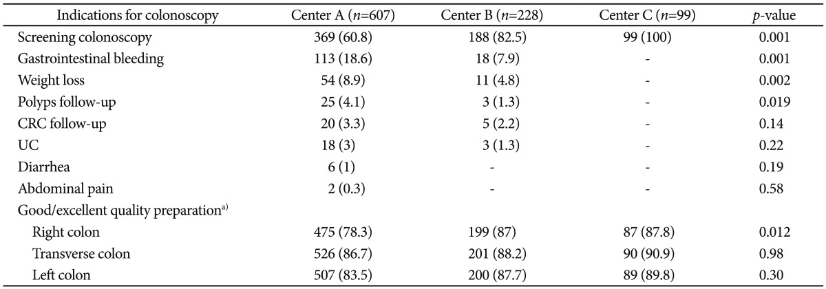
A total of 934 patients (542 women, 58%; and 392 men, 42%) were included. The mean age was 57.4±14.8 years. Retroflexion was successful in 917 patients (98.2%); retroflexion was attempted in all but two of the remaining 17 patients (1.8%) but could not be performed because of a contracted vault. Rectal retroflexion was not attempted in two patients who had severe ulcerative colitis. The number of patients according to center was as follows: center A, 607 (65%); center B, 228 (24.4%); and center C, 99 patients (10.6%). Indications for colonoscopy classified according to center are shown in Table 1. Good/excellent (2/3 points) quality of preparation of the right, transverse, and left colons was observed in 75.1%, 80.7%, and 78.6% of the patients, respectively. Data classified by centers are shown in Table 1.
Table 1.
Indications for Colonoscopy and Quality of Bowel Preparation according to Center

Values are presented as number (%).
CRC, colorectal cancer; UC, ulcerative colitis.
a)2/3 point in the Boston bowel preparation scale.
Lesions
Excluding internal hemorrhoids and polyps smaller than 5 mm (174 patients, 18.6%), distinct lesions in the anorectal area were found in 32 patients (3.4%). Of these, 10 (1%) were detected only on retroflexion (Table 2); 22 (2.4%), on both straight and retroflex views; and 0 (0%), only on straight view (Table 2). Of the 32 identified lesions, 16 were polyps, nine (0.9%) were angiodysplasias, six (0.6%) were ulcers, and one (0.1%) had a flat lesion (tubulovillous adenoma).
Table 2.
Type of Lesions Identified in the Rectal Vault Using Different Maneuvers

The mean size of the seven polyps detected only by retroflex view was 7.5 mm (range, 5 to 10), and all were sessile. All were detected in separate patients and none had high-grade dysplasia. One additional patient had a flat lesion in the distal rectum that was only detected by retroflex view (size, 10 mm).
Change in the diagnosis and therapeutic impact
The 10 patients (1%) in whom lesions were detected only by rectal retroflexion showed a therapeutic impact. In these patients, polyps were removed by snare polypectomy and submitted for histopathological analysis (Table 2).
Complications
Minor complications were reported in three patients (0.3%). This included anyone who required endoscopic treatment. All three cases consisted of erosions with minor bleeds that resolved spontaneously. No cases of rectal perforation were observed in this study.
Table 3 describes the success rate of rectal retroflexion, the changes in diagnosis, the therapeutic impact, and all reported complications classified according to the different centers.
Table 3.
Success Rate, Change in Diagnosis, Therapeutic Impact, and Complications of Rectal Retroflexion, Classified according to Original Center

Values are presented as number (%).
RR, rectal retroflexion.
DISCUSSION
In this prospective, multicenter, blind study, rectal retroflexion during colonoscopy had a limited effect on diagnosis and therapeutic impact. However, the rate of complications was low, as was the need for additional treatment.
In traditional practice, rectal retroflexion is considered as an important component of complete colonoscopy.7 However, its diagnostic yield and therapeutic impact have been questioned.4,6-8 The different conclusions reached by previous studies regarding the value of routine retroflexion could be related to the low prevalence of pathology detected only by retroflexion and not by the forward view. The interpretation of this low prevalence appears to depend on the authors of the studies5-8 and their previous experience.1-4 Cutler and Pop5 reported no adenomas detected only by retroflexion in 453 patients and questioned the value of routine retroflexion. Grobe et al.8 suggested that retroflexion was valuable in 75 patients but did not document a single adenoma detected only by retroflexion. Hanson et al.7 detected four adenomas in 526 patients that were visible only on rectal retroflexion, and one was a 15-mm tubulovillous adenoma. A single center study conducted by Saad and Rex11 showed the visualization of seven polyps only by retroflexion (only one tubular adenoma of 4 mm and six hyperplastic sessile polyps; 7/1,502 [0.46%]). Varadarajulu and Ramsey6 reported that among 590 patients (91% male), six had adenomas detected only on retroflexion. They stated that 50% of the distal rectal lesions were visible only on retroflexion. In our series, 31.2% (10/32) of all the lesions were identified only by retroflexion and 47% (8/17) of all the polyps were similarly identified. The results of the present study were similar to those reported by Varadarajulu and Ramsey.6
It is important to note that in the present study, data originated from three different centers in two different countries. Nevertheless, the results were consistent among the different centers and were in agreement with previous literature. As expected, the number of detected lesions increased according to sample size within different centers. Importantly, the results did not change according to the number of physicians participating in the maneuver of retroflexion; data from centers B and C (both private centers) were obtained from a single physician. In center C (university hospital), different physicians including senior residents and staff physicians participated. In the present study, the results were similar regardless of the variation in the economic status of the different populations or whether they were private practice patients or admitted to public hospitals, and regardless of the experience of the physicians performing the rectal retroflexion (Table 3). We were unable to evaluate whether the maneuver was well tolerated because colonoscopies were performed under sedation by an anesthesiologist.
One possible limitation of the present study was the number of observers. Although the same rectal retroflexion technique was used by all the physicians, endoscopists with less effective forward viewing techniques might expect a higher yield from retroflexion. However, no significant differences were observed independent of the physicians' experience (Table 3).
In conclusion, colonoscopic retroflexion in the rectum has little diagnostic yield and therapeutic impact. However, its low rate of major complications and the possible detection of lesions undetectable by straight viewing may justify its use.
Acknowledgments
We thank Michael Katz, Ph.D., for his encouragement and support in the writing of the final version of this paper.
Footnotes
The authors have no financial conflicts of interest.
References
- 1.Fu K, Ikematsu H, Sugito M, et al. Iatrogenic perforation of the colon following retroflexion maneuver. Endoscopy. 2007;39(Suppl 1):E175. doi: 10.1055/s-2007-966563. [DOI] [PubMed] [Google Scholar]
- 2.Chu Q, Petros JG. Extraperitoneal rectal perforation due to retroflexion fiberoptic proctoscopy. Am Surg. 1999;65:81–85. [PubMed] [Google Scholar]
- 3.Ahlawat SK, Charabaty A, Benjamin S. Rectal perforation caused by retroflexion maneuver during colonoscopy: closure with endoscopic clips. Gastrointest Endosc. 2008;67:771–773. doi: 10.1016/j.gie.2007.09.026. [DOI] [PubMed] [Google Scholar]
- 4.Quallick MR, Brown WR. Rectal perforation during colonoscopic retroflexion: a large, prospective experience in an academic center. Gastrointest Endosc. 2009;69:960–963. doi: 10.1016/j.gie.2008.11.011. [DOI] [PubMed] [Google Scholar]
- 5.Cutler AF, Pop A. Fifteen years later: colonoscopic retroflexion revisited. Am J Gastroenterol. 1999;94:1537–1538. doi: 10.1111/j.1572-0241.1999.01140.x. [DOI] [PubMed] [Google Scholar]
- 6.Varadarajulu S, Ramsey WH. Utility of retroflexion in lower gastrointestinal endoscopy. J Clin Gastroenterol. 2001;32:235–237. doi: 10.1097/00004836-200103000-00012. [DOI] [PubMed] [Google Scholar]
- 7.Hanson JM, Atkin WS, Cunliffe WJ, et al. Rectal retroflexion: an essential part of lower gastrointestinal endoscopic examination. Dis Colon Rectum. 2001;44:1706–1708. doi: 10.1007/BF02234394. [DOI] [PubMed] [Google Scholar]
- 8.Grobe JL, Kozarek RA, Sanowski RA. Colonoscopic retroflexion in the evaluation of rectal disease. Am J Gastroenterol. 1982;77:856–858. [PubMed] [Google Scholar]
- 9.Winawer SJ, Zauber AG, Ho MN, et al. Prevention of colorectal cancer by colonoscopic polypectomy. The National Polyp Study Workgroup. N Engl J Med. 1993;329:1977–1981. doi: 10.1056/NEJM199312303292701. [DOI] [PubMed] [Google Scholar]
- 10.Lai EJ, Calderwood AH, Doros G, Fix OK, Jacobson BC. The Boston bowel preparation scale: a valid and reliable instrument for colonoscopy-oriented research. Gastrointest Endosc. 2009;69(3 Pt 2):620–625. doi: 10.1016/j.gie.2008.05.057. [DOI] [PMC free article] [PubMed] [Google Scholar]
- 11.Saad A, Rex DK. Routine rectal retroflexion during colonoscopy has a low yield for neoplasia. World J Gastroenterol. 2008;14:6503–6505. doi: 10.3748/wjg.14.6503. [DOI] [PMC free article] [PubMed] [Google Scholar]


