Abstract
Background
Primary frozen shoulder (FS) is a painful contracture of the glenohumeral joint that arises spontaneously without an obvious preceding event. Investigation of the intra-articular and periarticular pathology would contribute to the treatment of primary FS.
Review of literature
Many studies indicate that the main pathology is an inflammatory contracture of the shoulder joint capsule. This is associated with an increased amount of collagen, fibrotic growth factors such as transforming growth factor-beta, and inflammatory cytokines such as tumor necrosis factor-alpha and interleukins. Immune system cells such as B-lymphocytes, T-lymphocytes and macrophages are also noted. Active fibroblastic proliferation similar to that of Dupuytren’s contracture is documented. Presence of inflammation in the FS synovium is supported by the synovial enhancement with dynamic magnetic resonance study in the clinical setting.
Conclusion
Primary FS shows fibrosis of the joint capsule, associated with preceding synovitis. The initiator of synovitis, however, still remains unclear. Future studies should be directed to give light to the pathogenesis of inflammation to better treat or prevent primary FS.
Introduction
Frozen shoulder (FS) is a common disorder in general orthopaedic practice, characterized by pain in the shoulder and limitation of glenohumeral motions. FS is a term coined by Codman in 1934 [1]. Synonyms include périarthrite scapulohumérale [2] and adhesive capsulitis [3]. In Japan, a term “goju-kata” (50-year-old-shoulder) has been used among the general public since the eighteenth century or before.
FS may arise spontaneously without an obvious preceding cause, or be associated with local or systemic disorders. Zuckerman proposed to classify FS into primary and secondary and subdivided secondary FS into intrinsic, extrinsic and systemic ones [4] (Table 1). The intrinsic category includes limitation of active and passive range of motions that occur in association with shoulder joint disorders, while the extrinsic category follows an identifiable abnormality outside the shoulder. The systemic category is associated with systemic disorders such as diabetes mellitus [4]. This classification is followed in this paper.
Table 1.
Classification of frozen shoulder
| Primary/idiopathic frozen shoulder |
| An underlying etiology or associated condition cannot be identified |
| Secondary frozen shoulder |
| An underlying etiology or associated condition can be identified |
| Intrinsic |
| In association with rotator cuff disorders (tendinitis and partial-thickness or full-thickness tears), biceps tendinitis, or calcific tendinitis |
| Extrinsic |
| In association with previous ipsilateral breast surgery, cervical radiculopathy, chest wall tumor, previous cerebrovascular accident, or more local extrinsic problems, including previous humeral shaft fracture, scapulothoracic abnormalities, acromioclavicular arthritis, or clavicle fracture |
| Systemic |
| Diabetes mellitus, hyperthyroidism, hypothyroidism, hypoadrenalism, etc. |
See Reference [4]
This review describes the pathological and immunohistochemical features of primary FS, as well as imaging findings that could represent the underlying pathology. This review also refers to possible concepts of pathogenesis of primary FS.
Pathology
Joint capsule and ligaments
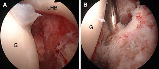
The main cause of painful restriction of movement in FS is an inflammatory contracture of the joint capsule. This can be observed during arthroscopic capsular release in patients with recalcitrant FS; one would see inflamed synovium most often in the rotator interval region and thickened joint capsule as it is divided (Fig. 1). Lundberg reported an increased amount of collagen in the joint capsule, and proposed that inflammation is an important event that leads to stiffness, pain, and capsular fibrosis [5]. Ozaki et al. [6] documented fibrosis, fibrinoid degeneration, and hyalinization in the rotator interval capsule and the coracohumeral ligament of the patients with recalcitrant shoulder stiffness. In an immunohistochemical study, Rodeo et al. [7] found type-III collagen in the anterosuperior capsule of FS, indicating new deposition of collagen. They also reported that cell and matrix staining for transforming growth factor (TGF)-beta, platelet-derived growth factor (PDGF), and hepatocyte growth factor was greater in FS than nonspecific synovitis, suggesting a fibrotic process in FS [7]. Presence of vimentin-positive cells confirms the fibrotic process in the joint capsule [8, 9]. As a result of progression of fibrosis, FS capsule has a greater stiffness than that of shoulders with rotator cuff tear, when measured with scanning acoustic microscopy [10].
Fig. 1.

Arthroscopic view of the right shoulder in a 57-year-old man with primary frozen shoulder. The arthroscope is inserted through the standard posterior portal. Inflamed synovium is noted in the anterosuperior region (a). Using an electric cautery, the anterior capsule is being divided (b). Note the thickened joint capsule. G glenoid fossa, LHB long head of biceps
Some investigators associated the fibrotic changes in FS to Dupuytren’s contracture [11, 12]. Investigation of the rotator interval capsule and coracohumeral ligament obtained from FS patients disclosed active fibroblastic proliferation accompanied by some transformation to myofibroblasts, but at least with inflammation and synovial involvement, which was very similar to those in Dupuytren’s disease [11, 12].
Synovium
Much work has been done to characterize the microscopic pathology and histochemical findings of the glenohumeral and subacromial synovium in FS. Kumagai et al. [13] reported the absence of multiplation of the superficial synovial layers and the absence of interleukin (IL)-1α-positive synoviocytes, and insisted that there is no inflammation in the synovium of primary FS. In contrast, Rodeo et al. [7] demonstrated a variety of inflammatory cytokines such as tumor necrosis factor (TNF)-alpha, IL-1 alpha, IL-1 beta, and IL-6 in the FS synovium, in addition to growth factors related to fibrotic process such as TGF-beta, PDGF, and fibroblast growth factors (aFGF, and bFGF). Similarly, both fibrinogenic (matrix metalloprotease <MMP> -3) and inflammatory (IL-6) cytokines are shown in the synovium [14]. Inflammatory cytokines are known to appear both in the glenohumeral and subacromial synovium [15]. Hand et al. [9] first documented the presence of immune system cells, i.e., B-lymphocytes, T-lymphocytes and macrophages, as well as the presence of mast cells, in the rotator interval synovium and capsule, suggesting an immunological response in FS. In addition, angiogenesis and neurogenesis are known to occur in the subsynovial layer [9, 16]. Molecules related with mechanical stress also appear in the FS synovium [17].
Imaging abnormalities
Imaging studies can represent, if not directly, pathology of primary FS. A plain example is the decrease of joint volume proved at arthrography, which indicates shortening of the joint capsule. Magnetic resonance imaging (MRI) can depict thickening of the joint capsule particularly in the axillary region [18, 19]. MRI also demonstrates thickening of the coracohumeral ligament [19]. MR arthrography may show obliteration of the subcoracoid fat triangle, resulting from shortening or fibrosis of the rotator interval capsule [19–21].
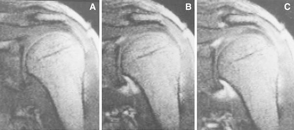
Alteration of the synovium can be shown by dynamic MRI enhanced with intravenous gadolinium administration. Using this technique, Tamai et al. [22] demonstrated a greater increase of signal intensity in the glenohumeral joint synovium in FS, compared to that of healthy volunteers or patients with subacromial impingent syndrome (Fig. 2). This indicates an increased perfusion of gadolinium from the vessels to the synovium, which most probably is the result of synovial inflammation in FS. They later reported that the increased signal intensity in the FS synovium could be reduced by intra-articular injection of corticosteroid or hyaluronate, associated with improvement of clinical scores [23, 24].
Fig. 2.

Dynamic magnetic resonance imaging of primary frozen shoulder. Serial gradient echo images (TR 45 ms; TE 10 ms; flip angle 40°) were obtained in an oblique coronal plane through the center of the humeral head before (a), and 55 s (b) and 121 s (c) following a bolus intravenous administration of Gd-DTPA. Note the marked enhancement in the glenohumeral joint and, to a lesser degree, in the subacromial region. Reproduced from Reference [24] with permission
The periarticular bony tissues also change in FS. In patients with a longstanding disease, bone atrophy of the humeral head is a common finding in a plain X-ray. The bone mineral density (BMD) of the humeral head was reported to be low as early as 2 months after the onset of symptoms [25]. The BMD usually returns to near normal with the improvement of clinical symptoms [26]. A bone scan generally shows positive, which indicates increased local blood flow in FS [27, 28].
Pathogenesis of primary FS
Most of the studies indicate that FS involves both synovial inflammation and capsular fibrosis. Since characteristically pain precedes stiffness in FS, it is most likely that inflammation evolves to fibrosis [9]. Cytokines such as TNF-alpha and ILs will produce synovitis in both the glenohumeral joint and the subacromial bursa, whereas matrix-bound TGF-beta may act as a persistent stimulus, resulting in capsular fibrosis [7].
The initiator of synovitis, however, remains still unclear. Based on the appearance of immune system cells, it is postulated that immunomodulated chronic inflammation may play some role in the pathogenesis of FS [9]. But the preceding immunological events are not known. Another possible initiator of synovitis is degeneration or injury of the rotator cuff tendon. Tendon fiber rupture, even if microscopic, may trigger induction of inflammatory mediators or fibrotic cytokines in the shoulder joint. This hypothesis, however, has not been established to date, whereas partial rotator cuff tear is known to accompany joint contracture [29]. In addition, there is no theory that explains why FS thaws spontaneously in most cases.
It is still uncertain whether FS is a process similar to Dupuytren’s contracture. There may be a failure of collagen remodeling in FS, which in part may result from a genetic failure to activate gelatinase A, or from elevation of the levels of the natural inhibitor of MMP’s in the joint capsule [12]. FS can be induced by administering a synthetic MMP inhibitor, suggesting that a decrease in MMP’s: MMP inhibitors ratio affects collagen turnover [30].
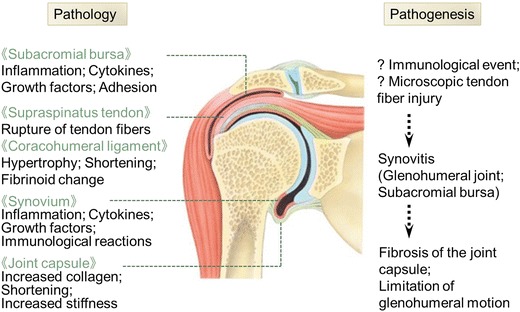
Figure 3 summarizes the pathological findings documented in the literature, and the possible pathogenesis of primary FS. Future studies should be directed to give light on the initiator of inflammation, as well as of fibrosis, with the final aim to better treat or prevent FS.
Fig. 3.

Pathology and pathogenesis of primary frozen shoulder. In the left of the scheme, the pathological findings documented in the literature are listed. In the right, the possible concept of the pathogenesis of primary FS is shown
Acknowledgments
The authors would like to thank Drs. Kazuo Tomizawa, Katsuhisa Yoshikawa, and Tadashi Yamamoto, Department of Orthopaedic Surgery, Dokkyo Medical Universtiy for their help in discussing the current issue and preparing the manuscript.
Conflict of interest
The authors declare that they have no conflict of interest.
Footnotes
This review article was presented at the 86th Annual Meeting of the Japanese Orthopaedic Association as an Instructional Lecture, Hiroshima, May 24, 2013.
References
- 1.Codman EA. The shoulder. New York: G. Miller & Co. Medical Publishers Inc.; 1934. p. 216–24.
- 2.Duplay ES. De la périarthrite scapulohumérale et des raideurs de l’epaule qui en son la consequence. Arch Gen Med. 1872;20:513–542. [Google Scholar]
- 3.Neviaser JS. Adhesive capsulitis of the shoulder. J Bone Jt Surg. 1945;27:211–222. [Google Scholar]
- 4.Zuckerman JD, Rokito A. Frozen shoulder: a consensus definition. J Should Elb Surg. 2011;20:322–325. doi: 10.1016/j.jse.2010.07.008. [DOI] [PubMed] [Google Scholar]
- 5.Lundberg BJ. The frozen shoulder: clinical and radiographical observations: the effect of manipulation under general anesthesia: structure and glycosaminoglycan content of the joint capsule: local bone metabolism. Acta Orthop Scand Suppl. 1969;119:1–59. [PubMed] [Google Scholar]
- 6.Ozaki J, Nakagawa Y, Sakurai G, Tamai S. Recalcitrant chronic adhesive capsulitis of the shoulder: role of contracture of the coracohumeral ligament and rotator interval in pathogenesis and treatment. J Bone Jt Surg Am. 1989;71-A:1511–5. [PubMed]
- 7.Rodeo SA, Hannafin JA, Tom J, Warren RF, Wickiewicz TL. Immunolocalization of cytokines and their receptors in adhesive capsulitis of the shoulder. J Othop Res. 1997;15:427–436. doi: 10.1002/jor.1100150316. [DOI] [PubMed] [Google Scholar]
- 8.Uhthoff HK, Boileau P. Primary frozen shoulder: global capsular stiffness versus localized contracture. Clin Orthop. 2007;456:79–84. doi: 10.1097/BLO.0b013e318030846d. [DOI] [PubMed] [Google Scholar]
- 9.Hand GC, Athanasou NA, Matthews T, Carr AJ. The pathology of frozen shoulder. J Bone Jt Surg Br. 2007;89-B:928–32. [DOI] [PubMed]
- 10.Hagiwara Y, Ando A, Onoda Y, Takemura T, Minowa T, Hanagata N, Tsuchiya M, Watanabe T, Chimoto E, Suda H, Takahashi N, Sugaya H, Saijo Y, Itoi E. Coexistence of fibrotic and chondrogenic process in the capsule of idiopathic frozen shoulders. Osteoarthr Cartil. 2012;20:241–249. doi: 10.1016/j.joca.2011.12.008. [DOI] [PubMed] [Google Scholar]
- 11.Bunker TD, Anthony PP. The pathology of frozen shoulder: a Dupuytren-like disease. J Bone Jt Surg Br. 1995;77-B:677–83. [PubMed]
- 12.Bunker TD, Reilly J, Baird KS, Hamblen DL. Expression of growth factors, cytokines and matrix metalloproteinases in frozen shoulder. J Bone Jt Surg Br. 2000;82-B:768–73. [DOI] [PubMed]
- 13.Kumagai J, Sano H, Sakurai M, Sato K, Sawai T. Arthroscopic and histologic findings of the frozen shoulder (authors’ translation). Seikei-Saigaigeka (Orthopaedic Surgery and Traumatology). 1994;37:1561–8 (in Japanese).
- 14.Krabbabe B, Ramkumar S, Richardson M. Cytogenetic analysis of the pathology of frozen shoulder. Int J Should Surg. 2010;4:75–78. doi: 10.4103/0973-6042.76966. [DOI] [PMC free article] [PubMed] [Google Scholar]
- 15.Lho Y-M, Ha E, Cho C-H, Song K-S, Min B-W, Bae K-C, Lee K-J, Hwang I, Park H-B. Inflammatory cytokines are overexpressed in the subacromial bursa of frozen shoulder. J Should Elb Surg. 2013;22:666–672. doi: 10.1016/j.jse.2012.06.014. [DOI] [PubMed] [Google Scholar]
- 16.Xu Y, Bonar F, Murrell GAC. Enhanced expression of neuronal proteins in idiopathic frozen shoulder. J Should Elb Surg. 2012;21:1391–1397. doi: 10.1016/j.jse.2011.08.046. [DOI] [PubMed] [Google Scholar]
- 17.Kanbe K, Inoue K, Inoue Y, Chen Q. Inducement of mitogen-activated protein kinases in frozen shoulders. J Orthop Sci. 2009;14:56–61. doi: 10.1007/s00776-008-1295-6. [DOI] [PMC free article] [PubMed] [Google Scholar]
- 18.Emig EW, Schweitzer ME, Karasick D, Lubowitz J. Adhesive capsulitis of the shoulder. MR diagnosis. Am J Roentgenol. 1995;164:1457–1459. doi: 10.2214/ajr.164.6.7754892. [DOI] [PubMed] [Google Scholar]
- 19.Gokalp G, Algin O, Yildirim N, Yazici Z. Adhesive capsulitis: contrast-enhanced shoulder MRI findings. J Med Imaging Radiat Oncol. 2011;55:119–125. doi: 10.1111/j.1754-9485.2010.02215.x. [DOI] [PubMed] [Google Scholar]
- 20.Mengiardi B, Pfirrmann CW, Gerber C, Hodler J, Zanetti M. Frozen shoulder: MR arthrographic findings. Radiology. 2004;233:486–492. doi: 10.1148/radiol.2332031219. [DOI] [PubMed] [Google Scholar]
- 21.Zhao W, Zheng X, Liu Y, Yang W, Amirbekian V, Diaz LE, Huang X. An MRI study of symptomatic adhesive capsulitis. PLoS One. 2012;7:e47277. doi: 10.1371/journal.pone.0047277. [DOI] [PMC free article] [PubMed] [Google Scholar]
- 22.Tamai K, Yamato M. Abnormal synovium in the frozen shoulder. A preliminary report with dynamic magnetic resonance imaging. J Should Elb Surg. 1997;6:534–543. doi: 10.1016/S1058-2746(97)90086-0. [DOI] [PubMed] [Google Scholar]
- 23.Tamai K, Yamato M, Hamada J, Mashitori H, Saotome K. Response of frozen shoulder to intraarticular corticosteroid and hyarulonate. A quantitative assessment with dynamic magnetic resonance imaging. Dokkyo J Med Sci. 1999;26:235–241. [Google Scholar]
- 24.Tamai K, Mashitori M, Ohno W, Hamada J, Sakai H, Saotome K. Synovial response to intraarticular injections of hyarulonate in frozen shoulder: a quantitative assessment with dynamic magnetic resonance imaging. J Orthop Sci. 2004;9:230–234. doi: 10.1007/s00776-004-0766-7. [DOI] [PubMed] [Google Scholar]
- 25.Shibuta H, Tamai K, Katsumoto H, Kosaki K. Osteopenia in the frozen shoulder (Authors’ translation) Katakansetsu (Should Jt) 1993;17:188–191. [Google Scholar]
- 26.Müller LP, Müller LA, Happ J, Kerschbaumer F. Frozen shoulder: a sympathetic dystrophy? Arch Orthop Trauma Surg. 2000;120:84–87. doi: 10.1007/pl00021222. [DOI] [PubMed] [Google Scholar]
- 27.Senocak O, Degirmenci B, Ozdogan O, Akalin E, Arslan G, Kaner B, Taşci C, Peker O. Technetium-99m human immunoglobulin scintigraphy in patients with adhesive capsulitis: a correlative study with bone scintigraphy. Ann Nucl Med. 2002;16:243–248. doi: 10.1007/BF03000102. [DOI] [PubMed] [Google Scholar]
- 28.Naka Y, Ito Y, Nakano Y, Manaka T, Nakamura N, Tomo H, Matsumoto I, Sakaguchi K, Takaoka K. Quantitative analyses of bone metabolism with bone scintigraphy in frozen shoulder (Authors’ translation) Katakansetsu (Should Jt) 2008;32:253–255. [Google Scholar]
- 29.Fukuda H. Partial-thickness rotator cuff tears: a modern view on Codman’s classic. J Should Elb Surg. 2000;9:163–168. doi: 10.1067/mse.2000.101959. [DOI] [PubMed] [Google Scholar]
- 30.Hutchinson JW, Tierney GM, Parsons SL, Davis TR. Dupuytren’s disease and frozen shoulder induced by treatment with a matrix metalloproteinase inhibitor. J Bone Jt Surg Br. 1998;80-B:907–8. [DOI] [PubMed]


