Abstract
Many genes, and particularly regulatory genes, are utilized multiple times in unrelated phases of development. For studies of gene function during embryogenesis there is often need of a method for interfering with expression only at a specific developmental time or place. Here we show that in sea urchin embryos cis-regulatory control systems which operate only at specific times and places can be used to drive expression of short designed sequences targeting given primary transcripts, thereby effectively taking out the function of the target genes. The active sequences are designed to be complementary to intronic sequences of the primary transcript of the target genes. In this work the target genes were the transcription factors alx1 and ets1, both required for skeletogenesis, and the regulatory drivers were from the sm30 and tbr genes. The sm30 gene is expressed only after skeletogenic cell ingression. When its regulatory apparatus was used as driver, the alx1 and ets1 repression constructs had the effect of preventing postgastrular skeletogenesis, while not interfering with earlier alx1 and ets1 function in promoting skeletogenic mesenchyme ingression. In contrast repression constructs using the tbr driver, which is active in blastula stage, block ingression. This method thus provides the opportunity to study regulatory requirements of skeletogenesis after ingression, and may be similarly useful in many other developmental contexts.
Keywords: Sea urchin embryo, Skeletogenic mesenchyme, Intron antisense RNA, Gene knockdown
Introduction
Unraveling the networks of regulatory gene interactions which control development requires technology for specific interference with gene expression. In frogs, fish, ascidians, sea urchins, and sea stars, i.e., in all non-amniote deuterostome research models for embryonic development, introduction of morpholino-substituted antisense oligonucleotides (MASO) into the egg has emerged as the method of choice. An intrinsic problem, due to their very effectiveness, is that MASOs obliterate whatever is the initial function of the transcription factor to which they are targeted, often compromising subsequent development. However, most regulatory genes have multiple successive functions (Howard-Ashby et al., 2006), and in a MASO-treated embryo these later roles can often not be studied. Here we show that in sea urchin embryos, this problem can be solved, and gene expression specifically eliminated at any desired time and place, by transcription of antisense intron RNA under control of a cis-regulatory module which operates at that time and place.
The approach that we describe is similar in principle to expression of siRNAs from vectors driven by cis-regulatory modules (Sutou et al., 2007), but in sea urchin embryos siRNAs fail to block gene expression. Because of their instability, simple antisense RNAs targeted to cytoplasmic message never remain at high concentration after injection, or if transcribed never accumulate to high concentration. The mRNAs encoding gene regulatory proteins, which are targets of particular interest, turn over at low rates in sea urchin embryos (t1/2 typically > several h), and despite very modest transcription rates, achieve concentrations of tens to hundreds of molecules per cell and sometimes more (Davidson, 1986; Bolouri and Davidson, 2003). The greatly enhanced stability of MASOs provides the solution which enables stoichiometrically favorable ratios of antisense to cytoplasmic regulatory mRNA to be attained, but with the disadvantage that only initial regulatory functions can be assessed.
The rationale for attempting to use antisense RNAs targeting intronic sequences is that intranuclear pre-mRNA transcripts are never present at very high concentrations. In sea urchin embryos there are typically only a few molecules of each pre-mRNA species per cell, for the reason that they are processed and turn over rapidly (t~20 min) (Davidson, 1986). Splice-blocking MASOs also prevent gene expression, and in fact prevent the appearance of mature mRNA, in sea urchin as in fish embryos (Imamura and Kishi, 2005; Dickey-Sims et al., 2005). Thus natural antisense RNAs designed similarly to block splicing might be effective as well, and these could be driven off cis-regulatory modules that would only function at given developmental stages and in given cells. The argument is that since the target nuclear pre-mRNAs are at low concentration, the additional stability afforded by the morpholino adduct which is required for cytoplasmic targets would be unnecessary for nuclear targets.
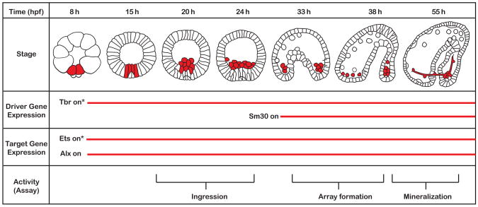
We chose the skeletogenic regulatory system of the sea urchin embryo as our test system. As summarized in Fig. 1, the skeletogenic cell lineage arises at the vegetal pole of the egg and its fate is specified early in cleavage by expression of a set of key regulatory genes, viz., alx1, tbr, and ets1, which are together derepressed exclusively in this lineage by operation of a double negative regulatory gate (Oliveri et al., 2002; Revilla-i-Domingo et al., 2007). The gene regulatory network (GRN) which includes this gate is now deeply understood (Revilla-i-Domingo et al., 2007; Oliveri et al, 2008; for always current version, see http://sugp.caltech.edu/endomes/). This network indicates the genomic code for all subsequent events of skeletogenic regulatory state specification downstream of the double negative gate, as well as for activation of skeletogenic gene batteries. As indicated in Fig. 1, until late blastula stage the 16 skeletogenic cells remain in the epithelial wall of the embryo, whereupon they ingress into the blastocoel (24 h) and commence skeletogenesis (>30 h). Prior to ingression they not only establish and lock down their state of specification, but also activate many skeletogenic differentiation genes, though biomineral deposition begins only after ingression. However, certain genes are activated only following ingression, one of which figures importantly in this work. This is sm30, which encodes a major protein of the biomineral structure (Frudakis and Wilt, 1995).
Fig. 1.
Time course of skeletogenesis and of relevant gene expressions. S. purpuratus embryo stages and time of development in hours post-fertilization are depicted relative to time of expression of the driver genes (tbr and sm30), target genes (ets1 and alx1). Skeletogenic lineage cells are shown in red. Zygotic tbr expression starts by 7–8 h post-fertilization, while sm30 expression begins around 30 h as illustrated by the red horizontal lines. The assay for ingression of skeletogenic cells occurs at 22–24 h, before the sm30 gene becomes active. The assays for skeleton formation (array formation and mineralization) are performed at 48 h, after sm30 expression turns on. Zygotic expression of ets1 and alx1 begin similarly to tbr at 7–8 h. Tbr and ets1 are additionally present as maternal message as indicated by the asterisks.
The alx1 and ets1 genes continue to be expressed actively after ingression, during the skeletogenic phase (Ettensohn et al., 2003; Rizzo et al., 2006). MASO directed against these genes and injected into eggs prevents ingression, and thus all subsequent events of skeletogenesis. These MASOs directly or indirectly break multiple GRN linkages, and in consequence the presumptive skeletogenic cells fail to become specified (op. cit.; Oliveri et al., 2008). While these same regulatory genes are very likely to be essential for the later functions of post-ingression skeletogenesis as well, their roles cannot be studied by use of alx1 or ets1 MASO, because the treated embryos completely lack ingressed skeletogenic cells. This is a specific example of the general problem adduced at the outset: what is needed is a means for extinguishing alx1 or ets1 expression in skeletogenic cells only after their ingression.
Materials and methods
Animal husbandry, embryos and microinjection
Adult Strongylocentrotus purpuratus were collected along the Southern California coast and maintained in 12°C seawater at Caltech’s Kerckhoff Marine Laboratory. Delivery of nucleic acid vectors was achieved by standard procedures. Gametes were harvested and eggs rinsed for one minute in 1 mM citric acid seawater and placed in seawater containing 300 mg/mL para-aminobenzoic acid. Approximately 1500 molecules of desired DNA construct (450 molecules for large vectors such as BACs) were injected along with a 6-fold molar excess of HindIII-digested carrier sea urchin DNA per egg in a 4pL volume of 0.12 M KCl. The DNA constructs, typically PCR products, were injected into eggs immediately following fertilization. Our injection solutions included both the antisense-expressing construct (either PCR product or linearized BAC vector) along with linearized Tbr BAC-GFP as a marker of incorporations plus excess HindIII-digested sea urchin genomic DNA as a carrier in a 0.12 M KCl solution. We followed injected embryos by observing GFP expression by fluorescent microscopy at various times post-fertilization. Embryos were placed on glass slides for fluorescent imaging with an AxioCam Mrm mounted on an AxoSkop 2 Plus (Zeiss).
Vector construction and constructs used
PCR (High-fidelity PCR kit, Roche, Indianapolis, IN) was used to amplify the desired cis-acting sequence, using a right (downstream) primer with a universal adaptamer tail. This PCR product was called the “driver PCR fragment.” In parallel, an oligomer was synthesized (Integrated DNA Technologies, Coralville, Iowa) called the “antisense oligo.” This was designed to contain the following in 5′ to 3′ order: the reverse complement SV40 polyadenylation sequence; the sense target site; and a universal adaptamer sequence. Fusion PCR was then performed combining the driver PCR fragment and antisense oligo as follows. Equal molar amounts of driver PCR fragment (desalted) and target oligo were added to 1X PCR mix containing buffer, dNTPs and enzyme to a final volume of 100 μL. The resulting reaction mix was then distributed among 10 PCR tubes and placed in a gradient thermal cycler with the following cycling protocol: 95°C for 20 sec, 54°C–62°C for 30 sec, 68°C for an appropriate time according to length of driver PCR fragment (~1 min per kb) for 12 to 15 cycles. Note that no additional primers were added for this reaction; the target oligo and driver PCR fragment in essence “prime” each other by annealing at the universal adaptamer sequence. A secondary reaction mix, this time containing outside primers, was added immediately following completion of the primary reaction and PCR was performed again with the same protocol for 25–30 additional cycles. PCR products were desalted by Qiagen Qiaquick columns and sequenced. Constructs used are as follows:
The following DNA oligonucleotides were used in aiRNA and sense control vector construction (all sequences given as ordered with IDT):
Alx1 i1 aiRNA (target sequence: 5′-CGCGGAAATGTGTTCACGTGGGAG-3′)
5′-ACGTCGTAGTAATAAACAGTAGTCGTAATAAAGTCATCAGTCAATAAACTCC CACGTGAACACATTTCCGCGGCCTCGATCTGCATAGCGATACAA-3′
Alx1 i1 sense
5′-ACGTCGTAGTAATAAACAGTAGTCGTAATAAAGTCATCAGTCAATAAACGCG GAAATGTGTTCACGTGGGAGGCCTCGATCTGCATAGCGATACAA-3′
Alx1 aiRNA (target sequence: 5′-GAGTTTACTTACACGTCGCTAAGC-3′)
5′-ACGTCGTAGTAATAAACAGTAGTCGTAATAAAGTCATCAGTCAATAAAGCTT AGCGACGTGTAAGTAAACTCGCCTCGATCTGCATAGCGATACAA-3′
Alx1 sense
5′-ACGTCGTAGTAATAAACAGTAGTCGTAATAAAGTCATCAGTCAATAAAGCTT AGCGACGTGTAAGTAAACTCGCCTCGATCTGCATAGCGATACAA-3′
Ets1 aiRNA (target sequence: 5′-CTTCGCGTTAGCCTGTAAGT-3′)
5′-ACGTCGTAGTAATAAACAGTAGTCGTAATAAAGTCATCAGTCAATAAACTTC GCGTTAGCCTGTAAGTGCCTCGATCTGCATAGCGATACAA-3′
Ets1 sense
5′-ACGTCGTAGTAATAAACAGTAGTCGTAATAAAGTCATCAGTCAATAAAACTT ACAGGCTAACGCGAAGGCCTCGATCTGCATAGCGATACAA-3′
cDNA preparation and quantitative real-time PCR (QPCR)
Embryos were collected at 24 h post-fertilization and pelleted in an Eppendorf 5415 D microcentrifuge for 4 min at 4000 rpm. As much seawater as possible was removed leaving no more than 50 μL, and embryos were lysed in 700 μL RLT buffer (Qiagen) with β-mercaptoethanol added. Total RNA was prepared using the Qiagen Rneasy Micro kit, including on column DNase I digestion, and eluted in 16 μL RNase-free water. Two μL of the eluate were set aside for later QPCR analysis as a reverse transcriptase-negative control, while the remainder was processed by the iScript cDNA Synthesis kit from BioRad in a 20 μL reaction volume according to the following reaction protocol: 5 min at 25ºC; 30 min at 42ºC; 5 min at 85ºC. Samples were then analyzed by QPCR using the iTaq SYBR Green Supermix with ROX from BioRad. In general, two embryo equivalents of cDNA solution were analyzed per well and reactions were run in quadruplicate wells in a 384-well plate at a final volume of 10μL; primers were at a final concentration of 250 nM. Samples were analyzed using an Applied Biosystems 7900 HT Fast Real-Time PCR System.
Results and discussion
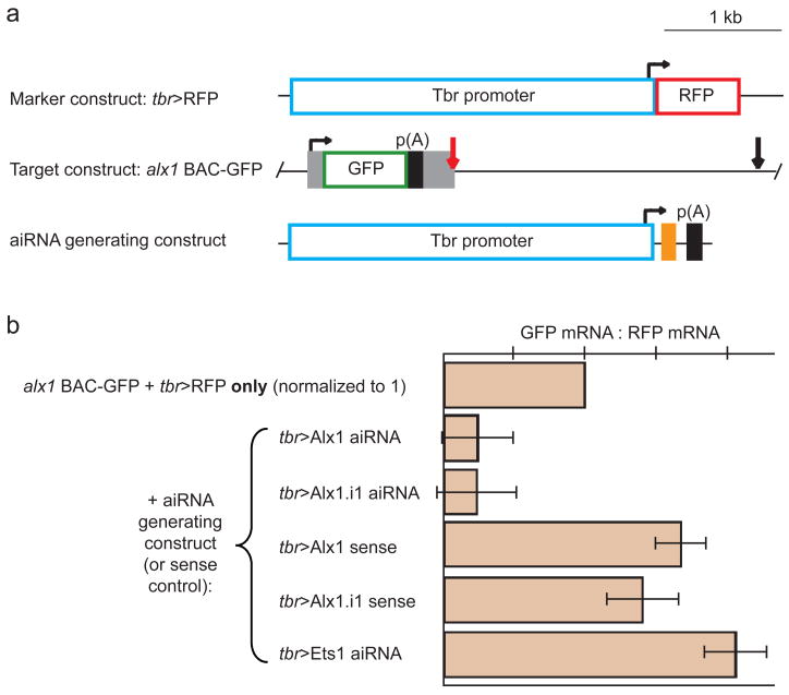
The following experiment shows the potential effectiveness and specificity of anti-intron antisense RNA (aiRNA), transcribed from a cis-regulatory expression vector, for blocking target gene expression. We made use of the fact that sea urchin eggs concatenate injected linear DNA, and whatever constructs are injected, stably incorporate these together into a blastomere chromosome, whereafter the exogenous concatenate replicates together with the host DNA (Livant et al., 1991; Revilla-i-Domingo et al., 2004; Arnone et al., 1997). Thus we injected a mixture of a marker construct, an aiRNA generating construct, and a target construct (Fig. 2A; a description of these and all other constructs mentioned in this paper are found in the Materials and methods section). The marker consisted of tbr cis-regulatory DNA driving an RFP gene as a reporter (tbr>RFP). It will express only in skeletogenic cells and will identify those cells which contain the exogenous mix of constructs. The target construct consisted of an alx1 BAC, containing its own endogenous cis-regulatory information as well as the complete gene, into the 5′UTR of which a GFP coding sequence had been inserted by homologous recombination (Kotzamanis and Huxley,). The aiRNA generating construct (tbr>aiRNA) consisted of the same tbr cis-regulatory sequence as in the marker construct, but here used to drive expression of the antisense transcripts. In one version the construct produced an antisense transcript targeting the intron1/exon1 splice junction of the alx1 gene (as would be targeted by a splice-blocking MASO); and in a second version, it produced an antisense transcript targeting an internal region of intron 1. These aiRNA transcripts were generated off 24 bp antisense oligonucleotides terminated with three tandem p(A) addition sites, cloned into the tbr expression vector. The results were monitored by QPCR measurement of GFP mRNA, normalized to the RFP mRNA in the same embryos (Fig. 2B). Both aiRNA constructs almost eliminated GFP mRNA production. Constructs generating sense rather than antisense transcripts of the same intronic sequences had no effect. Nor was expression of the alx1 BAC-GFP reporter affected by an aiRNA construct targeted against the ets1 gene, which is active in the same cells, excluding a non-specific interference with expression. We could not directly measure the effect of aiRNA constructs on endogenous alx1 transcripts in the same experiment by QPCR, due to the mosaic incorporation of the targeting vector, since about 3/4ths of the skeletogenic cells lack the exogenous DNA, and produce normal levels of alx1 in the same embryos. In contrast, since the constructs are co-incorporated, as noted above, in the alx1 BAC-GFP experiment in Fig. 2B, the three- to four-fold reduction in GFP transcript is the actual gene knockdown effect in those cells carrying the aiRNA construct. We may conclude: (i), that intranuclear stoichiometry does indeed appear to favor efficient target acquisition by endogenously produced antisense transcripts; (ii), that the interference with GFP production was not a general effect of interference with splicing machinery; (iii), that this interference operates on internal as well as junctional intronic sequences; (iv), that it causes destruction or inactivation of the whole target transcript since the target sequences are all downstream of the intact GFP sequence. In other words, it is likely the primary transcript is targeted for degradation. Though we expect that the p(A) sites would ultimately result in short aiRNA transcripts, we do not know whether the active inhibitory form is a longer readthrough pre-poly(A) RNA, or the terminated polyadenylated product.
Fig. 2.
Vector designs and specificity control experiment. (A) Expression constructs coinjected in control experiments. The “marker construct” consists of a 3.5 kb tbr promoter driving expression of RFP and serves as a marker of incorporation of the concatenate of injected constructs; SV40 polyadenylation sequences, black boxes as indicated; bent arrow, start of transcription. The “target construct” is an alx BAC with a GFP coding sequence inserted by homologous recombination into the 5′UTR and containing two target sites, one spanning the junction of exon1 and intron 1 (red arrow) and the other near the middle of intron 1 (black arrow); endogenous alx1 exons, gray boxes. Five different aiRNA generating constructs were individually tested, each using the 3.5 kb tbr promoter to drive expression of a 24-bp target sequence (orange box) as specified in (B). (B) Effects of aiRNA constructs on target construct expression. In all conditions the marker and target constructs were coinjected: the ratio of GFP mRNA to RFP mRNA was then measured by real-time quantitative PCR. When an aiRNA construct targeting either the exon1/intron1 junction of the alx1 gene or an internal region of the intron was also injected, the level of GFP transcripts relative to RFP fell, since the exon1/intron1 target sequence is present in the alx1 BAC-GFP vector but not the tbr>RFP construct. The comparable sense constructs for either of these targets, meanwhile, had no effect. To assess whether aiRNA vectors could adversely effect transcript splicing, we injected a vector driving expression of a sequence antisense to the exon1/intron1 junction of ets1, which is active in the same cells as alx1. This did not decrease GFP:RFP, indicating aiRNA constructs do not lead to general defects in splicing.
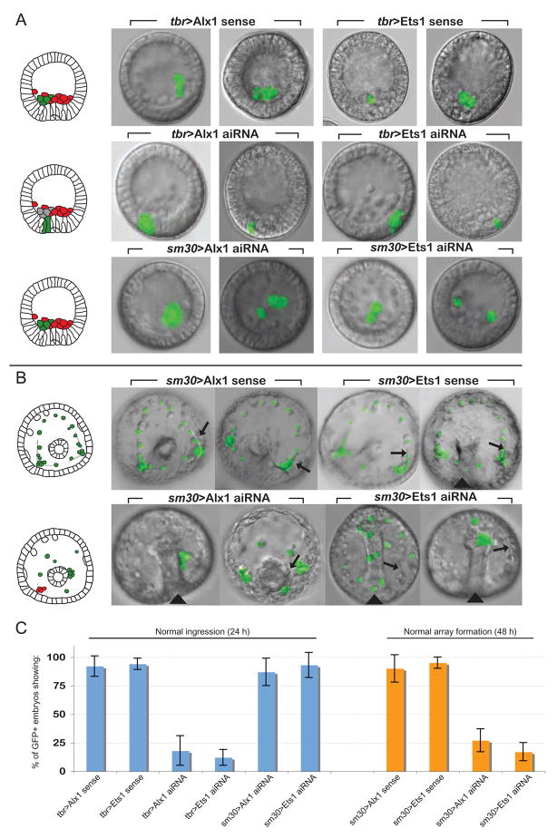
If the effect of an aiRNA construct is indeed the functional inactivation of the target transcript so that it cannot be expressed, then introduction of tbr>aiRNA against alx1 should produce the same morphological effect on the skeletogenic cells carrying it as does injection into the egg of MASO against alx1, since the tbr cis-regulatory control system initiates expression very early in development (Fig. 1). The alx1 MASO effect is total prevention of ingression (Ettensohn et al., 2003): alx1 regulates downstream differentiation genes required for this distinct function. The result of introducing tbr>aiRNA against alx1 (Fig. 3A, C) is indeed that in 80–90% of embryos bearing tbr>aiRNA targeted to alx1, no skeletogenic cells bearing the construct whatsoever emerge from the vegetal wall of the embryo, and in the remainder only a few do. The skeletogenic cells bearing the construct are marked by expression of tbr>GFP (i.e., the “marker” in these experiments is a tbr>GFP construct as opposed to the tbr>RFP construct used in Fig. 2 experiments). Expression of ets1 is also required for ingression, as shown by its MASO phenotype (op cit), and again, this phenotype is seen as well with tbr>aiRNA directed against an ets1 intron (Fig. 3A). Quantitatively, both aiRNA constructs are extremely effective in arresting ingression (Fig. 3C).
Fig. 3.
Effect of alx1 and ets1 aiRNA constructs on skeletogenic cell ingression and post-ingression function. (A) Ingression assay. The tbr> GFP construct was used as a marker. At 22–24 h post-fertilization embryos were analyzed for ingression of GFP-fluorescent cells. Embryos coinjected with tbr>aiRNA constructs targeting alx1 or ets1 showed minimal ingression of GFP+ cells; rather, in most embryos fluorescent cells remained entirely in the epithelial layer, illustrated in photographic images, and diagrammed in schematic at left (a commensurate number of mesenchymal cells, indicated in gray in the drawing, are presumably missing from the blastocoelar space but are difficult to assess); normally ingressed, nontransgenic skeletogenic mesenchymal cells in red. Embryos harboring sense control vectors showed a normal pattern of skeletogenic cell ingression, as did embryos injected with aiRNA vectors driven by the sm30 promoter. The sm30 promoter is not active until 27–30 h post-fertilization, after ingression has taken place. (B) Assay for array formation and biomineralization at 48 h post-fertilization. GFP+ cells were scored for normal array formation. As shown diagrammatically at left, a normal skeletogenic cell array forms a distinctive shape with biomineralized spicules apparent under polarized light (arrows in photos). Embryos injected with sense control constructs showed normal array formation by transgenic cells expressing GFP in the overwhelming majority of embryos. In embryos injected with sm30-driven aiRNA, by contrast, transgenic GFP expressing cells failed to form arrays or organized spicule rods. Arrowheads indicate blastopore. Incorporation of injected constructs is mosaic, and some skeletogenic cells do not incorporate the exogenous DNA. These do not express GFP and they are figured in red in the diagram. These cells participate in spicule formation, indicated by black arrows. (C) The percentage of GFP+-embryos showing normal ingression at 24 h post-fertilization (blue bars) or normal array formation at 48 h post-fertilization (orange bars) ± SEM following injection of various constructs as indicated.
We are now in a position to approach the problem outlined above: how to study late alx1 and ets1 function by knocking out expression in skeletogenic cells only after allowing these genes to function long enough to permit complete ingression. To this end we utilized the cis-regulatory system of the sm30 gene (George et al., 1991), which is turned on only after ingression. Sm30>aiRNA constructs targeted against the same intronic sequences of either alx1 or ets1 as in the tbr>aiRNA constructs were introduced, together with the tbr>GFP marker construct. Assuming that sm30 cis-regulatory control is sufficiently tight, the expectation is that there will be no expression of the aiRNAs prior to ingression and thus that neither construct will interfere with ingression. In fact, both sets of embryos displayed control levels of ingression (Fig. 3A). However, subsequent skeletogenesis was dramatically affected, though in a very specific way (Fig. 3B, C). In normal postgastrular embryos the skeletogenic cells migrate about the inner walls of the blastocoel, and then read signals displayed by the ectoderm cells, which specify the bilateral, branched form of the skeletal spicules. In response they arrange themselves in highly reproducible, ordered, linear arrays (Armstrong et al., 1993; cf. Fig. 3B controls). The cells then fuse laterally and secrete the skeleton into extracellular cables by which they are connected to one another. But the cells bearing sm30>aiRNA targeted to either alx1 or ets1 fail entirely to form these arrays, or to participate in secretion of organized spicule rods. The cells instead assume random positions on the inner wall of the blastocoel: thus they retain their motility, but it would appear that they have failed to respond to the spatial information presented on the blastocoel wall. That this information is being normally expressed in the same embryos can be seen by the presence of morphologically normal skeletal elements formed by cells not bearing the aiRNA constructs, i.e., not expressing GFP. Secondary skeletogenic cells were not observed up to 72 h post-fertilization. The basic biomineralization functions are also severely affected. Thus instead of all tbr>GFP cells producing biomineral as in controls, only about 3% of green cells in the aiRNA embryos are associated with rudimentary accumulations of biomineral, which can be detected in polarized light. In summary, the experiment shows that expression of alx1 and ets1 after ingression is required for alignment of the cells in response to ectodermal patterning information; whether these genes are needed for syncytial cable formation is moot since they never get in position to form linear cables. Both alx1 and ets1 are clearly required for completion of the skeletogenic program. These functions are consistent with the character of the gene regulatory network linkages set up by the time of ingression, which include, for both genes, inputs into signal receptors and into biomineralization differentiation genes (http://sugp.caltech.edu/endomes/). It is now clear that these regulatory linkages are set up to be utilized only after ingression, and that they and no doubt many others of similar nature are requisite for mature skeletogenic function.
Conclusions
We show here that in sea urchin embryos expression of RNA complementary to intronic sequence, under control of selected cis-regulatory modules, can be used to effect spatially and temporally targeted gene expression knockdown. The results of the ets1 and alx1 aiRNA experiments are exactly consistent with expectation from the model experiment that demonstrated aiRNA efficacy against the alx1 BAC GFP construct (Fig. 2B).
The mechanism by which these interference constructs work is not known. It is clearly distinct from that of classical RNAi since the latter causes destruction of target mRNAs in the cytoplasm, a process nucleated on the RNAi:3′trailer complex. Messenger RNA destruction mediated by RNAi is effected by cytoplasmic proteins (Hannon, 2002), while in our case the sequence targeted, i. e., the intron, exists only in the nucleus. Nor does the mechanism of interference with expression seem the same as that of splice-blocking morpholinos, even though we began this project with the thought that because of favorable stoichiometry we could duplicate the function of splice blocking morpholinos by use of endogenously synthesized antisense RNAs. Splice blocking morpholinos leave unspliced primary transcript fragments to accumulate in the nucleus where they are easily detected. But, as pointed out above, the aiRNA vectors apparently cause the destruction of the whole the transcript since even portions upstream of the targeted intron disappear (Fig. 2B). The actual mechanism by which formation of a 24 bp intron-antisense duplex effects primary transcript destruction will be most interesting to determine.
In the meantime, at the very least, this advance opens the way to exploration of a plethora of interesting problems in the regulatory control of postgastrular development and morphogenesis in the sea urchin embryo. The result will be to extend gene regulatory network analysis to the later development of this model embryonic system. The effectiveness of the method may depend on the intra-nuclear stoichiometric ratios of transcripts made on exogenous constructs to endogenous pre-mRNAs. Thus the generality of its application in other model organisms would need to be determined.
Table 1.
Constructs used
| Name | Driver promoter | Transcription product/Description |
|---|---|---|
| tbr>RFP (“Marker”) | Tbr | RFP, “Marker construct” in Fig. 2 |
| alx1 BAC-GFP (“Target”) | Alx1 | GFP, “Target construct” in Fig. 2 |
| tbr>Alx1.i1 aiRNA | Tbr | Antisense to alx1 central intronic sequence |
| tbr>Alx1.i1 sense | Tbr | Sense to alx1 central intronic sequence |
| tbr>GFP (“Marker”) | Tbr | GFP, Marker in Fig. 3 experiments |
| tbr>Alx1 aiRNA | Tbr | Antisense to alx1 exon1/intron1 junction |
| tbr>Ets1 aiRNA | Tbr | Antisense to ets1 exon1/intron1 junction |
| sm30>Alx1 aiRNA | Sm30 | Antisense to alx1 exon1/intron1 junction |
| sm30>Ets1 aiRNA | Sm30 | Antisense to ets1 exon1/intron1 junction |
| tbr>Alx1 sense | Tbr | Sense to alx1 exon1/intron1 junction |
| tbr>Ets1 sense | Tbr | Sense to ets1 exon1/intron1 junction |
| sm30>Alx1 sense | Sm30 | Sense to alx1 exon1/intron1 junction |
| sm30>Ets1 sense | Sm30 | Sense to ets1 exon1/intron1 junction |
Acknowledgments
The authors would like to thank R. Andrew Cameron for initial experiments and data analysis and Paola Oliveri for important insights, helpful suggestions and critical reading of the manuscript. J.S. offers thanks to Andy Ransick for embryo drawings and general help with embryology and microscopy. Research was supported by an NIH grant HD-37105.
Footnotes
Publisher's Disclaimer: This is a PDF file of an unedited manuscript that has been accepted for publication. As a service to our customers we are providing this early version of the manuscript. The manuscript will undergo copyediting, typesetting, and review of the resulting proof before it is published in its final citable form. Please note that during the production process errors may be discovered which could affect the content, and all legal disclaimers that apply to the journal pertain.
References
- Armstrong N, Hardin J, McClay DR. Cell-cell interactions regulate skeleton formation in the sea urchin embryo. Development. 1993;119:833–40. doi: 10.1242/dev.119.3.833. [DOI] [PubMed] [Google Scholar]
- Arnone MI, Bogarad LD, Collazo A, Kirchhamer CV, Cameron RA, Rast JP, Gregorians A, Davidson EH. Green Fluorescent Protein in the sea urchin: new experimental approaches to transcriptional regulatory analysis in embryos and larvae. Development. 1997;124:4649–59. doi: 10.1242/dev.124.22.4649. [DOI] [PubMed] [Google Scholar]
- Bolouri H, Davidson EH. Transcriptional regulatory cascades in development: initial rates, not steady state, determine network kinetics. Proc Natl Acad Sci USA. 2003;100:9371–9376. doi: 10.1073/pnas.1533293100. [DOI] [PMC free article] [PubMed] [Google Scholar]
- Davidson EH. Gene Activity in Early Development. 3. Academic Press; Orlando, FL: 1986. [Google Scholar]
- Davidson EH. The sea urchin genome: Where will it lead us? Science. 2006;314:939–40. doi: 10.1126/science.1136252. [DOI] [PubMed] [Google Scholar]
- Dickey-Sims C, Robertson AJ, Rupp DE, McCarthy JJ, Coffman JA. Runx-dependent expression of PKC is critical for cell survival in the sea urchin embryo. BMC Biol. 2005;3:18. doi: 10.1186/1741-7007-3-18. [DOI] [PMC free article] [PubMed] [Google Scholar]
- Ettensohn CA, McClay DR. Cell lineage conversion in the sea urchin embryo. Dev Biol. 1998;125:396–409. doi: 10.1016/0012-1606(88)90220-5. [DOI] [PubMed] [Google Scholar]
- Ettensohn CA, Illies MR, Oliveri P, De Jong DL. Alx1, a member of the Cart1/Alx3/Alx4 subfamily of Paired-class homeodomain proteins, is an essential component of the gene network controlling skeletogenic fate specification in the sea urchin embryo. Development. 2003;130:2917–28. doi: 10.1242/dev.00511. [DOI] [PubMed] [Google Scholar]
- Frudakis TN, Wilt F. Two cis elements collaborate to spatially repress transcription from a sea urchin promoter. Dev Biol. 1995;172:230–41. doi: 10.1006/dbio.1995.0018. [DOI] [PubMed] [Google Scholar]
- George NC, Killian CE, Wilt FH. Characterization and expression of a gene encoding a 30.6-kDa Strongylocentrotus purpuratus spicule matrix protein. Dev Biol. 1991;147:334–42. doi: 10.1016/0012-1606(91)90291-a. [DOI] [PubMed] [Google Scholar]
- Hannon G. RNA Interference. Nature. 2002;418:244–251. doi: 10.1038/418244a. [DOI] [PubMed] [Google Scholar]
- Howard-Ashby M, Materna SC, Brown CT, Tu Q, Oliveri P, Cameron RA, Davidson EH. High regulatory gene use in sea urchin embryogenesis: Implications for bilaterian development and evolution. Dev Biol. 2006;300:27–34. doi: 10.1016/j.ydbio.2006.10.016. [DOI] [PMC free article] [PubMed] [Google Scholar]
- Imamura S, Kishi S. Molecular cloning and functional characterization of zebrafish ATM. Int J Biochem Cell Biol. 2005;37:1105–16. doi: 10.1016/j.biocel.2004.10.015. [DOI] [PubMed] [Google Scholar]
- Kotzamanis G, Huxley C. Recombining overlapping BACs into a single larger BAC. BMC Biotechnol. 2004;4:1. doi: 10.1186/1472-6750-4-1. [DOI] [PMC free article] [PubMed] [Google Scholar]
- Livant DL, Hough-Evans BR, Moore JG, Britten RJ, Davidson EH. Differential stability of expression of similarly specified endogenous and exogenous genes in the sea urchin embryo. Development. 1991;113:385–98. doi: 10.1242/dev.113.2.385. [DOI] [PubMed] [Google Scholar]
- Oliveri P, Davidson EH. Gene regulatory network controlling embryonic specification in the sea urchin. Curr Opin Genet Dev. 2004;14:351–60. doi: 10.1016/j.gde.2004.06.004. [DOI] [PubMed] [Google Scholar]
- Oliveri P, Carrick DM, Davidson EH. A regulatory gene network that directs micromere specification in the sea urchin embryo. Dev Biol. 2002;246:209–28. doi: 10.1006/dbio.2002.0627. [DOI] [PubMed] [Google Scholar]
- Oliveri P, Tu Q, Davidson EH. Global regulatory logic for specification of an embryonic cell lineage. Proc Natl Acad Sci, USA. 2008 doi: 10.1073/pnas.0711220105. in press. [DOI] [PMC free article] [PubMed] [Google Scholar]
- Revilla-i-Domingo R, Minokawa T, Davidson EH. R11: a cis-regulatory node of the sea urchin embryo gene network that controls early expression of SpDelta in micromeres. Dev Biol. 2004;274:438–51. doi: 10.1016/j.ydbio.2004.07.008. [DOI] [PubMed] [Google Scholar]
- Revilla-i-Domingo R, Oliveri P, Davidson EH. A missing link in the sea urchin embryo gene regulatory network: hesC and the double-negative specification of micromeres. Proc Natl Acad Sci USA. 2007;104:12383–12388. doi: 10.1073/pnas.0705324104. [DOI] [PMC free article] [PubMed] [Google Scholar]
- Rizzo F, Fernandez-Serra M, Squarzoni P, Archimandritis A, Arnone MI. Identification and developmental expression of the ets gene family in the sea urchin (Strongylocentrotus purpuratus) Dev Biol. 2006;300:35–48. doi: 10.1016/j.ydbio.2006.08.012. [DOI] [PubMed] [Google Scholar]
- Sutou S, Kunishi M, Kudo T, Wongsrikeao P, Miyagishi M, Otoi T. Knockdown of the bovine prion gene PRNP by RNA interference (RNAi) technology. BMC Biotechnol. 2007;7:44. doi: 10.1186/1472-6750-7-44. [DOI] [PMC free article] [PubMed] [Google Scholar]


