. 2014 Feb 25;3:e01612. doi: 10.7554/eLife.01612

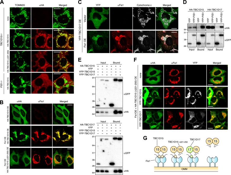
Figure 6. Identification of TBC1D17 as a Fis1 and TBC1D15 binding protein.

(A) TBC1D15−/− cells and those stably expressing HA-TBC1D15 WT and HA-TBC1D15 (Δ221-250), and FIS1−/− cells stably expressing HA-TBC1D15 WT were subjected to immunostaining with anti-TOMM20 and anti-HA antibodies. Scale bars, 10 μm. (B) HA-TBC1D15 WT or HA-TBC1D15 (Δ221-250) with or without Fis1 was transiently overexpressed (OE) in HeLa cells. Cells were subjected to immunostaining with anti-HA and anti-Fis1 antibodies. Scale bars, 20 μm. (C) YFP-TBC1D17 together with pcDNA vector or Fis1 was transiently overexpressed (OE) in HeLa cells. Cells were subjected to immunostaining with anti-Fis1 and anti-Cytochrome c antibodies. Scale bars, 20 μm. (D) YFP or YFP-Fis1 was co-overexpressed with HA-TBC1D15 or HA-TBC1D17 in HEK293 cells. The cell extracts were subjected to pull down assays with GFP-Trap. 5% input and bound fractions were analyzed by immunoblotting with anti-HA (upper panel) and anti-GFP (lower panel) antibodies. (E) YFP, YFP-TBC1D15, or YFP-TBC1D17 was co-overexpressed with HA-TBC1D15 (upper panel) or HA-TBC1D17 (lower panel) in HEK293 cells. The cell extracts were subjected to pull down assays with GFP-Trap. 5% input and bound fractions were analyzed by immunoblotting with anti-GFP and anti-HA antibodies. (F) HA-TBC1D15 (Δ221-250) together with Fis1 and YFP-TBC1D15 WT or YFP-TBC1D17 WT were transiently overexpressed (OE) in HeLa cells. Cells were subjected to immunostaining with anti-HA and anti-Fis1 antibodies. Images of HA and Fis1 staining were merged in the right panels. Scale bars, 20 μm. (G) Schematic model of Fis1, TBC1D15, and TBC1D17 binding. Homo- or hetero-dimer of TBC1D15 can interact with Fis1 dimer on the mitochondrial outer membrane (OMM).

DOI: http://dx.doi.org/10.7554/eLife.01612.017

Figure 6.

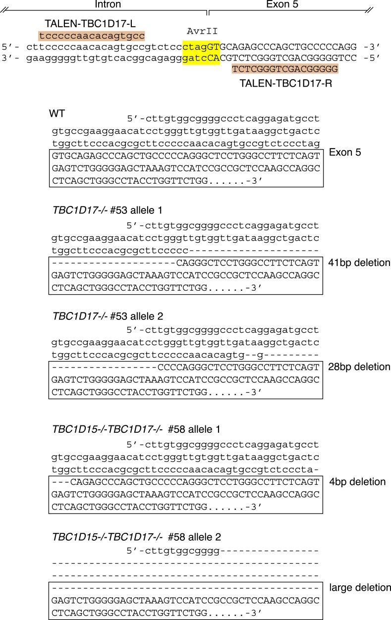
Figure 6—figure supplement 1. TBC1D17 gene knock out by TALENs.

Figure 6—figure supplement 1.

Highlighted TALEN-TBC1D17-L and TALEN-TBC1D17-R indicate TALEN binding pair that targets the intron–exon 5 junction of TBC1D17 gene. DNA sequencing confirmed nucleotide deletions in both alleles of TBC1D17−/− #74 clone and of TBC1D15/17 DKO #58 clone.
Figure 6—figure supplement 2. TBC1D17 is dispensable for normal mitochondria and peroxisome morphologies.

Figure 6—figure supplement 2.

(A) The indicated cell lines were analyzed by immunofluorescence microscopy using anti-Cytochrome c antibody for mitochondria and anti-PMP70 antibody for peroxisome staining. Images are displayed as z-stacks of 6 confocal slices. Magnified images are also shown for mitochondrial morphologies. Scale bars, 10 μm. (B) Quantification of mitochondrial morphologies in (A). Percentages of cells harboring fragmented, tubular or elongated mitochondria were shown. Tubular and elongated denote normal tubular mitochondria seen in WT cells and highly connected mitochondrial network, respectively. The error bars represent ±SD from three independent replicates. Over 50 cells were counted in each replicate.
Figure 6—figure supplement 3. YFP-LC3 accumulation in TBC1D17−/− and TBC1D15/17 DKO cells during mitophagy.

Figure 6—figure supplement 3.

(A) TBC1D17−/− and TBC1D15/17 DKO cells stably expressing YFP-LC3 and mCherry-Parkin were treated with valinomycin for 3 hr and subjected to immunofluorescence microscopy with anti-TOMM20 antibody. Scale bars, 10 μm. (B) YFP-LC3 morphologies in (A) were quantified. Percentages of cells harboring diffuse, punctate or accumulated YFP-LC3 are shown. The error bars represent ±SD from three independent replicates. Over 100 cells were counted in each well. (C) The indicated proteins were transiently overexpressed (OE) in TBC1D15/17 DKO cells stably expressing YFP-LC3 and mCherry-Parkin. The cells were treated with valinomycin for 3 hr followed by immunofluorescence microscopy with anti-HA antibody. Scale bars, 10 μm. (D) YFP-LC3 morphologies in (C) were quantified. The error bars represent ±SD from three independent replicates. Over 50 cells were counted in each well.