
Fig. 5.
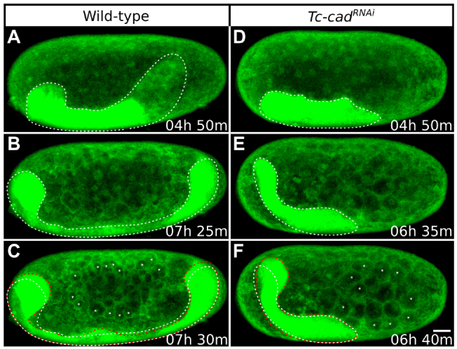
Patterns of Tribolium germband elongation in wild-type and Tc-cadRNAi embryos. Each column shows a single GAP43-YFP-labeled embryo at progressive time-points during stage 6. (A-C) Lateral views of the wild-type embryo also shown in Fig. 4F-J. (D-F) Lateral views of the Tc-cadRNAi embryo also shown in Fig. 4P-T. All images are average intensity projections with uniformly enhanced brightness/contrast to show the germband (outlined with dotted lines) and the membrane-bound yolk spheres. Panels are timed against the onset of stage 1. (A,D) Size and position of germbands immediately after serosa window closure. The germband is truncated posteriorly after Tc-cad RNAi. (B,E) The control germband grows considerably and extends round both poles. The Tc-cadRNAi germband fails to grow, but is displaced anteriorly with the head region curved around the anterior pole. (C,F) At the following time-point, both ends of the control embryo and the anterior end of the Tc-cadRNAi embryos move towards the dorsal side (previous position indicated with white dotted line and new position with red dotted line). This movement is accompanied by the appearance of extra membrane-bound yolk spheres (asterisks) compared with the previous time-point. Anterior is towards the left and dorsal towards the top. Scale bar: 50 μm.