Abstract
Pyoderma gangrenosum is a destructive inflammatory disease that commonly occurs in an idiopathic way. Its occurrence in the auricular area is very rare, although this fact does not seem to determine a different behavior of the disease with regard to ulcer aspects and response to treatment. The authors report the case of a patient with pyoderma gangrenosum affecting both earlobes. The patient responded well to treatment with oral prednisone and has not shown relapses after a six-month follow-up.
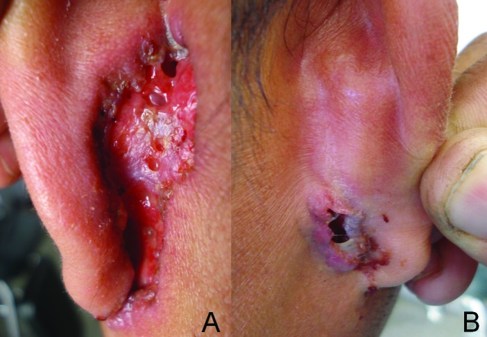
A 43-year-old male police officer presented with wounds affecting both his retroauricular areas and earlobes, which had developed from small erythematous papules into painful ulcers during the past 30 months. The two lesions had followed a parallel course in regard to time of appearance, site of injury, velocity of progression, and absence of remission since onset. Skin examination showed single bilateral ulcers; the biggest on the right side measuring 5.0x2.5cm. The lesions showed boggy granulating to purulent bases and erythematous to violaceous borders that at some areas were necrotic and undermined and at other areas showed slight infiltration (Figures 1A and IB). The patient denied presenting any systemic symptoms (i.e., fever, weight loss, asthenia, gastrointestinal or respiratory complaints). Moreover, there were neither personal nor family medical records related to the actual clinical picture.
Figure 1.
Aspect of pyoderma gangrenosum lesions before treatment. Extensive ulcer at the left retroauricular area showing an erythematous slightly infiltrated border and a boggy granulating base (A). Right retroauricular area showing a small ulcer with undermined necrotic borders (B)
The patient was subjected to immunological tests (i.e., purified protein derivative, venereal disease research laboratory, anti-human immunodeficiency virus 1/2, anti-hepatitis B surface antigen, anti-hepatitis B core, anti-Epstein-Barr virus, Chagas [ELISA]) and was diagnosed nonreactive. His blood count and serum protein levels were normal (hemoglobin 15.4g%; albumin 4.0g/dL; globulin 1.5g/dL). Histopathological analysis of ulcer border demonstrated hyperkeratosis and slight exocytose of neutrophils at the epidermis; a dense mixed infiltrate of polymorphonuclear leucocytes, neutrophils, and eosinophils, but few lymphocytes, occupying both papillary and reticular dermis. Vasculitis or vessel thrombosis was not seen in the specimen analyzed. Periodic acid-Schiff and Gomori-Grocott stains did not show any micro-organisms. Smear and culture for leishmaniasis were also negative. The diagnosis of an idiopathic pyoderma gangrenosum was then made following the exclusion of differential diagnosis that could lead to a similar clinical picture.
DISCUSSION
Pyoderma gangrenosum (PG), first described by Brunsting et al (1930), is a destructive inflammatory, noninfectious skin disease of chronic and recurrent evolution. It is characterized by painful nodules or pustules that progress and develop into ulcers with detached violaceous edges.1,2 PG affects mainly adults between 20 to 50 years of age and is considered by some authors to be more prevalent in women.1,3,4
Although PG pathogenesis is still unknown, dysregulation of the immune system, such as overexpression of growth factors and interleukins, especially interleukin (IL)-8 and tumor necrosis factor (TNF)-a, are thought to be involved in neutrophil dysfunction, which otherwise seems to play a crucial role in triggering the initial tissue aggression.2 PG frequently occurs in an idiopathic way; however, in 50 percent of cases, it is associated with neoplasm, drug intake, or system inflammatory illnesses, such as ulcerative colitis, Crohn’s disease, Behcet’s disease, Wegener’s granulomatosis, rheumatoid arthritis, and myeloproliferative diseases.1-5
There are no histopathological findings specific to PG, although a skin biopsy must always be performed to rule out other etiologies. Edema, massive neutrophilic infiltration, hemorrhage, and necrotic areas are usually seen with PG1,2,4 It is also common to find necrotizing vasculitis, leukocytoclasia, and intramural deposits of C3 fraction of complement (40% of cases).2
Any area of the body can be affected by PG, the most common site being the pretibial area, where the lesions may show bilateral involvement.4,5 The head and neck are also sites eventually affected by PG, although only a few cases of ear involvement have been reported to date, with none of them occurring bilaterally (Table 1). Iijima et al5 reported the case of a Japanese patient who presented a unilateral ulcer of PG at the left earlobe following a 10-year history of chilblains every winter. The patient had not presented with any signs of systemic comorbidities, similar to the case described herein. Also, he achieved a rapid response when treated with prednisolone at 40mg/day which allowed for a tapering of medication after the fifteenth day. However, 90 days after, the patient showed the appearance of new lesions on his back, folliculitis-like papules that progressed into ulcers, determining the necessity of increasing the daily prednisolone intake to achieve sustained clinical response.
TABLE 3.
Summary of cases of pyoderma gangrenosum reported to date involving the auricular area
| PYODERMA GANGRENOSUM | |||||
|---|---|---|---|---|---|
| Case | Gender | Age of onset (years) | Site of first involvement | Associated disease | Treatment |
| A6 | Female | 3 | Left retroauricle, neck, lower back, foot | Microcytic and hypochromicanaemia | PSL 1mg/kg/day Dapsone 2mg/kg/day |
| B7 | Male | 3.5 | Preauricle | Neonatal asphyxia, panhypopituitarism, AIDS | Dapsone 12.5mg/day |
| C6 | Male | 4 | Right retroauricle, neck, lower back | Microcytic and hypochromicanaemia | PSL 1mg/kg/day Dapsone 2mg/kg/day |
| D8 | Female | 23 | Auricle | Ulcerative colitis | Hydrocortisone 500mg/day |
| E9 | Female | 29 | Periauricle area, cheek oral mucosa, leg | None | Dapsone 100mg/day plus mPSL 100mg/day IV plus intralesional triamcinolone 20mg/week |
| F* | Male | 43 | Retroauricle (bilateral) | None | Clofazimine 100mg/day PS 60mg/day |
| G10 | Female | 58 | Auricle (conchae) | Perirectal fistula, ulcerative colitis, sclerosing cholangitis | Cyclosporine 10mg/kg/day |
| H5 | Male | 59 | Auricle (lobe) | None | PSL 40mg/day |
| I9 | Male | 64 | Retroauricle | Perirectal fistula Myelofibrosis, diabetes | PSL 80mg/day |
| J11 | Male | 65 | Auricle (pinna) | mellitus | PSL 40mg/day |
PSL=prednisolone; PS=prednisone; mPSL=methylprednisolone; *=case reported herein
Khandpur et al6 suggested the hypothesis of a familial predisposition for PG by reporting cases of extensive illness affecting two siblings ages 6 and 10 years old. The children presented with multiple lesions involving the retroauricular and cervical areas, trunks, and feet. The treatment with prednisolone at lmg/kg/day also initially determined disease remission after four weeks of usage. However, the suspension of the drug was followed by wound recurrence in both children. Nevertheless, the addition of dapsone at 2mg/kg/day to the previous regimen of prednisolone led to a long-lasting disease remission. The patients did not present with any comorbidities, except for anemia secondary to insufficient iron intake.
Many drugs have been used in the treatment of PG, although almost all the auricular PG cases previously reported did respond to the classical therapeutic strategy based on the use of corticoids or neutrophil chemotaxis inhibitors (Table 1). This fact must sustain the use of these drugs as first-choice in the treatment of auricular PG cases, although doctors must expect episodes of flare-up in some
patients, as usually observed in most cases of typical pyoderma. No cases of auricular PG treated with anti-TNF-α biologic agents have been reported. Thus, its value in managing refractory cases of auricular PG is unknown, as already shown in PG of other sites.12
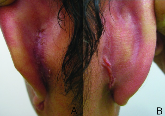
The patient described herein was initially given clofazimine (lOOmg/day) for a three-week period. As he did not show any clinical improvement, clofazimine was changed to prednisone at 60mg/day for a period of 30 days, followed by a long phase of dose tapering thereafter. Prednisone was impressively effective in our patient, as determined by progressive scarring of lesions, allowing its interruption after 13 months (Figures 2A and 2B). There was no recurrence or new lesion appearance since the start of prednisone to the end of a six-month treatment-free follow-up period.
Figure 2.
Cicatricial lesions after treatment. Retroauricular area showing atrophic (A) to slightly elevated scars (B)
From an additional analysis of cases listed in Table 1, it can be seen that PG affecting the auricular area has not shown gender or age predilection at the onset. Indeed, in 30 percent of the cases, it has been associated with inflammatory bowel diseases, although no final conclusions can be made about its early appearance as a predictor of systemic diseases.
Footnotes
DISCLOSURE:The authors report no relevant conflicts of interest.
REFERENCES
- 1.Ruocco E, Sangiuliano S, Gravina AG, et al. Pyoderma gangrenosum: an updated review. J Eur Acad Dermatol Venereol. 2009;23(9):1008–1017. doi: 10.1111/j.1468-3083.2009.03199.x. [DOI] [PubMed] [Google Scholar]
- 2.Wolff K, Goldsmith LA, Katz SI, et al., editors. Fitzpatrick’s Dermatology in General Medicine. New York: McGraw-Hill; 2010. 8th ed. [Google Scholar]
- 3.Santos M, Talhari C, Rabelo RF, et al. Pyoderma gangrenosum: a clinical manifestation of difficult diagnosis. An Bras Dermatol. 2011;86(1):153–156. doi: 10.1590/s0365-05962011000100025. [DOI] [PubMed] [Google Scholar]
- 4.Wollina U. Pyoderma gangrenosum—a review. Orphanet J Rare Dis. 2007;2:19. doi: 10.1186/1750-1172-2-19. [DOI] [PMC free article] [PubMed] [Google Scholar]
- 5.Iijima S, Ogawa T, Nanno Y, et al. Pyoderma gangrenosum first presenting as a recalcitrant ulcer of the ear lobe. Eur J Dermatol. 2003;13(6):606–609. [PubMed] [Google Scholar]
- 6.Khandpur S, Mehta S, Reddy BSN. Pyoderma gangrenosum in two siblings: a familiar predisposition. Pediatr Dermatol. 2001;18(4):308–312. doi: 10.1046/j.1525-1470.2001.01936.x. [DOI] [PubMed] [Google Scholar]
- 7.Paller AS, Sahn EE, Garen PD, et al. Pyoderma gangrenosum in pediatric acquired immunodeficiency syndrome. J Pediatr. 1990;117:63–66. doi: 10.1016/s0022-3476(05)82444-8. [DOI] [PubMed] [Google Scholar]
- 8.Lysy JL, Zimmerman J, Ackerman Z, et al. Atypical auricular pyoderma gangrenosum simulating fungal infection. J Clin Gastroenterol. 1989;11:561–564. doi: 10.1097/00004836-198910000-00014. [DOI] [PubMed] [Google Scholar]
- 9.Snyder RA. Pyoderma gangrenosum involving the head and neck. Arch Dermatol. 1986;122:295–302. [PubMed] [Google Scholar]
- 10.Shelly ED, Shelley WB. Cyclosporine therapy for pyoderma gangrenosum associated with sclerosing cholangitis and ulcerative colitis. J Am Acad Dermatol. 1988;18:1084–1088. doi: 10.1016/s0190-9622(88)70111-5. [DOI] [PubMed] [Google Scholar]
- 11.Oluwole M. Pyoderma gangrenosum: an unusual cause of periaural ulceration. Br J Clin Pharmacol. 1995;49:330–331. [PubMed] [Google Scholar]
- 12.Miller J, Yentzer B, Clark A, et al. Pyoderma gangrenosum: a review and update on new therapies. J Am Acad Dermatol. 2010;62:646–654. doi: 10.1016/j.jaad.2009.05.030. [DOI] [PubMed] [Google Scholar]


