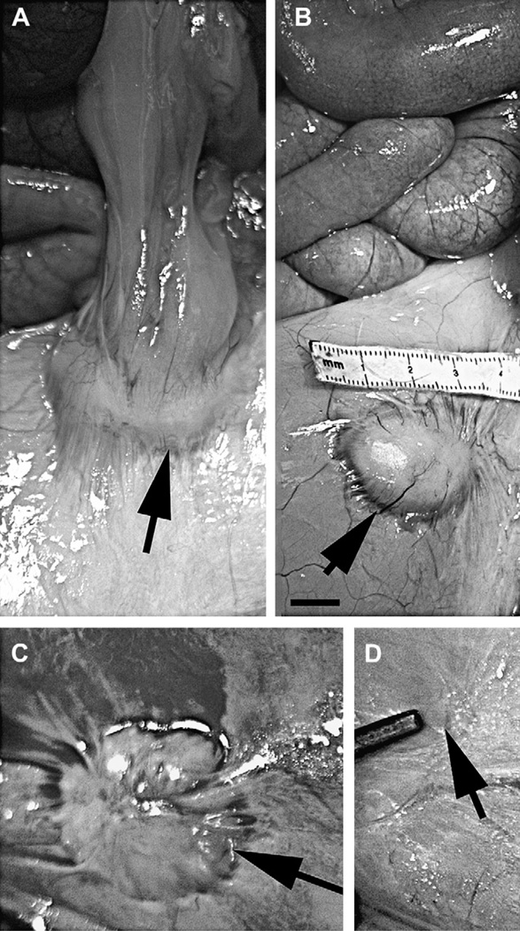
Fig. 1.
Residual bandage material remaining at the abdominal placement is encapsulated in a granuloma. Images of the viscera of four animals photographed at the end of the study at the time of necropsy are shown, depicting the size range of the granulomas (indicated by the arrows) localized in the serosa of the parietal peritoneum. The largest granuloma was approximately 4 cm while the smallest was almost undetectable. Contact between the peritoneum and the small intestine induced the formation of an adhesive bridge between the two sites. Scale bar = 1 cm.

